Article
4
Why I Don’t Like to Use My Eye: A Case of Anisometropia,
Anatomical Hypotropia, and Strabismic Amblyopia
Alicia Groce, OD, Nova Southeastern University College of Optometry, Class of 2014, Southern College of Optometry, Memphis, Tennessee
ABSTRACT
Background: Decreased vision, a common complaint in children, is often due to uncorrected refractive error. When glasses do not improve the vision completely, it is necessary to determine the cause.
Case Report: A four-year-old male presented for an evaluation of decreased vision especially with near work. Refraction revealed an anisometropic prescription of OD: -1.00-1.00x090, OS: +1.00 sph. After being corrected with glasses, the visual acuity was still reduced OD; stereopsis was also reduced. Follow up revealed a deep central suppression of the right eye, an anatomical vertical difference of 2-3mm between the left and right eyes, a 4∆ constant right hypotropia with left eye fixation preferred, and unsteady eccentric fixation of the right eye. The child was diagnosed with an anatomical right hypotropia, strabismic amblyopia, deep central suppression, and unsteady eccentric fixation OD. He was prescribed two hours of patching per day to improve the visual acuity and visual skills in the right eye.
Conclusion: This case shows the importance of patient follow up when there is decreased visual acuity and the role of evidence-based medicine in prescribing the most beneficial treatment.
Keywords: anisometropia, evidence-based medicine, hypotropia, strabismic amblyopia
Introduction
This case shows a step-by-step approach to treating a patient with reduced visual acuity that is not resolved by spectacle correction. It works through the differential diagnoses for reduced visual acuity and the importance of both subjective and objective testing in children to determine causation. This case demonstrates that with young children, several follow ups may be necessary to work through the differential diagnoses and to rule out pathology before making the diagnosis of amblyopia. The most recent research studies played an important role in developing the best course of treatment.
Case Report
A four-year-old male presented for an evaluation of decreased vision in the right eye
affecting both distance and near vision. His dad stated that the patient tired quickly with near work and covered the right eye when he complained of tiredness. His ocular and systemic histories were unremarkable except for seasonal allergies. His developmental history was unremarkable. He was of pre-school age and was in a mainstream program. He started reading early, and on the intake form, his dad checked off the following: Child tilts or turns their head when reading and assumes an awkward sitting position when reading.
monocular cues on the Lang stereopsis test. His cover test findings were ortho at distance and near, and his NPC was to the nose. His pupils, EOMs, confrontation fields, color vision, and slit lamp examination were all unremarkable. His IOP taken with the iCare tonometer was 12mmHg OD and 13mmHg OS. His DFE was unremarkable; the cup-to-disc ratio was 0.2 x 0.2 OD, OS. His refraction was done via dry and cycloplegic retinoscopy.
Dry Retinoscopy (fluctuating response) OD -3.00-1.00x180, 10/32- (20/64) OS -1.00 sph, 10/10 (20/20)
Cycloplegic Retinoscopy
OD -1.00-1.00x090
OS +1.00 sph
The dry retinoscopy was performed by a student, and the cycloplegic retinoscopy was performed by a staff doctor, which could explain why the dry retinoscopy axis for the right eye was the opposite of the cycloplegic. The findings were variable, so the patient was asked to return in two days to evaluate visual acuity, stereopsis, and keratometry before finalizing the prescription.
At the follow up, the autorefractor finding was similar to the dry retinoscopy: OD: -3.00-1.00x088, OS: +0.50 sph. The auto-keratometer findings were OD: 43.00/43.00, OS: 42.75/43.75. The spectacle prescription was based on the dry retinoscopy at this visit, which was the same as the cycloplegic retinoscopy at the previous visit (OD: -1.00-1.00x090, OS: +1.00sph). The patient was given that prescription and was instructed to wear the glasses full time. He was to return in three weeks to evaluate stereopsis and visual acuity.
At the three-week follow up, the aided visual acuity with Lea symbols was improved but still reduced in the right eye: OD: 20/50 and OS: 20/20 at near. His stereopsis was 200” with the Lang test, but he did not demonstrate any stereopsis with the Random Dot E. His cover test
was ortho at distance and near; a right head tilt was noted. Due to the low stereopsis, a Worth 4 dot was performed with the red lens over the right eye. The results showed three green dots at distance and intermediate in bright and dim illumination and four dots at near in bright and dim, indicating deep central suppression of the right eye. Due to the presence of suppression, reduced stereopsis, and reduced acuity OD, the patient was asked to return in 2-4 weeks for a micro/small angle constant strabismus workup and visual acuity check.
nasal eccentric fixation (EF), and the left eye had steady central fixation. The 2.5∆ of EF could be the reason why it was challenging to identify the deviation on unilateral cover test because it masked some of the deviation. The estimated acuity for 2.5∆ of EF is approximately 20/70,2 which is similar to the patient’s entering visual acuity. The father was educated on the diagnoses: anatomical right hypotropia, strabismic amblyopia, unsteady eccentric fixation OD, and central suppression OD. The father was advised to start patching two hours per day while the child was doing activities, log the hours, and return in one month.
Three weeks later, the patient’s father reported that patching occured two hours per day for a total of 45 hours of patching. The vision in the right eye was less blurry. His entering acuities and cover test findings remained the same. He was able to get 1200” of global stereopsis on Lang and 400” of local stereopsis on the stereo butterfly. Although in some of the earlier examinations limited global stereopsis was noted with Lang, it is unclear whether stereopsis was actually present, he was using monocular cues, or the correct answers were given away during test administration. Worth 4 dot was performed with the red lens over the right eye. The patient reported three green dots at seven feet in the light and four dots at all distances in the dark. Maddox rod was performed, and it was noted that with the patient’s head straight, the red line was seen above the white light (right hypo), but when the patient’s head was tilted, the red line was seen going through the light (ortho). Contrast sensitivity was administered to serve as a baseline against which to monitor changes at future visits. The contrast sensitivity function showed a visual acuity of OD: 20/100, OS: 20/20. The father was advised to keep patching two hours per day and to return to clinic in one month to monitor the stereopsis and visual acuity for improvement. Vision therapy was discussed as an option to train the visual skills
in the eyes and to promote binocularity once vision had improved.
The father called four months later and said that he thought that the patching was helping, but the patient was resistant. He asked if atropine could be substituted for the patch. A prescription for atropine ophthalmic eye drops 1%, to be instilled in the left eye on Saturday and Sunday each week, was issued. The father said he would bring the patient in for a follow up the following month to check visual acuity, stereopsis, and contrast sensitivity.
The patient was lost to follow up for four months, and when he returned to clinic, he indicated that his vision was similar in the two eyes. The father noted that the patient had been wearing his glasses full time, and he last instilled atropine seven days prior. The distance aided acuities were OD: 20/40+, OS: 20/20-, OU: 20/25+. The near aided acuities were OD: 20/30, OS: 20/20, OU: 20/20. The patient was able to appreciate all of the 800” global stereopsis targets and half of the 400” targets on the Preschool Stereopsis test. The patient had a right head tilt 95% of the time in the office. Visuoscopy revealed a slight unsteady superior EF in the right eye and steady central fixation in the left eye. The Amsler grid was normal. The refractive findings were as follows:
Dry Retinoscopy
OD -1.75-2.00x090 OS +1.75-0.50x180 Manifest Refraction 1: Dry
OD -1.00-1.50x090,
20/40-OS +1.00-0.50x090, 20/25+
Wet Retinoscopy
OD -1.00-1.00x090 OS +2.00-1.00x180
visual acuity, visual skills, and binocularity now that the vision had improved slightly. The patient’s parents decided to wait and think about it but wanted to continue the atropine. The father was told to continue instilling atropine in the left eye on the weekends and was asked to bring the patient back for a follow up in four to six weeks to check visual acuity, contrast sensitivity, stereopsis, 4 BO test, and correspondence using the Bagolini lenses. The patient never arrived for his follow up.
The father was contacted again six months later and was asked to bring the patient back for a follow up. The father reported that the patient’s vision seemed to be improving. He had been using the atropine every Saturday and Sunday as instructed until one month ago, when he discontinued use because the patient
was playing baseball on the weekends. The father asked if there was anything else that he could do at home. He was advised that he could do red/green activities (monocular in a binocular field). He was also educated that the patient could do in-office vision therapy as previously discussed.
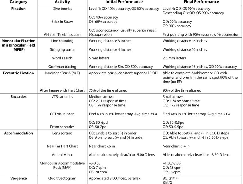
Several months later, the father contacted the clinic to inquire about starting vision therapy. The patient came in for a comprehensive examination to check his status before starting therapy. His entering distance and near acuities with Snellen had improved to OD: 20/30, OS: 20/20, OU: 20/20 at distance and OD: 20/25, OS: 20/20, OU: 20/20 at near. He had 400” of global stereopsis and 160” of local on the Preschool Stereo Test. He was no longer showing suppression with Worth 4 dot. His contrast Table 1: Vision Therapy Sessions 1-13
Category Activity Initial Performance Final Performance
Fixation Dive bombs
Stick in Straw
AN star (Telebinocular)
Level 1: OD 40% accuracy, OS 60% accuracy
OD: 40% accuracy OS: 60% accuracy
OD: poor accuracy (usually superior nasal), (-)suppression
Level 4: OD, OS 90% accuracy Descending O’s: OD, OS 90% accuracy
OD: 90% accuracy OS: 90% accuracy
Fast pointing with 90% accuracy, (-)suppression Monocular Fixation
in a Binocular Field (MFBF)
Line counting
Stringing pasta
Word search
Groffman tracing
Working distance 3 inches
Working distance 4 inches
5 mm letters
Working distance 5in, OD 50% accuracy
Working distance 16 inches
Working distance 16 inches
2.5 mm letters
Working distance 16 inches, OD 90% accuracy Eccentric Fixation Haidinger Brush (MIT)
After Image with Hart Chart
Appreciate brush, constant superior EF OD
75% of the time aligned
Able to complete Amblyomaze OD with pointer and brush in the same spot 90% of the time (no EF)
90% of the time aligned Saccades VTS saccades
CPT visual scan
Prism saccades
Medium arrows OD: 2.01 response time OS: 1.92 response time
Find 4 #’s in 150 letter array, Avg. time 3:04
OD: 50-6pd OS: 50-2pd
Small arrows
OD: 1.74 response time OS: 1.72 response time
Find 4#’s in 150 letter array, Avg. time 2.04
OD: 50-0.5pd OS: 50-0.5pd Accommodation Lens sorting
Near Far Hart Chart
Mental Minus
Monocular Accommodative Rock (MAR)
OD: Unable to sort (-) in order OS: Able to sort (+) and (-) in order
Near chart 7.5 in
Able to alternately clear/blur -5.00 D lens
+/-0.50 OD: 7 cpm OS: 20 cpm
OD: Able to sort (+) and (-) in 0.50 D steps OS: Able to sort (+) and (-) in 0.50 D steps
Near chart 3-4 in
Able to alternately clear/blur -3.50 D lens
+1.50/-3.00 OD: 13 cpm OS: 13 cpm Vergence Quoit Vectogram Appreciated SILO, float, parallax BO: 21/14
sensitivity had improved in the right eye and was only slightly reduced compared to the left eye. A right hypotropia was still noted but was intermittent, and he still showed about 2.5∆ of superior EF. His refraction remained stable, and his ocular health was normal. His dad was educated on the importance of in-office therapy and completing home activities to improve visual skills, improve visual acuity by reducing/ eliminating EF, and work on binocularity.
He started vision therapy once a week with home support. The details of each therapy session are outlined below in Table 1. After the ninth therapy session, the patient was fitted with contact lenses to help reduce the aniseikonia and improve binocularity. At the contact lens fitting, his prescription had changed to OD: -1.50-1.00x090, 20/25; OS: +1.00 sph, 20/20. The patient was given an updated glasses prescription and was fit in daily wear contact lenses: OD Dailies Aqua Comfort Plus -1.75-0.75x090, OS Focus Dailies +1.00 sph.
The patient was assessed after the thirteenth therapy session because he was moving out of town. At his assessment, his vision with contact lenses was 20/20- with isolated letters in the right eye and 20/20 with a full line of letters in the left eye. Visuoscopy revealed unsteady central fixation in the right eye. The patient was given activities to do at home as maintenance therapy (Near Far Hart Chart, Fast pointing, Mental Minus, MAR, BAR), as they could not find a doctor near them who offered vision therapy.
Discussion
When the patient first presented, the main concern was the uncorrected refractive error and anisometropic prescription. The patient was young and had a hard time responding to visual acuity and stereo tests. He was given a spectacle prescription based on the cycloplegic retinoscopy and was told to wear the glasses full time until the next visit to see whether visual acuity and stereopsis would
improve. When the child returned months later, the vision was still reduced, so further testing was performed to assess binocularity. His stereopsis was limited, and the Worth 4 Dot revealed deep central suppression of the right eye. At this point, a micro/small-angle constant strabismus was suspected to be the cause of the decreased acuity and suppression, and he was asked to come back for a workup.
concluded that the patient had an anatomical constant right hypotropia, strabismic ambly-opia OD, unsteady central fixation OD, and deep central suppression OD.
The gold standard for treating amblyopia is patching in conjunction with office-based vision therapy with home support. Patching allows the brain to recognize the signals being sent from the amblyopic eye, thus improving visual acuity and visual skills in that eye.4 Vision therapy is suggested in conjunction with patching to improve the visual skills and to equalize the two eyes, as well as to work on anti-suppression and binocularity.5 In this case, patching alone was prescribed initially to improve the visual acuity to a level sufficient for vision therapy to begin to work on binocularity.5 For moderate amblyopia (20/40-20/100), the Pediatric Eye Disease Investigator Group (PEDIG) found that 2 hours of patching was as effective as 6 hours of patching for improving visual acuity.4PEDIG also compared correcting patients with spectacles alone versus 2 hours of patching and found that patching improves visual acuity faster.6 In this case, the patient had already been corrected with spectacles for several months with little improvement. Based on the PEDIG studies, the patient was prescribed 2 hours of patching per day while doing near work such as reading or coloring to increase both the visual acuity and visual skills in the amblyopic eye. Studies have also found that the monocular spatial skills in the amblyopic eye can contribute to poor oculomotor skills due to bending of vertical lines and compression and expansion of horizontal lines.2 For this reason, vision therapy is an excellent addition to patching to provide patients with the best possible outcome after treatment.
At the follow up visit, the patient had been patching for three weeks. His visual acuity was unchanged, but he was able to get local stereopsis with the stereo butterfly and global stereopsis with Lang for the first time reliably.
His Worth 4 dot showed that he was no longer suppressing in the dark, so he went from deep to shallow central suppression. When evaluated with Maddox rod, he was aligned when his head was tilted but showed a hypodeviation when his head was straight, which explained his habitual posture. Contrast sensitivity testing showed a visual acuity of 20/100 in the right eye and 20/20 in the left eye. Contrast sensitivity testing was conducted based on study findings that contrast sensitivity is more sensitive in detecting small improvements in amblyopia than Snellen acuity.2,7
After patching for four months, the patient’s father requested that the patient be put on atropine instead of patching. The PEDIG group found that atropine is equally as effective as patching for moderate amblyopia, so atropine 1% was prescribed on Saturdays and Sundays in place of the patch.8,9 Ideally, he would be monitored for improvements in visual acuity, stereopsis, and contrast sensitivity every month and would start vision therapy to improve oculomotor skills, accommodative skills, and binocularity.5
The patient’s father stopped atropine on his own and said that he thought that the vision was improving, which was confirmed with an entering acuity of 20/40 in the right eye. Even though the vision was improving, the father was educated that there was still room for improvement and that the patient would benefit from an in-office therapy program. In the meantime, the father was told that he could work with the patient using red/green activities (monocular in a binocular field) to help strengthen the skills in the right eye.
superior EF, which was limiting the visual acuity, and his visual skills were not equal in the two eyes. The father was educated that the patient would greatly benefit from in-office vision therapy with home support. He was educated that in therapy the visual skills and EF would be addressed and that improving binocularity would be a goal. He was also given the option of enrolling in the ATS 18 study, which is looking at binocular computer activities for the treatment of amblyopia versus patching.10 This is a novel approach to vision therapy and is based on research that the non-amblyopic eye cortically inhibits signals from the amblyopic eye.11 A pilot study was conducted by Knox to evaluate dichoptic training as a means for binocular therapy to treat amblyopia. The study showed promising results for improvement in visual acuity and stereopsis in amblyopes.12
When the patient started vision therapy, the visual acuity in his amblyopic eye was 20/30, and he had eccentric fixation in the right eye. His father’s goal was to improve his visual acuity and binocular skills in order to help with schoolwork and sports. The therapy plan was to improve the visual skills in the right eye, to eliminate suppression, and then to work on the eccentric fixation. During the first several sessions of therapy, the patient worked on fixation skills and monocular in a binocular field activities (MFBF). Once his fixation had normalized and he was no longer suppressing, he started fast pointing activities and eccentric fixation activities using the Haidinger Brush (MIT). When the patient was fit with contact lenses, he improved rapidly. His vision improved to 20/25 in the right eye with the contact lenses, and within five sessions of therapy to work on eccentric fixation, he had improved to 20/20- and had obtained unsteady central fixation. The patient moved after his thirteenth session, but he was very excited about his progress and the improvement in his vision. He even
noted that he was doing better in baseball and was hitting the ball more often when he was up to bat.
Conclusion
This case shows the importance of following up on patients with reduced visual acuity. When children first present with reduced visual acuity, it is important to get an accurate refraction and to correct the refractive error, as uncorrected refractive error can cause impaired binocular findings. If a patient still shows reduced acuity and impaired stereopsis and binocular vision after being corrected, further investigation is required. It is important to rule out all other causes for reduced acuity before diagnosing amblyopia. Once all testing confirms the diagnosis, finding the appropriate treatment is very important. The PEDIG studies are a useful guide for determining the starting treatment for amblyopia. Vision therapy for amblyopes helps improve the visual skills in the amblyopic eye, promote binocularity, and improve binocular skills.
References
1. Caloroso E, Rouse M. Clinical Management of Strabismus. Boston, MA: Butterworth-Heinemann, 2007:22-3, 278-9. http://bit.ly/1ES0nVg
2. Bedell HD, Flom MC. Monocular spatial distortion in strabismic amblyopia. Invest. Ophthalmol Vis Sci 1981;20:263-8. http://1. usa.gov/1M1m5pj
3. Hess RF. On the relationship between strabismic amblyopia and eccentric fixation. Br J Ophthalmol 1977;61:767-73. http://bit.ly/1NlFkLU
4. Pediatric Eye Disease Investigator Group. A randomized trial of patching regimens for treatment of moderate amblyopia in children. Am J Ophthalmol 2003;121:603-11. http://bit. ly/1Jz7ErO
5. Lyon DW, Hopkins K, Chu RH, et al. Feasibility of a clinical trial of vision therapy for treatment of amblyopia. Optom Vis Sci 2013;90:475-81. http://1.usa.gov/1M1m9p1
7. Jindra LF, Zemon V. Contrast sensitivity testing: A more complete assessment of vision. J Cataract Refract Surg 1989;15(2):141-8. http://bit.ly/1QsD8lK
8. Pediatric Eye Disease Investigator Group. A randomized trial for atropine vs patching for treatment of moderate amblyopia in children. Arch Ophthalmol 2002;120:268-78. http://bit. ly/1i3bO1n
9. Pediatric Eye Disease Investigator Group. Atropine vs patching for treatment of moderate amblyopia: Follow-up at 15 years of age of a randomized clinical trial. JAMA Opthalmol 2014;132:799-805. http://bit.ly/1O5lwhv
10. Pediatric Eye Disease Investigator Group. Study of Binocular Computer Activities for Treatment of Amblyopia. http://pedig. jaeb.org/Studies.aspx?RecID=235. Last Accessed Nov 17, 2014. http://bit.ly/1iiE5ly
11. Hess RF. A binocular approach to treating amblyopia: Anti-suppression therapy. Optom Vis Sci 2010;87:697-704. http://bit.ly/1i3bYpN
12. Knox PJ. An exploratory study: prolonged periods of binocular stimulation can provide an effective treatment for childhood amblyopia. Invest Ophthalmol Vis Sci 2012;53(2):817-24. http://bit.ly/1Oi9gcs
Correspondence regarding this article should be emailed to Alicia Groce, OD at [email protected]. All statements are the author’s personal opinions and may not reflect the opinions of the the representative organizations, ACBO or OEPF, Optometry & Visual Performance, or any institution or organization with which the author may be affiliated. Permission to use reprints of this article must be obtained from the editor. Copyright 2015 Optometric Extension Program Foundation. Online access is available at www. acbo.org.au, www.oepf.org, and www.ovpjournal.org.