4 7 9 4 7 9 4 7 9 4 7 9 4 7 9
Instituto Dante Pazzanese de Cardiologia
Mailing address: Carlos A. C. Pedra - Instituto Dante Pazzanese de Cardiologia – Av. Dr. Dante Pazzanese, 500 - 04012-180 - São Paulo, SP, Brazil - Email: [email protected]
English version by Stela Maris C. e Gandour
Carlos A. C. Pedra, Luciano N. L. de Sousa, Simone R. F. F. Pedra, Waldinai P. Ferreira, Sérgio L. N. Braga, César A. Esteves, Maria Virgínia T. Santana, J. Eduardo Sousa, Valmir F. Fontes
São Paulo, SP - Brazil
New Percutaneous Techniques for Perforating the Pulmonary
Valve in Pulmonary Atresia with Intact Ventricular Septum
We report new percutaneous techniques for perfora-ting the pulmonary valve in pulmonary atresia with intact ventricular septum, in 3 newborns who had this birth defect. There was mild to moderate hypoplastic right ventricle, a patent infundibulum, and no coronary-cavitary communications. We succeeded in all cases, and no complications related to the procedure occurred. The new coaxial radiofrequency system was easy to handle, which simplified the procedure. Two patients required an additional source of pulmonary flow (Blalock-Taussig shunt) in the first week after catheterization. All patients had a satisfactory short-term clinical evolution and will undergo recatheterization within 1 year to define the next therapeutic strategy. We conclude that this technique may be safely and efficiently performed, especially when the new coaxial radiofrequency system is used, and it may become the initial treatment of choice in select neonates with pulmonary atresia and intact ventricular septum.
Even though pulmonary atresia with intact ventricular septum is an infrequent defect, accounting for less than 1% of congenital heart defects, it is the 3rd most frequent cyanotic heart
defect in the neonatal period 1. Its morphological spectrum is
broad with cases ranging from extremely hypoplastic tricuspid valves and right ventricles to ventricular cavities of almost nor-mal dimensions 2-4. Ebstein’s anomaly of the tricuspid valve and
extreme ventricular dilation have also been reported, and they are associated with a poor prognosis 5. In addition, communication
between the right ventricular cavity and the coronary arteries is a relatively common finding, which is sometimes associated with coronary circulation partially or totally dependent on the right
ventricle 6-8. Due to this anatomic heterogeneity, the therapeutic
algorithm should be individualized 6. The final objective is
always to attain biventricular correction with total separation between systemic and pulmonary circulations 6. However, this is
sometimes impossible, and correction with a 1½ ventricle or of the univentricular type (Fontan) is necessary 6-9. Cardiac
transplantation should also be considered for treating cases with severe stenoses or multiple interruptions in the coronary arteries and secondary left ventricular dysfunction 6,8. The initial
therapeutic approach in the neonatal period should, whenever possible (if the coronary circulation pattern allows), open the pulmonary valve to decompress the right ventricle and stimulate its growth 3,6-8. During the last decade, perforation of the
pul-monary valve to establish continuity between the right ventricle and the pulmonary artery with the aid of interventional catheteri-zation became a reality 10-15, even in Brazil 16. We report 2
per-cutaneous techniques of valve perforation, which were recently introduced into clinical practice, and their advantages and disadvantages are discussed.
Case report
We report the cases of 3 patients referred to our servi-ce from other neonatal units for investigation or treatment of cyanotic congenital heart defects. The clinical, echocar-diographic, and hemodynamic data, are listed in tables I, II, and III. It is worth noting that patient 2 had a previous diag-nosis of critical pulmonary stediag-nosis. All patients had mild to moderate cyanosis under continuous infusion of prosta-glandin; on auscultation, the 2nd cardiac sound was single
and low, and was followed by a mild systolic murmur in the dorsum. On chest X-ray, the cardiac silhouette was slightly enlarged, mainly because of the right atrium, and the pulmo-nary flow was reduced. The electrocardiogram showed sinus rhythm and left ventricular hypertrophy in all patients. QRS axes ranged from 90º to 120º. In regard to the echocardiographic findings, all patients had situs solitus, pulmonary atresia with imperforate pulmonary valve, and intact ventricular septum. The right ventricle was
4 8 0 4 8 0 4 8 0 4 8 0 4 8 0
poplastic with a significant reduction in the trabecular zone. The tricuspid and pulmonary valves also had varied degrees of hypoplasia (tab. II). No patient had communica-tion between the right ventricle and the coronary arteries. The pulmonary branches were of a good caliber, and pulmo-nary circulation was maintained through the arterial canal to the left. The patients were then referred to the catheteriza-tion laboratory for diagnostic testing and possible perfora-tion of the pulmonary valve. After a detailed explanaperfora-tion of the possible risks and benefits of the percutaneous proce-dure, the parents provided written consent. The hemody-namic study showed right ventricular systemic pressure le-vels equal to or above the systemic lele-vels in all patients (tab. III). Right ventriculography confirmed the echocardiogra-phic findings and absence of communication between the ri-ght ventricle and the coronary arteries (figs. 1 and 2). The in-fundibulum was slightly hypoplastic in patients 1 and 3 and almost normal in patient 2. The technique and results of per-cutaneous valve perforation are described for each patient.
Case 1 - A 5 Fr (Cook) right coronary Judkins catheter
was carefully positioned right below the valvar plane and monitored through fluoroscopy in the cranial posteroan-terior and lateral views (fig. 1 A and B). The hard tip of a 0.014” steerable angioplastic guidewire was manually molded accompanying the curvature of the right ventricular outflow tract, which was defined by angiography in the left
Table I – Clinical data of the patients
Patient Age Sex Weight Saturation
1 3 days Female 3.2kg 75%
2 5 days Female 3.0kg 85%
3 15 days Male 2.8kg 75%
Table II – Echocardiographic data
Patient TV (Z value) TI PuV RV Hypo
1 8.5mm (-1.7) Mild/Mod 7.0mm Moderate 2 9.0mm (-1,3) Mild/Mod 6.8mm Mild/Mod 3 7.0mm (-2,1) Mild/Mod 7.0mm Moderate
TV- diameter of the tricuspid valve; TI- degree of tricuspid insufficiency; PuV- diameter of the pulmonary valve; RV Hypo – degree of right ventricular hypoplasia; Mod- moderate
Table III - Data of the catheterization prior to valve perforation
Patient RV LV RV Hypo Infund. RV-Co (mmHg) (mmHg) Hypo Communic
1 60/6 41/6 Mild/Mod Mild No
2 50/6 50/6 Mild/Mod Normal No
3 60/7 50/6 Mod Mild No
RV – right ventricle; LV – left ventricle; RV Hypo – degree of right ventricular hypoplasia; Infund Hypo – degree of infundibular hypo-plasia; RV-Co Communic - communications between the right ventricle and the coronary arteries; Mod: moderate.
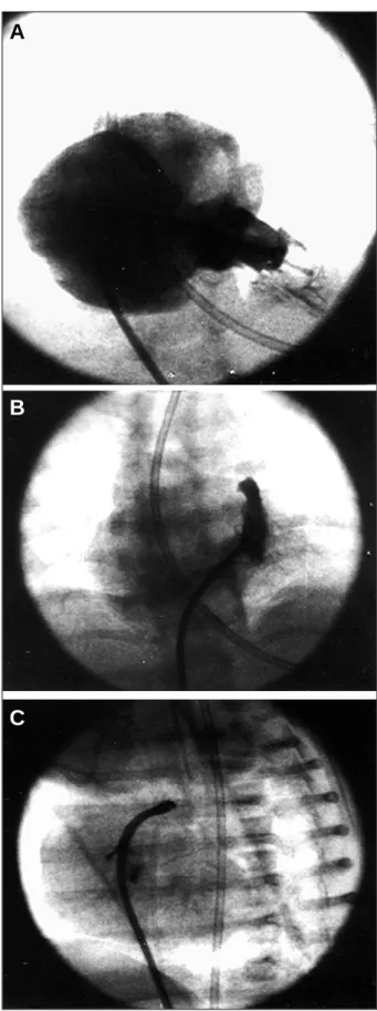
Fig. 1 - Case 1 - A) Right ventriculography in right cranial anterior oblique view. Note the right ventricle with mild to moderate hypoplasia and significant tricuspid in-sufficiency with mild opacification of the infundibular portion, without communica-tion between the coronary arteries and the right ventricle; B) selective manual in-jection into the infundibulum with mild hypoplasia showing unquestionable pulmo-nary atresia; C) infundibulum in left profile view. A minimum anterograde jet exists through the pulmonary valve after perforation with the hard tip of the steerable corona-ry guidewire. Note how the guidewire and the catheter follow the curvature of the in-fundibulum, leading to adequate perforation at an angle perpendicular to the valve.
A
B
4 8 1 4 8 1 4 8 1 4 8 1 4 8 1 profile view. This guidewire was then advanced through the
Judkins catheter and positioned right below the valvar pla-ne with its tip perpendicularly directed at the pulmonary leaflets. The appropriate positioning was confirmed by ma-nual injections of contrast medium through a Y system connected to the Judkins catheter, commonly used for coronary angioplasty. The pulmonary valve was then mechanically perforated, and the hard tip of the steerable guidewire was advanced. With test injections, perforation and appropriate positioning of the guidewire in the pul-monary trunk were confirmed, and no signs of perforation or contrast medium extravasation, or a combination of the two, were found (fig. 1 C). The guidewire was withdrawn, and the flexible and directional tip of another steerable guidewire (0.014”) was advanced through the same catheter, passing beyond the pulmonary valve through the orifice created by the hard tip of the previous guidewire. This guidewire was maneuvered through the arterial duat and positioned in the distal part of the descending aorta at the level of the
bifurcation of the iliac artery. A 3.0X20mm balloon catheter for coronary angioplasty (World-pass, Cordis) was advan-ced over the guidewire, positioned at the level of the valvar plane, and inflated under pressure monitoring with formati-on and disappearance of the sand-glass image (fig. 1 D). This catheter was then replaced by a 9X20 mm Tyshak II low-profile balloon catheter (Numed, Corwall, Canada) for completion of the pulmonary valvoplasty. The hemodyna-mic control revealed an infundibular gradient of 30 mm Hg and the absence of a gradient through the pulmonary valve (tab. IV). Control ventriculography showed reestablishment of right ventricle-pulmonary trunk continuity (fig. 1 E), with a dynamic infundibular reaction. Within the first 4 days after catheterization, we interrupted the infusion of prostaglandin but had to reinstate it due to systemic desatu-ration and hemodynamic lability. The patient was then referred for a right modified Blalock-Taussig shunt. The postoperative period was uneventful, with oxygen satura-tion maintained at around 80%-90% (patient extubated), ab-sence of metabolic acidosis, and normal levels of lactate. The patient was discharged after 10 days with an echocar-diogram showing anterograde flow through the pulmonary valve and a maximum systolic gradient of around 50 mm Hg, secondary to infundibular reaction. Moderate to severe pulmonary insufficiency and satisfactory flow through the Blalock-Taussig shunt existed. After a 7-month follow-up, the patient was clinically well, gaining weight, and his satu-ration was around 90%. The echocardiogram showed infun-dibular hypertrophy regression, a maximum systolic gradi-ent of 15mmHg through the right vgradi-entricular outflow tract, severe pulmonary insufficiency, and a normally functioning Blalock-Taussig shunt. The right ventricle and the tricuspid valve developed (Z value = -1). The patient now awaits reca-theterization to test occlusion of the shunt and of the oval foramen with new pressure measurements and calculation of cardiac output.
Case 2 – In this patient, we used a new coaxial
radiofre-quency system (Bayliss Medical Company, Mississauga, ON, Canada), designed by Nykanen, for perforation of the pulmonary valve (figs. 2A and B). As in the previous case, a 5 Fr. (Cook) right coronary Judkins catheter was positio-ned right below the valvar plane. The Nykanen radiofre-quency catheter (external diameter 0.024”, length 265cm) was advanced through the Judkins catheter and positioned below the pulmonary valve touching the leaflets. This ca-theter was connected to a generator source of radiofre-quency (BMC) and was programmed to apply 5W of
Fig. 1 - Case 1 - D) note the inflated coronary angioplastic balloon at the valvar level. The guidewire was left in the descending aorta through the arterial canal; E) right ventriculography in right cranial anterior oblique view after completion of the pul-monary valvoplasty. Note the anterograde flow through the valve with a reduction in the degree of tricuspid insufficiency. Dynamic infundibular reaction occurs.
D
E
Table IV – Hemodynamic data (in mm Hg) after pulmonary valve perforation
Patient PuA Infund RV
1 20-8 20-8 50-8
2 35-15 35-8 40-8
3 26-12 32-8 62-8
4 8 2 4 8 2 4 8 2 4 8 2 4 8 2
energy for 2s at most. The valve was perforated at the first attempt, with progression of the catheter to the pulmonary trunk with no need to advance it manually (fig. 2 C). Over the Nykanen radiofrequency catheter, a coaxial injectable
guide catheter [inner diameter 0.024”, outer diameter 0.035”, and length 145 cm (BMC)] was advanced to the pulmonary trunk, with confirmation of its position by manual injections of the contrast medium through the it (fig. 2 D). The radiofre-quency catheter was then replaced by a 0.014” steerable coronary guidewire that was advanced through the arterial duat and positioned in the distal part of the descending aorta. The coaxial injectable catheter was then withdrawn, and a 3.0X20 mm angioplastic balloon catheter (World-Pass, Cordis) followed by a 9X20mm Tyshak II low-profile balloon catheter (Numed) completed the pulmonary valvo-plasty (fig. 2 E). The hemodynamic control revealed an in-fundibular gradient of 5 mm Hg and the absence of a gra-dient through the pulmonary valve (tab. IV). Control ventri-culography showed the establishment of right ventricle-pul-monary trunk continuity (figs. 2 F and G), with a minimum dynamic infundibular reaction. The postcatheterization evolution was uneventful. Prostaglandin was suspended
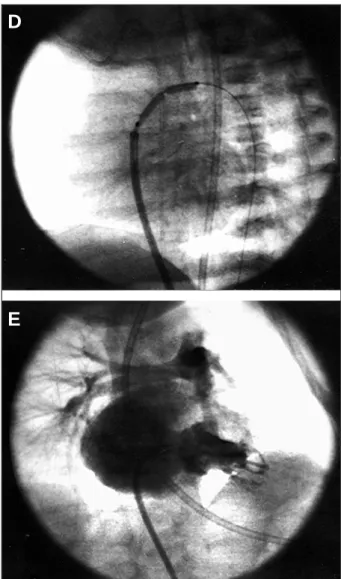
Fig. 2 - Case 2 - A and B) right ventriculography in cranial posteroanterior and left profile views: Note mild to moderate right ventricular hypoplasia with attenuation of the trabecular zone and well-formed infundibulum. Significant tricuspid insufficiency exists; B and C) in left profile view, note the radiofrequency catheter located right below the valvar plane, with immediate progression to the pulmonary trunk after application of radiofrequency energy.
A
B
C
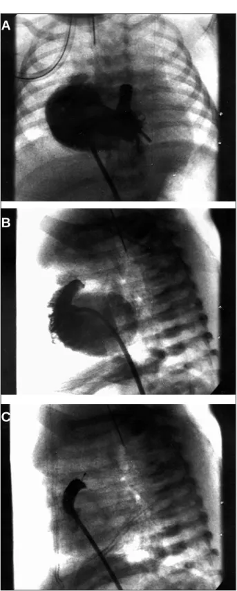
Fig. 2 - Case 2 - D) pulmonary trunk in posteroanterior view. The injectable guide ca-theter was advanced towards the pulmonary trunk over the Nykanen radiofrequency catheter, and its position was confirmed through a small injection of contrast medi-um, with mild opacification of the pulmonary arteries; E) after exchanging the radio-frequency catheter for a coronary guidewire and its progression to the descending aorta through the arterial canal, pulmonary valvoplasty was performed in a progres-sive way. Note the 9x20 mm Tyshak balloon being inflated at the level of the pulmo-nary valve for completion of the dilation.
D
4 8 3 4 8 3 4 8 3 4 8 3 4 8 3 after 4 days, saturation was maintained at around 80%
(patient extubated), no metabolic acidosis was observed, and lactate levels were normal. The patient was discharged after 8 days. His echocardiogram showed a nonobstructive anterograde flow through the pulmonary valve and a ma-ximum systolic gradient of around 15mmHg secondary to a mild infundibular reaction. Moderate to severe pulmonary insufficiency and minimum flow through the arterial duct existed. After a 3-month follow-up, the patient is clinically well, is gaining weight, and saturation is around 85%. The patient awaits a new echocardiogram and catheterization to define further management.
Case 3 – In this case, the procedure was performed in
the same way as in the previous patient. However, 2 appli-cations of 5W of energy during 2s were required for valvar perforation. A 2.5X20mm angioplasty balloon (World Pass, Cordis) was used, being followed by a 9X30mm Tyshak II low-profile balloon catheter (Numed) for completion of the pulmonary valvoplasty. Hemodynamic control revealed an infundibular gradient of 30mmHg and a valvar gradient of 6mmHg (tab. I). Control ventriculography showed the
es-tablishment of right ventricle-pulmonary trunk continuity, with an intense dynamic infundibular reaction, which led to the administration of 1mg/kg/day of propranolol. Five days after the procedure, we suspended prostaglandin, but had to reinstate it. A right Blalock-Taussig shunt was indicated. The patient HAO sepsis caused by Staphylococcus aureus, which responded well to antibiotics, and convulsive crises with the suspicion of paradoxical embolism secondary to deep venous thrombosis due to the presence of a central ve-nous catheter in the femoral vein. The postoperative echocar-diogram on the 30th day after catheterization revealed
regres-sion of the subpulmonary stenosis, a residual gradient of 9mmHg through the right ventricular outflow tract, and severe pulmonary insufficiency. The right ventricle developed, and the tricuspid ring diameter increased to 11mm (Z value =0). Despite the satisfactory flow through the surgical anastomo-sis, mild stenosis was detected in the right pulmonary artery at its insertion. Saturation in the postoperative period ranged from 80% to 90%.
Discussion
Opening of the right ventricular outflow tract through interventional catheterization is gaining acceptance as the initial therapeutic modality for patients with pulmonary atresia and intact ventricular septum 10-17. In patients who
have tripartite right ventricle 4, with a patent infundibulum 18,
moderate ventricular hypoplasia at most, and coronary cir-culation not depending on the right ventricle 6-8, this
techni-que promotes efficient decompression of the ventricular cavity, stimulating its growth 14,19. Therefore, some potential
surgical complications, which may occur in this type of di-sease, may be avoided. Right heart failure with systolic-dias-tolic dysfunction is not infrequent after opening of the right ventricular outflow tract with a transannular patch + ventri-culotomy, and it may hinder the anterograde flow to the lungs, worsening the right-to-left shunt through the oval foramen or the atrial septal defect 14,20,21. In addition,
reperfu-sion lereperfu-sions usually observed after the use of extracor-poreal circulation may be extremely noxious in these pati-ents with potential or real histological anomalies in the coro-nary arteries 1,14.
The percutaneous techniques already described in the literature for establishing the right ventricle-pulmonary trunk continuity include the use of laser or radiofrequency energy and mechanical perforation 10-17. Laser energy,
despi-te being the first to be used, especially by British groups 11,12,
was gradually abandoned due to its high cost, risks to the staff of the hemodynamics laboratory, and the difficulty of transporting it 17.
Mechanical perforation with the hard tip of a coronary guidewire was reported for the first time by Latson in 1991 10,
and since then occasional reports have been published 14.
Even though the low cost is an obvious advantage, this te-chnique has several potential problems. The catheter posi-tioned in the infundibulum right below the pulmonary valve tends to have its position modified due to rigidity of the
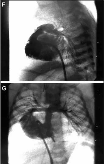
Fig. 2 - Case 2 -F and G) right ventriculography in posteroanterior and left profile views after the procedure: Note establishment of the right ventricle-pulmonary artery continuity with minimum sub-pulmonary reaction and reduction in tricuspid insufficiency.
F
4 8 4 4 8 4 4 8 4 4 8 4 4 8 4
hard tip of the guidewire when the latter is advanced 14. In
ad-dition, manual modeling of the hard tip may not be precise, leading to errors at the site of the transvalvular puncture 14.
In our first case, the pulmonary leaflets were very thin and offered no resistance to the progression of the hard tip of the guidewire. Thicker fibrotic valves may require greater strength for perforation, increasing the possibility of acci-dents and complications. In the event of accidental perfo-ration of the muscular portion of the right ventricular out-flow tract, or even of the pulmonary trunk, the occurrence of hemopericardium is a possibility. However, due to the reduced inner diameter of the guidewire (0.014”), sig-nificant bleeding is unlikely 14. Another limitation of this
technique is the need to withdraw the hard tip of the gui-dewire after the initial perforation, a new passage through the diminished orifice created being mandatory. This ma-neuver requires patience of the operator and is not always effective.
The use of radiofrequency energy for perforation of atretic pulmonary valves became a more popular method among pediatric interventionists because of its availability and more accessible costs in large centers 13-16. Even
thou-gh the most commonly used system is the Osypka 14, the
Nykanen system reported about in this article offers several advantages. First, it is a radiofrequency system specifically designed to perforate biological tissues, and it releases a hi-gh amount of energy in an extremely localized and specific location, therefore, against great impedance. Perforation is not caused by an increase in local temperature, but secon-dary to an alteration in intracellular electrical charges, resul-ting in a very well localized cellular necrosis 22,23. The
Osyp-ka system was not originally designed for this purpose. Be-cause it is used for ablation of arrhythmias, the system functions with low impedance, causing tissue lesions due to the generation of local heat. Consequently, the lesion is more superficial and extensive, which may be an advantage in cases of ablation, but not when the major objective is to obtain a localized perforation. Even though our team has al-ready successfully used the Osypka system for pulmonary valve perforation in case 1 16, in another case (unpublished
data), we could not pass beyond the valve with the guidewi-re after the initial perforation. Because the Nykanen system is coaxial, such a limitation does not exist. After an initial perforation, the radiofrequency catheter serves as a sup-port for the advancement of the coaxial injectable guide theter to the pulmonary trunk. Then the radiofrequency ca-theter itself may be maneuvered to pass beyond the arterial ducts and reach the descending aorta, or it may be replaced by a steerable coronary guidewire, as performed in both ca-ses. This provides more control, safety, and rapidity to the procedure, avoiding the need to pass beyond the pulmona-ry valve again after the initial perforation, which is a funda-mental advantage in the management of a cyanotic and cri-tically ill newborn infant. In addition, the system is easy to use and handle for the professional used to interventions in pediatric cardiology. The cost of the catheters and system may be a limiting factor in our environment.
After percutaneous valvar opening, the next step in the therapeutic algorithm of this defect is the discontinua-tion of prostaglandin, which should be tried within the first week after catheterization 14. Systemic saturation above
75%-80% and the absence of metabolic acidosis with nor-mal levels of lactates usually indicate satisfactory antero-grade pulmonary flow, which depends directly on improve-ment in right ventricular compliance. This phenomenon takes variable amounts of time to establish, and reliable pre-dictors that this moment occurs or actually will occur do not exist 19. Theoretically, significant hypoplastic ventricles are
less compliant and do not manage to maintain an effective anterograde pulmonary flow. This sometimes is not confir-med in clinical practice, and to our surprise, suspension of prostaglandins is possible even under unfavorable anato-mic conditions. This fact suggests that other mechanisms, in addition to the degree of ventricular hypoplasia, are impli-cated in diastolic dysfunction, including subclinical ische-mia and myofibrillar disarray intrinsic to the underlying defect 1-3. In addition, defining the degree of right
ventricu-lar hypoplasia is difficult in clinical practice. To this end, so-me authors use the Z value of the tricuspid valve 6, while
others use a mere subjective estimate 17. If no success is
ob-tained in the initial suspension of prostaglandin, theoreti-cally the prolonged administration of this drug, including its oral administration, is an alternative, but not feasible in our environment. Others suggest stent implantation in the arte-rial canal in the initial valve perforation procedure 15. From
our point of view, if the patient does not respond well to the attempt to suspend prostaglandins after 1 week, the Bla-lock-Taussig shunt is indicated because it is simpler and has more predictable postoperative results. This type of thera-peutic algorithm in stages reflects another advantage of the initial approach by the percutaneous route in these pati-ents. Surgery would only be indicated after failure in sus-pending the infusion of prostaglandin. In centers where surgical opening of the right ventricular outflow tract is the initial therapeutical option, the medical team faces the cli-nical dilemma of defining, beforehand, which patient will re-quire an additional source of pulmonary flow in the same surgical stage. They risk performing an unnecessary shunt or having to perform it in a second stage, increasing the morbidity and mortality inherent in performing 2 surgical procedures in a critically ill patient.
Both percutaneous and surgical valvar openings, mainly when the latter is accomplished by transannular patch, result in severe pulmonary insufficiency as observed in our patients. This finding has always been classically considered benign in nature. However, recent evidence sug-gests that pulmonary insufficiency may be extremely no-xious to the right ventricle, with a negative impact both on systolic and diastolic functions, mainly if accompanied by residual obstruction of the outflow tract 20,21,24. Therefore,
strict follow-up of right ventricular function is crucial in these patients.
pul-4 8 5 4 8 5 4 8 5 4 8 5 4 8 5 monary atresia and an intact ventricular septum should
undergo serial echocardiographies and a new catheteriza-tion at the approximate age of 12 months 15,25. On that
occa-sion, the degree of growth and improvement in the diastolic function of the right ventricle should be evaluated, and the cardiac output after temporary occlusion of the oval fora-men or of the atrial septal defect should be calculated, as should the Blalock-Taussig shunt, if present. If cardiac output is maintained after test occlusion of the defects, the-se may be occluded in the same procedure by the percuta-neous route with different intravascular devices 15,25,
ad-vancing the perspective that some patients with such a complex anomaly may be entirely treated without surgery 15.
If the degree of development of the right ventricle is not sa-tisfactory with a reduction in cardiac output, hypotension (>20% of the base line), and a significant increase in right atrial pressure (>15mmHg) after test occlusion 15,25, the case
should be individualized and the patient should be treated according to an algorithm for univentricular correction or that for a 1.5 ventricle 6,9.
In summary, we report the cases of 3 patients with pulmonary atresia and intact septum, in which the pulmo-nary valve was opened in the neonatal period through a percutaneous route with new techniques that proved ef-fective. The coaxial system of perforation using radiofre-quency designed by Nykanen is easy to handle, adding simplification and increased safety to a procedure formerly considered high risk.
Acknowledgments
We thank Bayliss Medical Company, represented by Mr Naheed Visram, for performing the 2 procedures reported.
Addendum
After writing this article, we learned of another newborn infant who had pulmonary atresia and intact ven-tricular septum with clinical, morphological, and functional characteristics similar to those of the above reported pati-ents and who underwent an attempt at valvar perforation with the hard tip of the steerable coronary guidewire, be-cause the radiofrequency system was not yet definitively available in our service on that occasion. In this latter case, the infundibulum had mild to moderate hypoplasia and the pulmonary valve was thicker and probably fi-brotic, which made mechanical perforation unfeasible. Unintentional perforation of the right ventricular outflow tract occurred twice with minimum extravasation of the contrast medium to the pericardium, not causing tampo-nade or hemodynamic impairment. The newborn infant was maintained on prostaglandin infusion with oximetric, metabolic, and hemodynamic stability, and was referred for surgical pulmonary valvotomy and a right Blalock-Taussig shunt 2 days after catheterization. The patient HAO low cardiac output that did not respond to volume
and usual vasoactive drugs, and died on the 7th
posto-perative day. This outcome confirms the previous com-ments made in this article.
1. Freedom RM, Mawson MB, Yoo SJ, Benson LN. Pulmonary atresia and intact ventricular septum. In: Freedom RM, Mawson MB, Yoo SJ, Benson LN, editors. Congenital Heart Disease. Textbook of Angiography. Armonk, NY. Futura Pu-blishing Co., Inc., 1997: 617-65.
2. Freedom RM, Perrin D. The right ventricle: morphological consideration. In: Freedom RM, ed. Pulmonary Atresia with Intact Ventricular Septum. New York: Futura Publishing Co. Inc., 1989; 53-76.
3. Freedom RM, Wilson G, Trusler GA, Williams WJ, Rowe RD. Pulmonary atresia and intact ventricular septum: a review of the anatomy, myocardium and factors in-fluencing right ventricular growth and guidelines for surgical intervention. Scand J Thorac Cardiovasc Surg 1983; 17: 1-28.
4. De Leval M, Bull C, Stark J, Anderson RH, Macartney FJ. Pulmonary atresia and intact ventricular septum. Surgical management based on a revised classification. Circulation 1982; 66: 272-80.
5. Freedom RM, Benson LN, Smallhorn JF. Pulmonary atresia and intact ventricular septum. In: Freedom RM, Benson LN, Smallhorn JF, ed. Neonatal Heart Disease. London: Springer-Verlag, 1992: 289-92.
6. Hanley FL, Sade RM, Blackstone MD, Kirklin JW, Freedom RM, Nanda NC. Outcomes in pulmonary atresia and intact ventricular septum. A multiinstitutio-nal study. J Thorac Cardiovasc Surg 1993; 105: 406-27.
7. Mainwaring RD, Lamberti JL. Pulmonary atresia with intact ventricular septum: surgical approach based on ventricular size and coronary anatomy. J Thorac Car-diovasc Surg 1993; 106: 733-8.
8. Giglia TM, Jenkins KJ, Matitiau A, et al. Influence of right size heart on outcome in pulmonary atresia and intact ventricular septum. Circulation 1993; 88: 2248-56. 9. Reddy VM, Mc Elhinney DB, Silverman NH, Marianeschi SM, Hanley FL. Partial biventricular repair for complex congenital heart defects: an intermediate option for complicated anatomy or functionally borderline right complex heart. J Thorac Cardiovasc Surg 1998; 116: 21-7.
10. Latson LA. Nonsurgical treatment of a neonate with pulmonary atresia and intact
References
ventricular septum by transcatheter puncture and ballon dilation of the atretic valve membrane. Am J Cardiol 1991; 68: 277-9.
11. Qureshi SA, Rosenthal E, Tynan M, Anjos R, Baker EJ. Transcatheter laser-assis-ted balloon pulmonary valve dilation in pulmonic valve atresia. Am J Cardiol 1991; 67: 428-31.
12. Parsons JM, Rees MR, Gibbs JL. Percutaneous laser valvotomy with balloon di-latation of the pulmonary valve as a primary treatment for pulmonary atresia. Br Heart J 1991; 66: 36-8.
13. Rosenthal E, Qureshi SA, Chan KC, et al. Radiofrequency-assisted balloon dila-tation in patients with pulmonary valve atresia and intact ventricular septum. Br Heart J 1993; 69: 347-51.
14. Justo RN, Nykanen DG, Williams WG, Freedom RM, Benson LN. Transcatheter perforation of the right outflow tract as initial therapy for pulmonary valve atre-sia and intact ventricular septum in the newborn. Cathet Cardiovasc Diagn 1997; 40: 408- 13.
15. Alwi M, Geetha K, Bilkis AA, et al. Pulmonary atresia and intact ventricular septum percutaneous radiofrequency-assisted valvotomy and balloon dilation versus surgical valvotomy and Blalock Taussig shunt. J Am Coll Cardiol 2000; 35: 468-76.
16. Fontes VF, Esteves CA, Braga SL, et al. Atresia pulmonar com septo íntegro. Per-furação com radiofreqüência. Arq Bras Cardiol 1995; 64: 231-3.
17. Gibbs JL, Blackburn ME, Uzun D, Dickinson DF, Parsons JM, Chatrath RR. Laser valvotomy with ballon valvuloplasty for pulmonary atresia with intact ven-tricular septum: five years experience. Heart 1997; 77: 225-8.
18. Pawade A, Capuani A, Penny DJ, Karl TR, Mee RB. Pulmonary atresia with intact ventyricular septum: surgical management based on right ventricular infundibu-lum. J Cardiac Surg 1993; 8: 371-83.
4 8 6 4 8 6 4 8 6 4 8 6 4 8 6
20. Redington AN, Penny D, Rigby ML, Hayes A. Antegrade diastolic pulmonary artery flow as a marker of diastolic restriction after complete repair of pulmonary atresia with intact ventricular septum and critical pulmonary valve stenosis. Cardiol Young 1992; 2: 382-6.
21. Norghard G, Gatzoulis MA, Moraes F, et al. Relationship between type of outflow tract repair and posroperative right ventricular diastolic physiology in tetralogy of Fallot. Circulation 1996; 94: 3276-80.
22. Weaver JC. Electroporation: a general phenomenon for manipulating cells and tissues. Jour Cell Biochemistry 1993; 51: 426-35.
23. Tsong TY. Electroporation of cell membranes. Biophysics J 1991; 60: 297-306. 24. Ilbawi MN, Iddriss FS, De Leon SY, et al. Factors that exaggerate the deleterious effects of pulmonary insufficiency on the right ventricle after tetralogy repair. Sur-gical implications. J Thorac cardiovasc Surg 1987; 93: 36-44.
25. Keane JF. Specific lesions: How to catheterize tetralogy of Fallot, Pulmonary atresia with IVS, Single ventricle (pre and post Fontan), others. In: Lock JE, Keane JF, Perry SB, editors. Diagnostic and Interventional Catheterization in Congenital Heart Disease. 2nd edition. Boston: Kluwer Academic Publishers,
2000: 322-4.
Editor da Seção de Fotografias Artísticas: Cícero Piva de Albuquerque
Correspondência: InCor - Av. Dr. Enéas C. Aguiar, 44 - 05403-000 - São Paulo, SP - E-mail: dclcicero@incor. usp.br