Exploring Biological Motion Processing in
Parkinson
’
s Disease Using Temporal Dilation
Ruihua Cao1, Xing Ye2, Xingui Chen2, Long Zhang2, Xianwen Chen2, Yanghua Tian2, Panpan Hu2*, Kai Wang2*
1Department of Geriatric Medicine, Anhui Provincial Hospital, Hefei, Anhui Province, China,2Department of Neurology, the First Affiliated Hospital of Anhui Medical University, Hefei, Anhui Province, China
*[email protected](KW);[email protected](PH)
Abstract
Biological motion (BM) perception is the compelling ability of the visual system to perceive complex animated movements effortlessly and promptly. A recent study has shown that BM can automatically lengthen perceived temporal duration independent of global configuration. The present study aimed mainly to investigate this temporal dilation effect of BM signals in Parkinson’s disease (PD) patients. We used the temporal dilation effect as an implicit mea-sure of visual processing of BM. In all, 32 PD patients (under off-therapy conditions) and 32 healthy controls (HCs) participated in our study. In each trial, an upright BM sequence and an inverted BM sequence were presented within an interval in the center of the screen. We tested both canonical and scrambled BM sequences; the scrambled ones were generated by disturbing the global configuration of the canonical ones but preserving exactly the same local motion components. Observers were required to make a verbal two-alternative forced choice response to indicate which interval (the first or the second) appeared longer. Statistical analy-ses were conducted on the points of subjective equality (PSEs). We found that the temporal dilation effect was significantly reduced for PD patients compared with HCs in both canonical and scrambled BM conditions. Moreover, no temporal dilation effects of scrambled BM were shown in both early- and late-stage PD patients, while the temporal dilation effect of canonical BM was relatively preserved in the early stages.
Introduction
Johansson (1973) first demonstrated the phenomenon of biological motion (BM) perception [1]. He showed that a set of twelve moving light points attached to the joints of the body suf-ficed to create a rich perception of a moving human figure. However, when the point-light dis-plays are presented upside down, adequate perception is strongly impaired in a manner called the“inversion effect”[2–4]. BM provides socially relevant information that includes the iden-tity of the moving stimulus, his or her actions, intentions, and even emotions. The human visual system is fine-tuned to detect and extract socially relevant information from BM rapidly and effortlessly. It has been suggested that performance on BM tasks may serve a hallmark of social cognition [5]. Therefore, a deficit in BM processing may have wide-ranging conse-quences for social perception and interpersonal functioning.
OPEN ACCESS
Citation:Cao R, Ye X, Chen X, Zhang L, Chen X, Tian Y, et al. (2015) Exploring Biological Motion Processing in Parkinson’s Disease Using Temporal Dilation. PLoS ONE 10(9): e0138502. doi:10.1371/ journal.pone.0138502
Editor:Marina A. Pavlova, University of Tuebingen Medical School, GERMANY
Received:November 18, 2013
Accepted:August 31, 2015
Published:September 18, 2015
Copyright:© 2015 Cao et al. This is an open access article distributed under the terms of theCreative Commons Attribution License, which permits unrestricted use, distribution, and reproduction in any medium, provided the original author and source are credited.
Funding:This work was supported by grants from the Key Project of the National Natural Science Foundation of China (91232717), the National Basic Research Program of China (2011CB707805), the National Natural Science Foundation of China (31000503) (http://www.nsfc.gov.cn/Portal0/ default152.htm). The funders had no role in study design, data collection and analysis, decision to publish, or preparation of the manuscript.
BM links the perception of local motion (carried by the motion of those points over time) with the perception of global form (carried by the position of points on the body), which are two qualities that involve largely different cortical processing streams [6]. The views on contri-butions of form and motion to the vivid perception of point-light displays have been controver-sial. While some studies claim that global form cues are critical, others emphasize the role of local motion signals [7–9].
Recently, a study claimed that BM could automatically lengthen perceived temporal dura-tion independent of global configuradura-tion in healthy adults [10]. In this study, the authors adopted a duration discrimination paradigm and found that an upright BM sequence was per-ceived significantly longer than its inverted counterpart of the same physical duration. This temporal dilation could be extended to spatially scrambled biological sequences that shared the same local motion components as the canonical ones but without the gestalt of a global figure, which suggests that local BM is sufficient to trigger a special mechanism underlying temporal encoding of human motion.
The special mechanism remains unclear. However, the neural energy hypothesis has suggested that the perceived duration is a signature of the amount of neural energy required to represent a stimulus [11], which is in agreement with the findings that neural substrates associated with BM processing are at least partly different from those processing inverted stimuli. Neuroimaging studies have established the view that the posterior superior temporal sulcus (pSTS) plays a key role in processing BM [12–15]. Other regions involved include the fusiform body area (FBA) in the lateral fusiform gyrus and the extrastriate body area (EBA) in the occipital cortex [16–17].
Bradykinesia and motor slowness of Parkinson’s disease (PD) patients have been at the fore-front of shaping the hypothesis that the inability to recognize the actions of others, a BM pro-cessing deficit, occurs in PD [18]. Moreover, PD is not just a motor disorder. Cognitive impairment, including social cognition, is frequent even in early-stage PD without dementia [19]. Much work has been done on the neurophysiological changes that take place in the brain. As PD is characterized by depletion of dopaminergic input from the substantia nigra to the stri-atum [20], therefore PD offers an ideal model to explore whether the basal ganglia and dopami-nergic transmitter system are also related to the mechanisms underlying BM processing.
The dopamine depletion is clearly related to dysfunction of prefrontal cognitive areas through three different frontostriatal circuits: the dorsolateral circuit, the orbital circuit, and the anterior cingulate circuit [21]. In the early stages of PD, dopamine depletion largely affects the most dor-solateral portion of the head of the caudate nucleus in the dordor-solateral frontostriatal circuit, largely preserving the processes based on the orbital frontostriatal circuit. With the progression of PD, the prefrontal cortex is directly affected by the neuropathology [22]; dopamine depletion within the striatum also affects the orbital frontostriatal circuit producing related dysfunction.
In the present study, we aimed to explore BM processing in PD patients. We investigated the temporal dilation effect of visual processing of BM signals in PD patients, which is a novel approach to studying BM processing. Although time perception per se may be impaired in PD patients, cognitive millisecond time processing is thought to be spared, which is the basis of our experimental design [23]. As BM signals automatically lengthen their perceived temporal duration, we can examine BM processing of observers by testing their temporal performance. We adopted 2 duration discrimination tasks to compare the temporal dilation effect of BM sig-nals between PD patients and healthy controls (HCs). Experiment 1 adopted scrambled BM sequences (upright and inverted) and experiment 2 adopted canonical ones (upright and inverted). The inverted ones were used to trigger the temporal dilation effect, and the scram-bled ones were representative of local BM. The present study aimed to compare BM processing in patients with PD and healthy participants. We also discuss possible underlying mechanisms related to the basal ganglia and dopaminergic transmitter system.
Materials and Methods
2.1. Ethics statement
Written informed consent was obtained from all of the participants, and the study was con-ducted according to the principles expressed in the Declaration of Helsinki and was approved by the Ethics Committee of Anhui Medical University.
2.2. Participants
The participants in this study were 32 right-handed patients (as determined by scores of the Chinese hand preference questionnaire, which was revised according to the Edinburgh hand-edness test) affected by idiopathic PD, as well as 32 right-handed HCs of comparable age and education. An expert neurologist diagnosed the PD patients according to the London Brain Bank Criteria [24].
Exclusion criteria for PD patients were as follows: (1) patients who presented with a history of other neurological or psychiatric illnesses such as depression, cerebral infarction, or migraine, (2) dementia based on clinical examination or a Mini Mental State Examination (MMSE) score24 [25], (3) the use of active central nervous system therapies other than levo-dopa and levo-dopamine agonists, alcohol, or other substance abuse or dependence, and (4) deficits in seeing and hearing. PD patients were untreated or treated. Those treated had not taken med-icine other than levodopa, and they participated in the experiment during an off-therapy con-dition, withdrawing pharmacological therapy no later than the evening before [26–27].Table 1
shows demographic and clinical characteristics.
Table 1. Demographic Data, Clinical characteristics and Neuropsychological Findings of PD and HCs (Mean±Standard Deviation).
PD HCs t Pa
Number 32 32 -
-Age (years) 60.47±8.99 59.94±9.27 0.233 0.817
Gender (M/F) 18/14 20/12 0.773 0.442
Education Background (year) 7.13±4.50 7.63±3.61 0.490 0.626
Disease duration (years) 2.356±1.917 - -
-onset side (left/right) 12/20 - -
-Hoehn and Yahr stage
Stage 1 9 - -
-Stage 2 11 - -
-Stage 3 11 - -
-Stage 4 1 - -
-Therapy conditon (treated/untreated) 13/19 - -
-Levodopa equivalent daily dose (mg/day) 0.313±0.167 - -
-MMSE score (out of 30) 27.22±1.88 27.59±1.78 0.821 0.415
VFTb 11.63±2.30 12.91±2.19 2.284 0.026
DS(f)b(out of 8) 5.72±1.11 6.25±0.95 2.052 0.044
DS(b)b(out of 7) 3.59±0.88 4.28±0.89 3.119 0.003
PD = Parkinson’s Disease; HCs = Healthy controls; DS (b) = Digital Span (backward); VFT = verbalfluency task; DS (f) = Digital Span (forward); MMSE = mini-mental state examination.
aAnalyzed by two-sided independent-samplest-tests. bIndicates a signi
ficant effect of group (P<0.05).
2.3. Neuropsychological background tests
The neuropsychological background tests used were the MMSE, digit span (DS), and verbal fluency test (VFT). DS and VFT are considered sensitive to frontal lobe dysfunction. The Ham-ilton depression scale (HAMD) was also administered to evaluate possible depression.
2.4. Stimuli
Stimuli were generated and displayed using MATLAB (Mathworks) and the Psychophysics Toolbox extension [28]. The canonical point-light BM videos were adopted from Vanrie and Verfaillie [29]. They were originally created using a motion capture system and then synthe-sized by computer programs. In the scrambled BM sequences, the starting positions of each point were randomly displaced from their veridical positions about a central axis within the region identical in size to the canonical BM sequences, and the local motion cues were pre-served. Only the global configuration information was entirely disrupted, so that familiar limb sequences were more difficult to identify. Inverted BM counterparts (canonical and scrambled) were derived by vertically mirror-flipping all of the motion sequences.
2.5. Experimental procedure
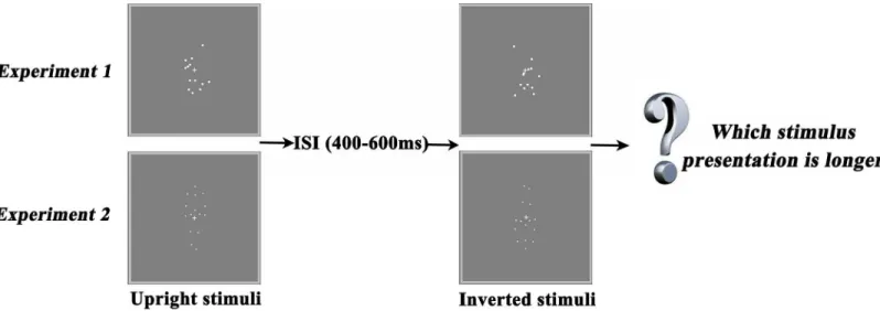
Experiment 1 adopted scrambled BM sequences (upright and inverted) and experiment 2 adopted canonical ones (upright and inverted).Fig 1represents the task used in our study. Sti-muli were white on a gray background, and observers viewed them from approximately 80 cm away. In each trial, 2 stimuli (e.g., an upright canonical BM sequence and an inverted canonical BM sequence) were presented within an interval in the center of the screen, such that the dots subtended approximately 4.0° × 6.8° in visual angle. One of the stimuli (the upright or inverted figure) was randomly selected to be presented for 1000 ms; the other was displayed for 100 ms, 400 ms, 700 ms, 1000 ms, 1300 ms, 1600 ms, or 1900 ms, which resulted in a total of 7 test con-ditions. Thus, the difference between the presentation durations of the two stimuli (upright vs. inverted) was -900 ms, -600 ms, -300 ms, 0 ms, 300 ms, 600 ms, or 900 ms. To avoid a potential interference effect, a blank interval with a randomized duration of 400–600 ms was inserted
Fig 1. Task design.Experiment 1 employed scrambled biological motion (BM) sequences. Either an upright or an inverted scrambled BM stimulus was presented for an interval, followed by an interstimulus interval (ISI) and then the other stimulus. Experiment 2 employed canonical BM sequences. The presentation order of the two stimuli was randomized across trials.
between displays of the two stimuli. The presentation order of the two stimuli and the initial frame of the point-light display for each test stimulus were also randomized across trials (see
S1andS2Video Clips).
Observers were required to make a verbal two-alternative forced choice to judge, as accu-rately as possible, which interval (the first or the second) was longer, regardless of what type of stimulus was shown. Participants were explicitly told not to count aloud or subvocally and that neither stimulus order nor content predicted stimulus presentation duration. The next trial started only after observers made their choice for the previous one.
To ensure that every observer was naive to the nature of the scrambled sequences, all observers were assigned to experiment 1 before experiment 2, since the canonical one may have introduced the concept of a human figure and had an impact on the subsequent scram-bled one. After both experiments, we asked each subject what he or she could recognize from the sequences. Each experiment consisted of 70 trials, with 10 trials for each test condition. Thus, each observer performed 140 trials. A rest break was provided after every 20–30 trials.
2.6. Data analysis
The results of each individual observer from this two-alternative forced-choice task were fitted with a Boltzmann sigmoid function (Eq 1).Fig 2depicts this function, in which the x-axis shows the difference between the presentation durations of the two stimuli (upright vs. inverted), ranging from -900 ms to +900 ms; the y-axis shows the proportion of“long” responses to upright stimuli. The statistical analyses were conducted on the point of subjective equality (PSE) and difference limen (DL). PSE referred to the point at which observers per-ceived the two stimuli equal in terms of the presentation duration, and it was estimated by the midpoint of the Boltzmann function:
fðxÞ ¼1=ð1þexp½ðx x0Þ=oÞ ð1Þ
A negative PSE indicated that when observers perceived two stimuli having as the same duration, the upright stimulus was presented for less time than the inverted counterpart in physical duration (i.e., temporal dilation); a positive PSE indicated the reverse (i.e., temporal compression). The DL was estimated by the interquartile range of the fitted function, and it was used to measure the temporal discrimination sensitivity [30].
Fig 2. Psychometric functions in two conditions.(A) Psychometric function in the scrambled biological condition: the X-axis shows the deviation in the durations of the two stimuli (upright minus inverted), ranging from -900 ms to +900 ms; the Y-axis shows the proportion of“long”responses to upright stimuli. The arrow with the dashed line indicates the PSE, and the arrows with solid lines indicate the DL. A negative PSE indicates a temporal dilation effect for upright stimuli. (B) Psychometric function in the canonical biological condition. The psychometric functions shown represent the means of these individual functions.
The Shapiro-Wilk test was used to determine the normality of the data. The demographic and neuropsychological background data of the two groups were examined by independent-samplest-tests (two-sided). The one-samplet-test (two-sided) was used to detect whether the PSE was different from zero, and the Analysis of Variance (ANOVA) was used to detect overall main effects and interactions.
Results
3.1. Demographic data, PD-related clinical characteristics, and
neuropsychological findings
As analyzed by two-sided independent-samplest-tests, the demographic data, including age, gender, and educational levels, did not show significant differences between the two groups (P>0.05) (seeTable 1). A two-sided independent-samplest-test revealed that MMSE scores did not significantly differ between the two groups (P>0.05); however, VFT and DS (DS [f] and DS [b]) scores were significantly different between the two groups (allP<0.05) (see
Table 1). Disease severity was assessed using the Hoehn–Yahr Scale (H&Y) [31], which is a commonly used system for describing the progression of PD symptoms and the relative levels of disability, including stages 1 to 5.
3.2. Duration discrimination task findings of 2 groups
Table 2shows the PSEs and DLs for PD patients and HCs in the canonical and scrambled BM conditions.Fig 2presents the fitted summary psychometric functions for the groups and tasks.
In the scrambled BM condition, all of the participants reported that they did not recognize any clue of a human figure when observing the stimuli, which included not only the inverted ones, but the upright ones as well. While in the canonical BM condition, all of the participants reported that they recognized walking human figures (upright or inverted) when observing the stimuli.
For HCs, a two-sided one-samplet-test revealed a significant negative PSE in the scrambled BM condition [t(31) = 3.619,P= 0.001] and in the canonical BM condition [t(31) = 8.842, P= 0.000], which suggests that BM signals lengthen their perceived temporal duration inde-pendent of global form (as demonstrated by Wang and Jiang [2012]).
For PD patients, there was no significant negative PSE [t(31) = 0.232,P= 0.818] in the scrambled BM condition, suggesting that local BM processing may be impaired in PD patients. In the canonical BM condition, PD patients perceived upright BM sequences as significantly longer than its inverted counterpart [t(31) = 2.731,P= 0.010]. However, when compared with HCs, we found a significant difference between the two groups [t(62) = 2.223,P= 0.031], which indicates that such a temporal dilation effect of the upright BM sequences is preserved but scaled down. A two-way mixed ANOVA with group (PD patients vs. HCs) as between
Table 2. Point of subjective equality (PSE) and difference limen (DL) for Parkinson’s disease (PD; n = 32) and control (n = 32) groups across canonical and scrambled BM conditions (Mean±Standard Deviation)
Condition Group PSE DL
Canonical BM PD -0.143±0.296* 2.178±1.319
Control -0.279±0.178 2.027±0.941
Scrambled BM PD 0.008±0.193* 2.323±1.261
Control -0.123±0.193 2.064±0.920
*In comparison with healthy controls: analyzed by two-sided independent-samplest-tests,P<0.05.
factor and task (canonical vs. scrambled BM condition) as within factor was performed on the PSEs. Analysis revealed significant main effects of group [F(1, 62) = 11.780,P= 0.001] and task [F(1, 62) = 15.481,P= 0.000]. There was no task × group interaction [F(1, 62) = 0.003,
P= 0.954] (seeFig 3).
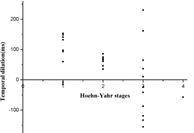
In addition, we found that PSEs in the canonical BM condition and Hoehn–Yahr stages were significantly correlated for PD patients (r= 0.495,P= 0.004) (seeFig 4). We further divided PD patients into 2 subgroups according to their Hoehn-Yahr stages; stage 1–2 (20 patients) belonged to early-stage PD and stage 3–4 (12 patients) belonged to late-stage PD.
For late-stage PD patients, results showed that the PSEs were not different from zero in both canonical and scrambled BM conditions [t(11) = 0.274,P= 0.789;t(11) = 0.372,
P= 0.717, respectively], indicating deficits in BM processing. For early-stage PD patients, two-sided one-samplet-tests revealed a significant negative PSE for the canonical BM condition [t (19) = 7.273,P= 0.000001], but not for the scrambled BM condition [t(19) = 0.034,P= 0.973]. In addition, no significant differences were found between HCs and early-stage PD patients [t (50) = 0.653,P= 0.517], which suggests that scrambled BM processing was impaired and canonical BM processing was relatively preserved early in the course of PD.
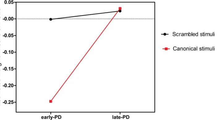
Another two-way mixed ANOVA with subgroup (early- vs. late-stage PD) as between factor and task (canonical vs. scrambled BM condition) as within factor was carried out on the PSEs for PD patients. The analysis showed significant main effects of subgroups [F(1, 30) = 6.300, P= 0.015], and a significant task × group interaction [F(1, 30) = 4.407,P= 0.040] (seeFig 5).
A two-sided independent-samplesttest revealed that the differences between early-stage PD patients and late-stage PD patients were significant in the canonical BM condition [t(30) = 2.858,P= 0.008], and no differences were found between the two subgroups in the scrambled BM condition [t(30) = 0.347,P= 0.731].
Fig 3. Estimated marginal means of points of subjective equality (PSEs) in experiments 1 and 2.Effects of group [Parkinson’s disease (PD) patients vs. health controls (HCs)] and task [canonical vs. scrambled biological motion (BM) condition] on PSEs and their interactions.
A task × onset sides (sides of motor disorders at disease onset) ANOVA showed no signifi-cant main effect of onset sides [F(1, 30) = 0.186,P= 0.668]. A similar ANOVA failed to find a significant difference between medicated and non-medicated patients [F(1, 30) = 0.003, P= 0.955]. No differences were found between males and females in experiments 1 and 2 [F(1, 62) = 0.988,P= 0.324;F(1, 62) = 1.652,P= 0.204, respectively]. Temporal discrimination sen-sitivities (i.e., DL) of the observers were analyzed with a task × group ANOVA. There was no effect of task and group [F(1, 62) = 0.209,P= 0.649;F(1, 62) = 1.070,P= 0.303, respectively].
Fig 6shows decreases of the temporal dilation effect with disease stage increasing in canoni-cal BM and scrambled BM conditions.
Discussion
This study compared BM processing between patients with PD and healthy participants. A negative PSE indicated temporal dilation, and we used this effect as an implicit measure of visual processing of BM. Therefore, differences in PSE measures between PD patients and HCs may suggest a BM processing deficit in PD patients. We studied both canonical and scrambled BM sequences.
We found that the temporal dilation effect was significantly reduced for PD patients com-pared with HCs in both canonical and scrambled BM conditions. In the early stages of the dis-ease, the effect was relatively preserved in the canonical BM condition. However, given that the temporal dilation effect in the canonical BM condition was significantly correlated with Hoehn-Yahr stages, we believe that impaired temporal performance may occur earlier than the data suggests since the sample size was limited.
Fig 4. Group scatter plot of the distribution of the temporal dilation effect at different Hoehn-Yahr stages of Parkinson’s disease (PD) patients.
The reduced temporal dilation effect of visual processing of BM signals agrees with a previ-ous study that demonstrated impairments in perceptual sensitivity to kinematic invariants in PD. In this study, the invariant properties characterizing motion perception in neurologically healthy individuals (the“two-thirds power law”) lost their constraints on motion perception in PD patients [32].
As PD is characterized by tremor, rigidity, bradykinesia, and postural abnormalities, the reduced temporal dilation effect of BM processing found in our study supports coupling between action and perception. Ample evidence suggests action-perception coupling in
Fig 5. Estimated marginal means of points of subjective equality (PSEs) for Parkinson’s disease (PD) patients in experiments 1 and 2.Effects of subgroup [early-stage vs. late-stage PD] and task [canonical vs. scrambled biological motion (BM) condition] on PSEs and their interactions.
doi:10.1371/journal.pone.0138502.g005
Fig 6. Decreases of the temporal dilation effect with disease stage increasing in canonical BM and scrambled BM conditions.Deficits in the temporal dilation effect in the canonical BM condition correlated with Hoehn-Yahr stages; patients in stages 3–4 were most involved, while deficits in the scrambled BM condition happened much earlier, independent of disease severity. Error bars indicate standard error.
humans and nonhuman primates [33–38]. One possible mechanism is called“mirror neuron system”(MNS); a group of neurons that were originally discovered in the premotor cortex of monkeys' discharges when a movement is executed and when the same movement is observed [39]. Previous studies have shown that action observation can have a positive additional impact on rehabilitation of motor deficits after stroke by activating motor areas containing MNS [40–
42]. Two recent LFP studies in PD patients suggested that basal ganglia might be engaged by activity of the human MNS [43–44], which may contribute to impaired temporal performance in PD patients. Pavlova et al. also proposed that there might be an inherent brain network responsible for the coupling; BM perception does not necessarily positively correlate with motor ability or experience per se [45].
In our study, the temporal dilation effect in the scrambled BM condition was impaired early in the course of the disease, while the effect in the canonical BM condition was relatively pre-served in the early stages. Thus, the different patterns of impairment may indicate different mechanisms underlying local and global BM processing, which agrees with the theoretical model by Giese and Poggio [46]. Observed differences between canonical and scrambled BM processing are in accordance with previous fMRI studies [47–50]. Downing et al. observed a stronger activation for canonical BM displays in the EBA than for scrambled controls with identical motion signals [47]. In addition, Grossman and Blake found a slight increase in acti-vation for BM over the scrambled control in the EBA and a strong and significant difference in the STS [48].
Underlying pathophysiological mechanisms may include deficits due to dopamine depletion in frontostriatal circuits in PD patients. Evidence suggests that dopamine depletion is involved in dynamic visuospatial processing [43,51–52]. Deficits in DS and VFT found in our study indicated frontal lobe dysfunction. Moreover, the progressive pathophysiological changes that occur in frontostriatal circuits in PD patients may account for the successive damage of tempo-ral dilation effects in scrambled and canonical BM conditions. In our study, only the“off” levo-dopa condition was completed. Further studies are needed to compare both“on”and“off” levodopa conditions to establish the influence of dopaminergic involvement.
In our study, we investigated visual processing of BM signals in PD patients using the tem-poral dilation effect. However, time perception per se may be impaired in PD patients. Previous findings have suggested that the cerebellum may contribute to the precise timing of salient events in the millisecond range, which determines the duration of a stimulus independently from basal ganglia activity or the onset and end of movements [53–55]. Findings from recent studies [23,56] agree with the observation that PD patients are not impaired in cognitive milli-second time processing. The cerebellum is also thought to be engaged in the neural network dedicated to visual processing of body motion through a structural pathway between the right posterior STS and the left cerebellum [57]. Increasing evidence suggests that the cerebellum participates in the pathophysiology of PD; major roles may include pathological and compen-satory effects [58–59].
Conclusion
In summary, the temporal dilation effect was significantly reduced in PD patients compared with HCs, which may indicate deficits in BM processing.
Supporting Information
S1 File. Supplementary data.Raw data from the present study. (XLS)
S1 Video Clip. BioMotion.This video clip presents the canonical biological motion sequences used in experiment 2.
(GIF)
S2 Video Clip. BioMotionScramble.This video clip presents the scrambled biological motion sequences used in experiment 1.
(GIF)
Author Contributions
Conceived and designed the experiments: RHC XY YHT KW. Performed the experiments: RHC XWC PPH. Analyzed the data: RHC XGC. Contributed reagents/materials/analysis tools: LZ. Wrote the paper: RHC.
References
1. Johansson G (1973) Visual perception of biological motion and a model for its analysis. Percept Psy-chophys 14: 201–211.
2. Sumi S (1984) Upside-down presentation of the Johansson moving light-spot pattern. Perception 13: 283–286. PMID:6514513
3. Pavlova M, Sokolov A (2000) Orientation specificity in biological motion perception. Percept Psycho-phys 62: 889–899. PMID:10997036
4. de-Wit LH, Lefevre CE, Kentridge RW, Rees G, Saygin AP (2011) Investigating the status of biological stimuli as objects of attention in multiple object tracking. PLoS One 6: e16232. doi:10.1371/journal. pone.0016232PMID:21483844
5. Pavlova MA. (2012) Biological motion processing as a hallmark of social cognition. Cereb Cortex 22:981–95. doi:10.1093/cercor/bhr156PMID:21775676
6. Beintema J. A. and Lappe M. (2002) Perception of biological motion without local image motion. PNAS 99:5661–5663. PMID:11960019
7. Troje NF, Westhoff C (2006) The inversion effect in biological motion perception: evidence for a "life detector"? Curr Biol 16: 821–824. PMID:16631591
8. Chang DH, Troje NF (2008) Perception of animacy and direction from local biological motion signals. J Vis 8: 3 1–10.
9. Wang L, Zhang K, He S, Jiang Y (2010) Searching for life motion signals. Visual search asymmetry in local but not global biological-motion processing. Psychol Sci 21: 1083–1089. doi:10.1177/ 0956797610376072PMID:20581341
10. Wang L, Jiang Y (2011) Life motion signals lengthen perceived temporal duration. Proc Natl Acad Sci U S A 109: E673–677.
11. Eagleman DM, Pariyadath V (2009) Is subjective duration a signature of coding efficiency? Philos Trans R Soc Lond B Biol Sci 364:1841–1851. doi:10.1098/rstb.2009.0026PMID:19487187 12. Akiyama T, Kato M, Muramatsu T, Saito F, Nakachi R, Kashima H. (2006) A deficit in discriminating
gaze direction in a case with right superior temporal gyrus lesion. Neuropsychologia 44: 161–170. PMID:16005033
13. Allison T, Puce A, McCarthy G. (2000) Social perception fromvisual cues: Role of the STS region. Trends Cogn Sci 4: 267–278. PMID:10859571
15. Pinsk MA, Arcaro M, Weiner KS, Kalkus JF, Inati SJ, Gross CG, et al. (2009) Neural representations of faces and body parts in macaque and human cortex: A comparative FMRI study. Journal of Neurophys-iology 101: 2581–2600. doi:10.1152/jn.91198.2008PMID:19225169
16. Downing PE, Chan AW, Peelen MV, Dodds CM, Kanwisher N. (2006) Domain specificity in visual cor-tex. Cerebral Cortex 16: 1453–1461. PMID:16339084
17. Schwarzlose RF, Baker CI, Kanwisher N. (2005) Separate face and body selectivity on the fusiform gyrus. Journal of Neuroscience 25: 11055–11059. PMID:16306418
18. Poliakoff E. Representation of action in Parkinson's disease: imagining, observing, and naming actions. J Neuropsychol 2013; 7: 241–254. doi:10.1111/jnp.12005PMID:23320735
19. Dubois B, Pillon B. Cognitive deficits in Parkinson’s disease. J Neurol 1997; 244:2–8. PMID:9007738 20. Kish SJ, Shannak K, Hornykiewicz O. (1988) Uneven pattern of dopamine loss in the striatum of
patients with idiopathic Parkinson’s disease. Pathophysiologic and clinical implications. N Engl J Med 318: 876–80. PMID:3352672
21. Bernheimer H, Birkmayer W, Hornykiewicz O, Jellinger K, Seitelberger F (1973) Brain dopamine and the syndromes of Parkinson and Huntington. Clinical, morphological and neurochemical correlations. J Neurol Sci 20: 415–455. PMID:4272516
22. Scatton B, Rouquier L, Javoy-Agid F, Agid Y (1982) Dopamine deficiency in the cerebral cortex in Par-kinson disease. Neurology 32: 1039–1040. PMID:7202156
23. Koch G, Costa A, Brusa L, Peppe A, Gatto I, Torriero S, et al. (2008) Impaired reproduction of second but not millisecond time intervals in Parkinson's disease. Neuropsychologia 46: 1305–1313. doi:10. 1016/j.neuropsychologia.2007.12.005PMID:18215403
24. Daniel SE, Lees AJ (1993) Parkinson's Disease Society Brain Bank, London: overview and research. J Neural Transm Suppl 39: 165–172. PMID:8360656
25. Folstein MF, Robins LN, Helzer JE (1983) The Mini-Mental State Examination. Arch Gen Psychiatry 40: 812. PMID:6860082
26. Marsili L, Agostino R, Bologna M, Belvisi D, Palma A, Fabbrini G, et al. (2014) Bradykinesia of posed smiling and voluntary movement of the lower face in Parkinson’s disease. Parkinsonism Relat Disord 20:370–375. doi:10.1016/j.parkreldis.2014.01.013PMID:24508573
27. Koch G, Oliveri M, Brusa L, Stanzione P, Torriero S, Caltagirone C. (2004) High-frequency rTMS improves time perception in Parkinson disease. Neurology 63:2405–2406. PMID:15623713 28. Brainard DH (1997) The Psychophysics Toolbox. Spat Vis 10: 433–436. PMID:9176952
29. Vanrie J, Verfaillie K (2004) Perception of biological motion: a stimulus set of human point-light actions. Behav Res Methods Instrum Comput 36: 625–629. PMID:15641407
30. Wearden JH, Ferrara A (1996) Stimulus range effects in temporal bisection by humans. Q J Exp Psy-chol B 49: 24–44. PMID:8901385
31. Hoehn MM, Yahr MD (1967) Parkinsonism: Onset, progression, and mortality. Neurology, 17, 427–
442. PMID:6067254
32. Dayan E, Inzelberg R, Flash T. (2012) Altered perceptual sensitivity to kinematic invariants in Parkin-son's disease. PLoS One 7:e30369. doi:10.1371/journal.pone.0030369PMID:22363430
33. Casile A, Giese MA (2006) Nonvisual motor training influences biological motion perception. Curr Biol 16: 69–74. PMID:16401424
34. Reed CL, Farah MJ (1995) The psychological reality of the body schema: a test with normal partici-pants. J Exp Psychol Hum Percept Perform 21: 334–343. PMID:7714475
35. Hommel B, Musseler J, Aschersleben G, Prinz W (2001) The Theory of Event Coding (TEC): a frame-work for perception and action planning. Behav Brain Sci 24: 849–878. PMID:12239891
36. Prinz W (1997) Perception and Action Planning. European Journal of Cognitive Psychology 9: 129–
154.
37. Decety J, Grezes J (1999) Neural mechanisms subserving the perception of human actions. Trends Cogn Sci 3: 172–178. PMID:10322473
38. Iacoboni M, Woods RP, Brass M, Bekkering H, Mazziotta JC, Rizzolatti G. (1999) Cortical mechanisms of human imitation. Science 286: 2526–2528. PMID:10617472
39. Kato Y, Muramatsu T, Kato M, Shibukawa Y, Shintani M, Mimura M. (2011) Magnetoencephalography study of right parietal lobe dysfunction of the evoked mirror neuron system in antipsychotic-free schizo-phrenia. PLoS One 6: e28087. doi:10.1371/journal.pone.0028087PMID:22132217
41. Ertelt D, Hemmelmann C, Dettmers C, Ziegler A, Binkofski F. (2012) Observation and execution of upper-limb movements as a tool for rehabilitation of motor deficits in paretic stroke patients: protocol of a randomized clinical trial. BMC Neurol 12:42. doi:10.1186/1471-2377-12-42PMID:22708612 42. Bhasin A, Padma Srivastava MV, Kumaran SS, Bhatia R, Mohanty S. (2012) Neural interface of mirror
therapy in chronic stroke patients: a functional magnetic resonance imaging study. Neurol India 60:570–576. doi:10.4103/0028-3886.105188PMID:23287316
43. Alegre M, Rodriguez-Oroz MC, Valencia M, Perez-Alcazar M, Guridi J, Iriarte J, et al. (2010) Changes in subthalamic activity during movement observation in Parkinson's disease: is the mirror system mir-rored in the basal ganglia? Clin Neurophysiol 121:414–425. doi:10.1016/j.clinph.2009.11.013PMID: 20006544
44. Marceglia S, Fiorio M, Foffani G, Mrakic-Sposta S, Tiriticco M, Locatelli M, et al. (2009) Modulation of beta oscillations in the subthalamic area during action observation in Parkinson's disease. Neurosci-ence 161:1027–1036. doi:10.1016/j.neuroscience.2009.04.018PMID:19364520
45. Pavlova M, Staudt M, Sokolov A, Birbaumer N, Krägeloh-Mann I. (2003) Perception and production of biological movement in patients with early periventricular brain lesions. Brain 126:692–701. PMID: 12566289
46. Giese MA, Poggio T (2003) Neural mechanisms for the recognition of biological movements. Nat Rev Neurosci 4:179–192. PMID:12612631
47. Downing PE, Jiang Y, Shuman M, Kanwisher N (2001) A cortical area selective for visual processing of the human body. Science 293:2470–2473. PMID:11577239
48. Grossman ED, Blake R (2002) Brain areas active during visual perception of biological motion. Neuron 35:1167–1175. PMID:12354405
49. Thompson JC, Clarke M, Stewart T, Puce A (2005) Configural processing of biological motion in human superior temporal sulcus. J Neurosci 25:9059–9066. PMID:16192397
50. Peelen MV, Downing PE (2005) Selectivity for the human body in the fusiform gyrus. J Neurophysiol 93:603–608. PMID:15295012
51. Bodis-Wollner I. (2003) Neuropsychological and perceptual defects in Parkinson’s disease. Parkinson-ism 9(suppl 2): S83–S89.
52. van der Hoorn A, Renken RJ, Leenders KL, de Jong BM. (2014) Parkinson-related changes of activa-tion in visuomotor brain regions during perceived forward self-moactiva-tion. PLoS One. 9:e95861. doi:10. 1371/journal.pone.0095861PMID:24755754
53. Spencer RM, Zelaznik HN, Diedrichsen J, Ivry RB (2003) Disrupted timing of discontinuous but not con-tinuous movements by cerebellar lesions. Science 300: 1437–1439. PMID:12775842
54. Buhusi CV, Meck WH (2005) What makes us tick? Functional and neural mechanisms of interval tim-ing. Nat Rev Neurosci 6: 755–765. PMID:16163383
55. Ivry RB, Spencer RM, Zelaznik HN, Diedrichsen J (2002) The cerebellum and event timing. Ann N Y Acad Sci 978: 302–317. PMID:12582062
56. Spencer RM, Ivry RB (2005) Comparison of patients with Parkinson's disease or cerebellar lesions in the production of periodic movements involving event-based or emergent timing. Brain Cogn 58: 84–
93. PMID:15878729
57. Sokolov AA, Erb M, Grodd W, Pavlova MA. (2014) Structural Loop Between the Cerebellum and the Superior Temporal Sulcus: Evidence from Diffusion Tensor Imaging. Cereb Cortex 24:626–632. doi: 10.1093/cercor/bhs346PMID:23169930
58. Wu T, Hallett M. (2013) The cerebellum in Parkinson’s disease. Brain 136:696–709. doi:10.1093/ brain/aws360PMID:23404337