Pleomorphic Adenoma of the Tongue Base:
Case Report and Review
Luiz Augusto Nascimento
1Thais Gonçalves Pinheiro Vilela
1,21Division of Head and Neck Surgery, Department of Otolaryngology – Head and Neck Surgery of the Brasilia University Hospital, Federal University of Brasilia, Brasilia/DF, Brazil
2Department of Otolaryngology, The Medicine School of the University of São Paulo, São Paulo/SP, Brazil
Int Arch Otorhinolaryngol 2014;18:328–331.
Address for correspondence Luiz Augusto Nascimento, MD, PhD, Department of Otolaryngology–Head and Neck Surgery, Federal University of Brasilia, SGAN 605, Av. L2 Norte, 70840-901 Brasilia/DF, Brazil (e-mail: [email protected]).
Introduction
Pleomorphic adenoma, also known asmixed tumor, is the
most common benign tumor of the major and minor salivary glands.1–3 Adenoid cystic carcinoma is the most common malignant tumor of this region, including the tongue.4The literature indicates an overall ratio of1:6 for
benign/malig-nant lingual salivary gland tumors.2More than 74% of the cases of pleomorphic adenoma arise in the major salivary gland,3and it is predominantly found in the parotid gland. The palate is the most common site in the minor salivary glands.5 The occurrence of pleomorphic adenoma of the
tongue base is very rare and very few cases have been reported in the literature.1,3
The authors report a rare case of pleomorphic adenoma of the tongue base causing dysphagia and dyspnea and present a review of the literature.
Case Report
A 55-year-old woman presented to the head and neck surgery service complaining of a cervical mass that had been growing for 3 years. At the moment of presentation, she had dyspha-gia, severe dyspnea, and difficulty in talking. She had Keywords
►
adenoma
►
pleomorphic
►
head and neck
neoplasms
►
tongue neoplasms
►
oropharyngeal
neoplasms
Abstract
Introduction
Pleomorphic adenoma, also known as
mixed tumor
, is the most common
benign tumor of the major and minor salivary glands. The occurrence of pleomorphic
adenoma of the tongue base is very rare, and very few cases have been reported in the
literature.
Objective
The authors present a rare case of pleomorphic adenoma of the tongue
base and a review of the literature.
Case Report
A 55-year-old woman had an extensive cervical mass, with little pain, from
the submental level to the level below the hyoid bone. Fiberoptic endoscopic
examina-tion showed an extensive mass at the base of the tongue with considerable reducexamina-tion in
the airway. Magnetic resonance image scan revealed a contrast-enhancing mass of
heterogeneous density over the base of the tongue of 8
8
7 cm and a reduction of
the hypopharyngeal airway. Biopsy of the lesion was performed along with a
tracheos-tomy due to the bulging tongue base and acute respiratory failure. Histologic
examination with an immunohistochemistry study revealed a diagnosis of pleomorphic
adenoma. The excision of the tumor was performed by a lateral pharyngotomy
approach and the total mass was excised.
Conclusion
The authors consider the rarity of this case and show that this is the 11th
and the largest pleomorphic adenoma reported in the English-language medical
literature.
received February 26, 2013 accepted May 3, 2013 published online November 5, 2013
DOI http://dx.doi.org/ 10.1055/s-0033-1351683. ISSN 1809-9777.
Copyright © 2014 by Thieme Publicações Ltda, Rio de Janeiro, Brazil
Case Report
important pain and eventual oral bleeding during deglutition. The patient reported smoking 15 cigarettes (withoutfilter) per day for 30 years. There was no history of alcoholism and no visible pulsations could be seen in the mass.
Physical examination revealed an 8-cm mass in major
diameter in the midline at cervical level I (►Fig. 1). The patient presented lockjaw. Fiberoptic endoscopic examina-tion showed a big mass at the tongue base with tissue necrosis and a substantial reduction of the airway.
Magnetic resonance image scan revealed a contrast-en-hancing mass of 887 cm of heterogeneous density over
the base of the tongue and a reduction of the hypopharyngeal airway (►Fig. 2).
Biopsy of the lesion was performed along with a tracheos-tomy due to the bulging tongue base and acute respiratory failure. A nasoenteral feeding tube was placed.
Histologic examination revealed the possibility that it was a mixed tumor of a malignancy type of pleomorphic carcinoma
ex adenoma, low grade, without a definitive conclusion due to fragmentation of the material. Immunohistochemically, the cells were positive for glialfibrillary acid protein, favoring a diagnosis of pleomorphic adenoma.
Fig. 1 Patient with an extensive submental mass of afirm elastic consistency.
Fig. 2 Magnetic resonance image: coronal (A) and axial (B) cut showing extensive tumor mass at the base of the tongue with extension to the hypopharynx and cervical level I.
Fig. 3 Epithelial cells (blue arrow) and myoepithelial cells (yellow arrow; hematoxylin-eosin stain, original magnification400).
Fig. 4 Glialfibrillary acid protein immunoreactivity.
International Archives of Otorhinolaryngology Vol. 18 No. 3/2014
Subsequently, excision of the mass was performed under general anesthesia using a lateral pharyngotomy approach, and the total mass was excised with a clear cleavage plan with the neighboring structures. The histologic examination of the mass confirmed the diagnosis (►Figs. 3and4). The postop-erative period was uneventful and the patient was success-fully decannulated on the 50th postoperative day and she is free of disease to date.
Discussion
The majority of salivary gland neoplasms are benign and pleomorphic adenoma is the most common. Tumors of the salivary glands comprise 3% of all neoplasms.4The incidence of salivary gland neoplasms in minor glands varies from 9 to 22%. Approximately 8% of pleomorphic adenomas involve the minor salivary glands. The majority involve the palate, fol-lowed by the lips and maxillary sinus.1,3,5Involvement of the base of the tongue is rare.1,2 Malignant tumors such as adenocarcinoma, adenoid cystic carcinoma, and mucoepider-moid carcinoma involve the tongue more frequently.1–3
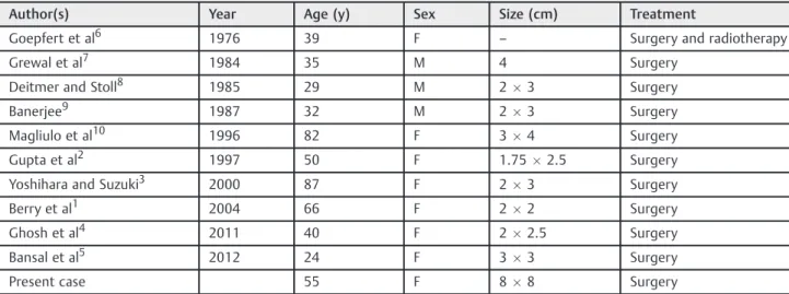
Only 10 other cases of pleomorphic adenoma involving the tongue base have been reported in the English-language literature (►Table 1). Patients’ages ranged from 24 to 87 years old (average of 49) and the male-to-female ratio was 3:8, including the present case.
These tumors are slow-growing and sometimes the treat-ment may be late.2Dysphagia is the most frequent initial symptom, and some of the tumors are detected on routine physical examinations by general practitioners.3,4
Treatment is primarily surgical irrespective of the site of origin. Resection of the tumor with an adequate margin is essential to avoid recurrence, although these tumors are well encapsulated.1Recurrence is uncommon and may be attrib-uted to partial excision or a multifocal origin of the tumor.2,4 Some studies report a recurrence rate of 6% in patients with benign minor salivary gland tumors.1 Surgical approaches vary according to the size and site of the tumor: transoral, combined transoral-transcervical, transmandibular, and
transpharyngeal. Transmandibular can be lip splitting, man-dibular swing, or median labiomanman-dibular glossotomy. Trans-pharyngeal can be either suprahyoid or transhyoid pharyngotomy or by lateral pharyngotomy.5When the tumor is malignant with extensive invasions into surrounding tis-sues, the latter two approaches are recommended.3
The origin of the pleomorphic adenoma is myoepithelial cells and intercalated duct cells.2The histopathologic appear-ance of a pleomorphic adenoma is mainly composed of epithelial and myoepithelial elements, with a variety of patterns ending up embedded in mucopolysaccharide stro-ma. Fibrosis of the surrounding salivary parenchyma forms a capsule, usually false.1Pleomorphic adenoma of the minor salivary gland is known to have more cellular and fewer mesenchymal components. In cases of the elderly, malignant degeneration to carcinoma ex pleomorphic adenoma must be taken into consideration.3
Conclusion
This is the 11th case of pleomorphic adenoma involving the tongue base that has been reported in the English-language literature and the biggest one.
References
1 Berry S, Tay H, Puentes CP. Pleomorphic adenoma of the base of the
tongue. Ear Nose Throat J 2004;83:646–648, 648
2 Gupta AK, Singhal SK, Mann SBS, Bapuraj JR, Saran RK.
Pleomor-phic adenoma presenting as a base of tongue mass. J Laryngol Otol 1997;111:1177–1178
3 Yoshihara T, Suzuki S. Pleomorphic adenoma of tongue base causing
dysphagia and dysphasia. J Laryngol Otol 2000;114:793–795
4 Ghosh SK, Saha J, Chandra S, Datta S. Pleomorphic adenoma of the
base of the tongue: a case report. Indian J Otolaryngol Head Neck
Surg 2011;63(Suppl 1):113–114
5 Bansal S, Kalsotra G, Mohammed AW, Bahl A, Gupta AK.
Pleomor-phic adenoma of base of tongue: is midline mandibulotomy necessary for approaching benign base tongue lesions? Case Rep Otolaryngol 2012;2012:851501
Table 1 Reported cases of pleomorphic adenoma of tongue base
Author(s) Year Age (y) Sex Size (cm) Treatment
Goepfert et al6 1976 39 F – Surgery and radiotherapy
Grewal et al7 1984 35 M 4 Surgery
Deitmer and Stoll8 1985 29 M 23 Surgery
Banerjee9 1987 32 M 23 Surgery
Magliulo et al10 1996 82 F 34 Surgery
Gupta et al2 1997 50 F 1.752.5 Surgery
Yoshihara and Suzuki3 2000 87 F 23 Surgery
Berry et al1 2004 66 F 22 Surgery
Ghosh et al4 2011 40 F 22.5 Surgery
Bansal et al5 2012 24 F 33 Surgery
Present case 55 F 88 Surgery
International Archives of Otorhinolaryngology Vol. 18 No. 3/2014
Pleomorphic Adenoma of the Tongue Base Nascimento, Vilela
6 Goepfert H, Giraldo AA, Byers RM, Luna MA. Salivary gland tumors of the base of the tongue. Arch Otolaryngol 1976;102:
391–395
7 Grewal DS, Pusalkar AG, Phatak AM. Pedunculated pleomorphic
adenoma of the tongue base manifesting with dysponea. A case
report. J Laryngol Otol 1984;98:425–427
8 Deitmer T, Stoll W. [Rare tumors of the base of the tongue and their
therapy]. HNO 1985;33:366–369
9 Banerjee S. Benign pleomorphic adenoma of the base of the
tongue. J R Coll Surg Edinb 1987;32:164–165
10 Magliulo G, Terranova G, Cristofari P. Pleomorphic adenoma
of the tongue base. Ann Otol Rhinol Laryngol 1996;105:835–837
International Archives of Otorhinolaryngology Vol. 18 No. 3/2014