Original Research
Upper Lip Pleomorphic Adenoma: Comparison of Reported Cases
between 1990 and 2012
Hamed Mortazavi
1, Somayeh Alirezaei
1, Saranaz Azari-Marhabi
1,
Maryam Baharvand
1, Majid Eshghpour
21
Department of Oral Medicine, Dental School, Shahid Beheshti University of Medical Sciences,
Tehran, Iran
2
Dental Research Center, Faculty of Dentistry, Mashhad University of Medical Sciences,
Mashhad, Iran
Received 16 April 2013 and Accepted 27 June 2013
Abstract
Introduction: Pleomorphic adenoma of the upper lip is a rare entity and its diagnosis requires a high index of suspicion. The aim of this study was to review the reported cases of pleomorphic adenoma (PA) of the upper lip. Methods: This study was performed on the basis of the clinical features of 10 well-documented reported cases of the upper lip pleomorphic adenomas from 1990 to 2012 which have been indexed in the PubMed. The search strategy based on MeSH keywords included "salivary gland tumor", "salivary gland cancer", "pleomorphic adenoma", and "mixed tumor". In the clinical records the following data have been considered: Age, sex, clinical view, complaint time, site, size, texture, pathological view, treatment, recurrence rate, symptom and follow-up period. Results: Of the 11 reported cases of PA, 7 (63.7%) were female and 4 (36.3%) were male, with age ranging from 12 to 65 years. 50% of the patients were between 35 and 55 years of age. Only 3 (27.2%) of cases were younger than 20. The main clinical presentation of lesion in all cases was a submucosal nodule. There was a large interval between the first symptoms and diagnosis. The size of the lesions were between 1 and 3 cm. Involvement of the right half of the upper lip was more common than the left side. 63.6% of the lesions showed a firm consistency and bone formation was seen in one (9.09%) case. The follow-up period ranged from 5 to 48 months. There was no evidence of recurrence in any of the reported cases. Conclusion: Although rare, pleomorphic adenoma should be considered as a differential diagnosis for the swellings in the upper lip. PA has a potential for malignant transformation.
Therefore this entity should be evaluated carefully by all clinicians.
Key words: Mixed tumor, pleomorphic adenoma, salivary gland cancer, salivary gland tumor.
---
Mortazavi H, Alirezaei S, Azari-Marhabi S, Baharvand M, Eshghpour M. Upper Lip Pleomorphic Adenoma: Comparison of Reported Cases between 1990 and 2012. J Dent Mater Tech 2013; 2(4): 125-9.Introduction
suspicion. Therefore the aim of this study was to review of the upper lip PAs in the well-documented reported cases from 1990 to 2012.
Materials and Methods
This study was performed on the basis of the clinical features of 10 well-documented full text reported cases of the upper lip pleomorphic adenomas between 1990 and 2012 which have indexed in the PubMed. The search strategy based on MeSH keywords included "salivary gland tumor", "salivary gland cancer", "pleomorphic adenoma", and "mixed tumor". In the clinical records, the following data was considered: age, sex, clinical view, complaint duration, site, size, texture, pathologic view, treatment, recurrence rate, symptoms and follow-up period. For the studies performed before 1990, we were unable to find the necessary information. Also some of these reports were not in English. Therefore, these studies were excluded.
Results
Of the 11 full text reported cases of PA, 7 (63.7%) were female and 4 (36.3%) were male. The female to male ratio was about 2:1. Although the patients' age ranged from 12 to 65 years, about half of them were between 35 and 55 and only 3 (27.2%) individuals were younger than 20. The main clinical presentation of the lesion in all cases was an asymptomatic nodule. Furthermore, all reported cases were encapsulated and surgery was the accepted treatment. There was a large time gap of 12 to 96 months (mean: 54 months) between the onset of first symptoms and making the diagnosis. The size of the lesions was between 1 and 3 cm, with the average of 2 cm. Involvement of the right half of the upper lip was more common than the left. 63.6% of the lesions had a firm consistency and bone formation was seen in one (9.09%) case. All cases (100%) were capsulated and surgical excision was the treatment of choice. The follow-up period ranged from 5 to 48 months with the average of 28 months. There was no evidence of recurrence in any reported cases. Additional data were summarized in Table 1.
Table 1. Clinical data of 10 upper lip pleomorphic adenomas in the English-language literature indexed in the PubMed from 1990-2012 Follow-up period (month) Texture Size (cm) Site Complaint duration (month) Sex Age (year) No. of cases Author 16 Firm 1.5×2 upper lip upper lip/right 24 female 65 1 Kataria 2
2011 12 rubbery 3×3 18-19 male 33 2 Ali 24
2011 -Firm 1.8×1.5 upper lip/right 84 Male 35 3
Küçük 8 2011 12 rubbery 1.5×2 upper lip/left 12 Female 55 4
Debnath 5 2010 48 Firm 1×1.3 upper lip 120 Female 51 5
Pons Vicente 1 2008 12 Rubbery 1.5×2 upper lip/right 12 Male 12 6
Lotufo 6 2008 26 Firm -upper lip 120 Female 40 7
Asuquo 21 2009 5 Firm 1×3 upper lip 24 Female 15 8
Jorge 3 2002 39 Firm 1×3 upper lip 12 Female 18 9
Jorge 3 2002 48 Hard 1×1 upper lip/left 24 Female 53 10
Hamakawa 22 1997 - Firm - upper lip/left 96 Male 61 11
Report of a New Case
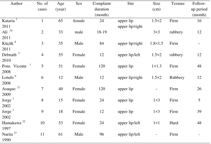
A 20-year-old female patient attended to the Oral Medicine Department, Dental School, Shahid Beheshti University of Medical Sciences complaining of a swelling in her upper lip. Her history revealed a mass appeared in the left half of her upper lip about 12 months ago which gradually increased in size. The lesion was asymptomatic and did not cause any functional limitation or disability for the patient. Clinical examination showed a freely movable mass of firm consistency with the size of about 1.5×1.8cm. The overlying mucosa was smooth and intact with a pinkish color (Fig. 1). The medical history was unremarkable. Based on the clinical examination the diagnosis of a salivary gland tumor or a benign mesenchymal tumor was made.
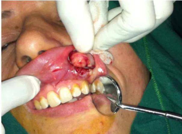
Under local anesthesia, the tumor was completely removed by an intraoral excisional biopsy, a common surgical procedure (Fig. 2). Histopathologic examination of the specimen revealed an encapsulated tumor composed of a mixture of glandular epithelium and myoepithelial cells within a mesenchymal-like stroma. The epithelium was arranged in ducts, cystic structures and sheets. There were also nests of epithelial cells with squamous metaplasia and keratin pearl formation. Sheets of plasmacytoid, epitheloid and a few clear myoepithelial cells in a chondroid, mucoid and hyalinized stroma were seen as well. There was no evidence of malignancy. The histopathologic diagnosis of pleomorphic adenoma was established. Subsequent follow-up after 1 year revealed no signs of recurrence.
Figure 1. Clinical presentation of a patient with pleomorphic adenoma
Figure 2. Total surgical excision of the tumor
Discussion
Definition and EtiopathogenesisPleomorphic adenoma or benign mixed tumor is the most common salivary neoplasm, accounting for 40% of all minor salivary gland tumors (8,9). Tumors arising from the minor salivary glands are uncommon clinical entities and comprise 10-25% of all salivary gland neoplasms. Malignant transformation may occur in as many as 5% of cases (5,9). The main etiopathogenesis of PA remains unclear. Cytogenic and molecular studies have described that it is of epithelial origin with chromosomal abnormalities at 8q12 and 12q15 (5,6). Exposure to ionizing radiation has been the only known risk factor for cancers of the salivary glands (9-11).
Clinical and Pathological Aspects
benign lesions predominate on the upper lip. This finding may be related to the differences in embryonic development between the lower and upper lips (2,7). There is a large time interval between the first symptoms and diagnosis of PA, ranging from 2 days to 15 years (3).
Histologically, PA of the extra-major salivary glands is similar to that in the major salivary glands and is composed of a mixture of epithelial and stromal elements. There are three major histologic subtypes; myxoid (80% stroma), cellular (myoepithelial predominant), and mixed (classic) (9,15). Ductal epithelial glands make up glandular and cystic structures of various sizes. Myoepithelial cells are responsible for the production of extra cellular matrix with chondroid, collagenous, mucoid and osseous stroma (3,5). Myxochondroid changes are the most frequent change in the stroma. The cartilaginous differentiation areas are commonly observed in immunohistochemical evaluations in tumors arising from the parotid glands. The ductal epithelial element is positive with keratin and EMA and also, the myoepithelial component stains positively with actin, myosin, keratin, other smooth muscle-specific proteins, fibronectin and S-100 (5,8). A capsule is usually seen in PA. However, it may be incomplete or show infiltration by the tumor cells. This lack of complete encapsulation is more common for minor salivary gland neoplasms. The capsule forms as a result of the fibrosis of the surrounding parenchyma (16).
Diagnosis and Differential Diagnosis of PA The diagnosis of minor salivary gland tumors is based on the clinical history and physical examination, supported by complementary techniques including computerized tomography (CT), magnetic resonance imaging (MRI), sialography, fine needle aspiration biopsy (FNAB) and incisional biopsy. CT scanning is the best for bony involvement and MRI is better for displaying soft tissue invasion or perineural spread. However, the combination of these methods helps making a tentative diagnosis (1,3). Differential diagnosis of intraoral pleomorphic adenoma includes other minor salivary gland tumors (mucoepidermoid carcinoma, myoepithelioma, basal cell adenoma) and benign and malignant mesenchymal neoplasms (lipoma, neurofibroma, rabdomyo and sarcoma) (1,3,17-19).
Treatment and Prognosis
Pleomorphic adenomas are usually treated by surgical excision. Superficial parotidectomy and total parotidectomy were suggested for lesions in the superficial and deep lobes of the parotid gland, respectively. Submandibular gland tumors are best treated by total removal of the gland with the neoplasm. Minor salivary gland tumors are usually excised with a safe margin. It is important to know that an inadequate
resection or rupture of the capsule can lead to local recurrence (2,8,9,21).
The prognosis of PA is excellent, with a cure rate of 95%. In addition, tumors with a weak or negative staining for PCNA and P.53 have better prognosis (3,19). According to the literature since 1939, recurrences occur in 2-44% of pleomorphic adenomas. The risk for recurrence seems to be lower for tumors of the minor salivary glands. Inadequate surgery was reported to be the main cause of recurrence (8,9,20-24). Furthermore, it is noted that the chances of recurrence are higher when PA occurs before 30 years of age (6).
Conclusion
Although rare, pleomorphic adenoma should be considered as a differential diagnosis of swellings in the upper lip. This lesion, in most frequent cases, is asymptomatic and the patient may not be aware of its existence and is discovered accidentally by a dentist. Also, it is important to know that pleomorphic adenoma can transform to a malignant lesion.
References
1. Pons Vicente O, Almendros Marqués N, Berini
Aytés L, Gay Escoda C. Minor salivary gland
tumors: A clinicopathological study of 18 cases.
Med Oral Patol Oral Cir Bucal 2008;13:E582-8.
2. Kataria SP, Tanwar P, Sethi D, Garg M.
Pleomorphic adenoma of the upper lip. J Cutan
Aesthet Surg 2011;4:217-9.
3. Jorge J, Pires FR, Alves FA, et al. Juvenile
intraoral pleomorphic adenoma: report of five
cases and review of the literature. Int J Oral
Maxillofac Surg 2002;31:273-5.
4. Williamson JJ, Meskin LH. Pleomorphic adenoma
of the upper lip. Oral Surg Oral Med Oral Pathol
1965;20:771-5.
5. Debnath SC, Adhyapok AK. Pleomorphic
adenoma (benign mixed tumor) of the minor
salivary glands of the upper lip. J Maxillofac Oral
Surg 2010;9:205-8.
6. Lotufo MA, Júnior CA, Mattos JP, França CM.
Pleomorphic adenoma of the upper lip in a child. J
Oral Sci 2008;50:225-8.
7. Bernier JL. Mixed tumors of the lip. Ann
8. Küçük U, Tan S. Pleomorphic adenoma of the
upper lip. Turk Patoloji Derg 2011;27:73-6.
9. Neville BW, Damm DD, Allen CM, Bouquot JE.
Oral and maxillofacial pathology. St. Louis: W. B.
Saunders, 2009.
10. Otoh EC, Johnson NW, Olasoji H, Danfillo IS,
Adeleke OA. Salivary gland neoplasms in
Maiduguri, north-eastern Nigeria. Oral Dis
2005;11:386-91.
11. Beal KP, Singh B, Kraus D, Yahalom J, Portlock
C, Wolden SL. Radiation-induced salivary gland
tumors: a report of 18 cases and a review of the
literature. Cancer J 2003;9:467-71.
12. Yih WY, Kratochvil FJ, Stewart JC. Intraoral
minor salivary gland neoplasms: review of 213
cases. J Oral Maxillofac Surg 2005;63:805-10.
13. Toida M, Shimokawa K, Makita H, Kato K,
Kobayashi A, Kusunoki Y. Intraoral minor
salivary gland tumors: a clinicopathological study
of 82 cases. Int J Oral Maxillofac Surg
2005;34:528-32.
14. Bradley P, McClelland L, Mehta D. Paediatric
salivary gland epithelial neoplasms. ORL J
Otorhinolaryngol Relat Spec 2007;69:137-45.
15. Ogawa Y, Toyosawa S, Ishida T, Ijuhin N. Keratin
14 immunoreactive cells in pleomorphic adenomas
and adenoid cystic carcinomas of salivary glands.
Virchows Arch 2000;437:58-68.
16. Varghese BT, Sebastian P, Abraham EK, Mathews
A. Pleomorphic adenoma of minor salivary gland
in the parapharyngeal space. World J Surg Oncol
2003;25:2.
17. Dalati T, Hussein MR. Juvenile pleomorphic
adenoma of the cheek: a case report and review of
literature. Diagn Pathol 2009; 22:32.
18. Dhanuthai K, Boonadulyarat M, Jaengjongdee T,
Jiruedee K. A clinico-pathologic study of 311
intra-oral salivary gland tumors in Thais. J Oral
Pathol Med 2009;38:495-500.
19. Maynard JD. Management of pleomorphic
adenoma of the parotid. Br J Surg 1998;75:305-8.
20. Krolls SO, Boyers RC. Mixed tumors of salivary
glands. Long-term follow-up. Cancer
1972;30:276-81.
21. Asuquo ME, Otei OO, Ekpo R, Abang I, Adams
U, Bassey EE. Salivary gland tumor of the lip:
report of two cases and literature review. Cent Afr
J Med 2009;55:43-6.
22. Hamakawa H, Takarada M, Ito C, Tanioka H.
Bone-forming pleomorphic adenoma of the upper
lip: report of a case. J Oral Maxillofac Surg
1997;55:1471-5.
23. Narita H, Kobayashi T, Kanzaki T. Pleomorphic
adenoma of the lip. J Dermatol 1990;17:710-2.
24. Ali I, Gupta AK, Singh S. Pleomorphic adenoma
of the upper lip. Natl J Maxillofac Surg
2011;2:219-21.
Corresponding Author:
Majid Eshghpour Faculty of Dentistry
Vakilabad Blvd, Mashhad, Iran P.O. Box: 91735-984
Tel: +98-511-8829501 Fax: +98-511-8829500