Genes of
Yersinia pseudotuberculosis
IP32953 at 28
6
C
and 3
6
C
Eveliina Palonen*, Miia Lindstro¨m, Reija Karttunen, Panu Somervuo, Hannu Korkeala
Department of Food Hygiene and Environmental Health, Faculty of Veterinary Medicine, University of Helsinki, Helsinki, Finland
Abstract
Yersinia pseudotuberculosis is a significant psychrotrophic food pathogen whose cold tolerance mechanisms are poorly understood. Signal transduction systems serve to monitor the environment, but no systematic investigation of their role at cold temperatures inY. pseudotuberculosishas yet been undertaken. The relative expression levels of 54 genes predicted to encode proteins belonging to signal transduction systems inY. pseudotuberculosisIP32953 were determined at 28uC and 3uC by quantitative real-time reverse transcription-PCR. The relative expression levels of 44 genes were significantly (p,0.05) higher at 3uC than at 28uC. Genes encoding the two-component system CheA/CheY had the highest relative expression levels at 3uC. Mutational analysis revealed thatcheAis important for growth and motility at 3uC. The relative expression level of one gene,rssB, encoding an RpoS regulator, was significantly (p,0.05) lower at 3uC than at 28uC. The results suggest that several signal transduction systems might be used during growth at low temperature, and at least, CheA/CheY two-component system is important for low-temperature growth.
Citation:Palonen E, Lindstro¨m M, Karttunen R, Somervuo P, Korkeala H (2011) Expression of Signal Transduction System Encoding Genes of Yersinia pseudotuberculosisIP32953 at 28uC and 3uC. PLoS ONE 6(9): e25063. doi:10.1371/journal.pone.0025063
Editor:Ulrich Dobrindt, Universita¨t Mu¨nster, Germany
ReceivedMarch 30, 2011;AcceptedAugust 26, 2011;PublishedSeptember 20, 2011
Copyright:ß2011 Palonen et al. This is an open-access article distributed under the terms of the Creative Commons Attribution License, which permits unrestricted use, distribution, and reproduction in any medium, provided the original author and source are credited.
Funding:This work was supported by the Finnish Centre of Excellence in Microbial Food Safety Research of the Academy of Finland (grants 118602, 141140) http://www.aka.fi/fi/A/; the Doctoral Program of the Faculty of Veterinary Medicine of the University of Helsinki http://www.vetmed.helsinki.fi/; the Finnish Veterinary Foundation http://www.sell.fi/elainlaakariliitto/linkkeja_ja_tietolahteita/suomen_elainlaaketieteen_saatio_/; the Walter Ehrstro¨m Foundation http:// www.maitohygienialiitto.fi/walter_ehrstrom_alku.html; the Medical Fund of the University of Helsinki http://www.helsinki.fi/rahastot/lahjoitusrahastot/ laaketieteen_rahasto.htm. The funders had no role in study design, data collection and analysis, decision to publish, or preparation of the manuscript.
Competing Interests:The authors have declared that no competing interests exist.
* E-mail: [email protected]
Introduction
Yersinia pseudotuberculosis is an important food-borne pathogen capable of growing at refrigeration temperatures and under modified atmospheres [1]. Typical symptoms of yersiniosis resulting from mesenteric lymphadenitis of the small intestine, such as fever and acute abdominal pain [2], are often mistaken for appendicitis and have led to unnecessary appendectomies [3]. Apart from infections due to contaminated drinking water [4,5],Y. pseudotuberculosishas caused outbreaks through contaminated fresh produce stored at low temperature [6–8]. While low temperature efficiently controls the growth of many pathogenic bacteria, it readily favors the growth ofY. pseudotuberculosis[9]. Although, little is known about the cold tolerance mechanisms ofY. pseudotuber-culosis (reviewed in [1]), key changes reported to occur in
Enterobacteriaceaeduring adaptation to growth at cold temperatures include an increase in low-melting-point lipids in cell membranes and accumulation of compatible solutes in the cells [1].
To survive, bacteria must sense changes in temperature and other extrinsic circumstances. Two-component signal transduction sys-tems are widespread among bacteria and help them monitor and adapt to changes in their extra- or intracellular environment [10]. A classical two-component system consists of a sensor histidine kinase and a response regulator located in the cell membrane and cytoplasm, respectively [10]. In response to a specific stimulus, histidine kinase autophosphorylates. Subsequently, the phosphoryl
group is transferred to the response regulator, which activates and binds to the DNA, resulting in changes in transcription [10]. A variant of the classical two-component system is a multistep phosphorelay consisting of a hybrid histidine kinase, a histidine phosphotransferase, and a response regulator [10]. In response to a stimulus, hydrid histidine kinase autophosphorylates. This is then followed by intramolecular transmission of the phosphoryl group to the response regulator-like receiver domain of the hydrid histidine kinase. The histidine phosphotransferase further transfers the phosphoryl group to the response regulator [10]. The number of two-component systems varies between bacteria [11]. While
Mycoplasma genitaliumhas none,Synechocystis sp.has 80 two-component proteins [11]. Predicted byin silicoanalysis, theY. pseudotuberculosis
strain IP32953 has 24 complete signal transduction systems and 5 orphan hybrid histidine kinases or response regulators [12].
predicted response regulators inY. pseudotuberculosisin resistance to conditions faced in the gastrointestinal tract of the host [26]. The mutants were screened for susceptibility to inorganic and organic acids, high salinity, polymyxin B, hydrogen peroxide, and sodium choleate. Tolerance to one type of stress was altered in four mutants (rcsB,ntrC,rstA, andyfhA) and to several types of stresses in four mutants (ompR, arcA, phoP, and pmrA). Furthermore, ompR,
phoP,rstA, andyfhAwere shown to play roles in virulence [26]. The roles of the signal transduction systems ofY. pseudotuberculosisat low temperature remain unknown.
As refrigeration is the most important preservation method used in the modern food industry, adaptation to low temperature is a key for bacteria to survive in the food chain. Y. pseudotuberculosis
tolerates cold well and has an advantage over most mesophilic bacteria in foods. However, the mechanisms underlying the cold tolerance of Y. pseudotuberculosis remain poorly understood. A systematic investigation of all the signal transduction systems ofY. pseudotuberculosisat low temperature would provide information on how these key sensory and regulatory elements are involved in cold tolerance. The aim of this study was to monitor the expression of all the predicted signal transduction system encoding genes inY. pseudotuberculosis IP32953 at 3uC relative to their expression at 28uC using quantitative real-time reverse transcription-PCR (RT-qPCR). Several signal transduction systems seemed to be involved in adaptation to cold temperature. Results were confirmed by selective mutants and their phenotypical characterization.
Materials and Methods
Bacterial strain and growth conditions
Y. pseudotuberculosis strain IP32953 is a clinical isolate from a human patient and completely sequenced [27]. The strain was gratefully received from Dr. Elisabeth Carniel (Institut Pasteur, Paris, France).Y. pseudotuberculosiswas grown in Luria-Bertani (LB) broth with shaking or on LB agar plates (BD, Franklin Lakes, New Jersey, USA) at 28uC or 3uC representing the optimum growth temperature [28] and a stressful temperature allowing detectable growth, respectively. Escherichia coli strain comparable to DH5a
(Sigma-Aldrich Co., St. Louis, Missouri, USA) was grown at 37uC with shaking in LB broth or on LB agar (BD) supplemented with 1% glucose (Sigma-Aldrich Co.), 100mg/ml ampicillin
(Sigma-Aldrich Co.), 25mg/ml chloramphenicol (Sigma-Aldrich Co.) or 50mg/ml kanamycin (Sigma-Aldrich Co.) when appropriate.
RNA isolation
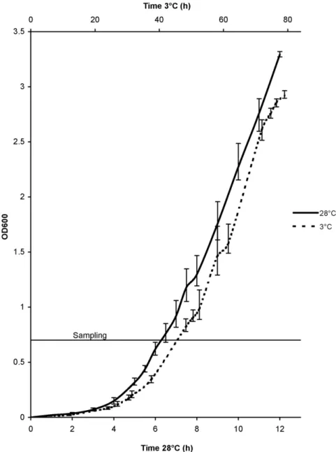
Y. pseudotuberculosisIP32953 was grown at 28uC on LB agar for 24 h. Three colonies (biological replicates) were inoculated and grown separately in LB broth overnight, inoculated into fresh LB (1:100), and grown at 28uC or 3uC to an early logarithmic growth phase (OD600of 0.7, corresponding to 10
8
viable cells/ml, Figure 1). Samples for total RNA extraction were collected by mixing 2610 ml of bacterial culture with a cold phenol-ethanol mixture (1:9) and kept on ice for 30 min. Samples were subsequently centrifuged at 2uC at 50006gfor 15 min, and the resulting cell
pellets were stored at270uC until RNA isolation. The total RNA was isolated using an RNeasy Midi kit (Qiagen GmbH, Hilden, Germany) with on-column DNase digestion using an RNase-free DNase set (Qiagen GmbH) according to the manufacturer’s instructions. An additional DNase treatment was performed with a DNA-free kit (Applied Biosystems, Foster City, California, USA) according to the manufacturer’s instructions. RNA concentration and quality were measured with a Nanodrop ND-1000 spectro-photometer (Thermo Fisher Scientific Inc., Waltham, Massachu-setts, USA). An A260/A280ratio of 2.0 (varied between 2.06–2.13)
was considered pure RNA. RNA integrity was examined using an Agilent 2100 Bioanalyzer (Agilent Technologies Inc., Santa Clara, California, USA). RNA was stored at270uC until use.
Reverse transcription
Each RNA sample was reverse transcribed into cDNA in duplicate (RT replicates) using a Dynamo cDNA Synthesis kit (Finnzymes Oy, Espoo, Finland) according to the manufacturer’s instructions. RNA was incubated for five min at 65uC to destabilize secondary structures. The total reaction volume for reverse transcription was 20ml and contained 300 ng of random hexamers, 900 ng of RNA,
and 2ml of M-MuLV RNase H+reverse transcriptase. The reactions were incubated at 25uC for 10 min, at 37uC for 30 min, and at 85uC for 5 min. To control DNA contamination, minus-RT controls were prepared from RNA samples by adding all the reaction components except the reverse transcriptase. The cDNAs were stored at220uC before use in RT-qPCR.
RT-qPCR
RT-qPCR was performed using a Dynamo Flash SYBR Green qPCR kit (Finnzymes Oy) according to the manufacturer’s instructions. Primers were designed for the predicted signal transduction system genes of IP32953 [12,29], the reference gene 16S rRNA gene (Table 1), and the ibpA encoding a heat shock protein (GenBank accession number BX936398) by using Primer3 software [30] (http://frodo.wi.mit.edu/primer3/) (Table S1). The total reaction volume was 20ml, including 4ml of template cDNA and 0.5mM of each primer. PCR runs were performed with a
Rotor-Gene 3000 Real Time Thermal Cycler (Qiagen GmbH). The amplification protocol consisted of initial denaturation at 95uC for 7 min, 40 cycles of denaturation at 95uC for 10 s, annealing at 60uC for 15 s, extension at 72uC for 20 s, and a final extension at 60uC for 1 min. Fluorescence data were acquired at the end of each extension step. After each run, a melt curve analysis was performed by raising the temperature at a rate of 0.5uC/5 s from 60uC to 98uC to confirm specificity. Minus-RT controls yielded no specific products, thus indicating no DNA contamination. Dilution series of pooled cDNA originating from the RT replicates of the biological replicates at 28uC and 3uC were amplified in triplicate (PCR replicates) to determine standard curves and thus the amplification reaction efficiencies for each primer pair. The reaction efficiency, based on the dilution series described above, was determined for 16S rRNA gene for four dilution series, and for the genes encoding signal transduction systems andibpAfor the dilution series from the cDNAs to be used in subsequent runs of the gene. For each primer pair, threshold fluorescence levels were set automatically with Rotor-Gene 3000 software, and reaction efficiencies were calculated as 10(21/M)
-1, where M is the slope of the straight line from a semilogarithmic plot of the quantification cycle (Cq) as a function of the cDNA concentration using Rotor-Gene 3000 software. Reaction efficiencies for 16S rRNA gene and signal transduction system encoding genes appear in Table S2 and Table S3, reaction efficiency ofibpAwas 0.92.
R~ 1
zEgeneDCq,gene(calibrator{sample)
1zE16SrRNAgene DC
q,16SrRNAgene(calibrator{sample) [31], where
Egene is the amplification reaction efficiency of a signal
tran-sduction system gene or ibpA transcript, E16S rRNA gene is the amplification reaction efficiency of 16S rRNA gene transcripts,
DCq,geneis the Cqdeviation between calibrator and sample for the signal transduction system gene or ibpA transcript, and DCq,16S
rRNA geneis the Cqdeviation between calibrator and sample for the 16S rRNA gene transcripts. The resulting Rs were averaged for RT replicates. The student’s t-test (Microsoft Excel) was performed for the biological replicates to test the differences between the relative expression levels of genes that encode signal transduction systems andibpAat 3uC and 28uC.
Mutagenesis
A cheA30–31::Ltr KanR mutant (hereafter called cheA30) was created using the TargeTron Gene Knockout System (Sigma-Aldrich Co.) following manufacturer’s instructions. All the primers used in constructing and confirming mutants are listed in Table S4. Briefly, re-targeting PCR was performed with primerscheA30
-Figure 1. Growth curves ofYersinia pseudotuberculosisIP32953 at 36C and 286C.
doi:10.1371/journal.pone.0025063.g001
Table 1.Verification of the use of 16S rRNA gene as a reference gene at low temperature inYersinia
pseudotuberculosisIP32953.
Parameter between 36C and 286C Value
Arithmetic mean (Cq) 15.82
Geometric mean (Cq) 15.81
Average deviation 0.47
CV(% Cq)a 2.96
IBS,cheA30-EBS1d,cheA30-EBS2 and EBS Universal. Resulting PCR product was digested and ligated into plasmid pACD4K-C, and transformed into E. coli by heat shock. The re-targeted plasmid clone was isolated by using a GeneJET Plasmid Miniprep Kit (Fermentas International Inc., Burlington, Ontario, Canada) and sequenced to confirm correct sequence with T7 primer. Electrocompentent Y. pseudotuberculosis IP32953 was made as described previously [32] and plasmid pAR1219 (Sigma-Aldrich Co.) was transformed into Y. pseudotuberculosis IP32953 using 0.1 cm cuvettes with 25mF, 200Vand 1.8 kV. Subsequently, cells were incubated in super-optimal broth with catabolite repression (SOC) (Sigma-Aldrich Co.) for 3 hours and plated on LB agar with ampicillin. Electrocompentent Y. pseudotuberculosis IP32953 containing pAR1219 was made as described above and the re-targeted pACD4K-C was introduced into the cells. After 3-hour SOC incubation, cells were plated on LB agar with ampicillin and chloramphenicol. After 24 hours, colonies from the LB agar plate were inoculated and grown overnight in LB broth with ampicillin and chloramphenicol. Cells were inoculated into fresh LB broth (1:50) with ampicillin and chloramphenicol and grown to an OD600 of 0.2. Expression and insertion of the intron were induced by adding 0.5 mM isopropylb-D-thiogalactoside (IPTG) (Sigma-Aldrich Co.) and incubation was continued overnight. Cells were centrifuged and resuspended into fresh LB. After 3 hours incubation, cells were plated on LB agars containing kanamycin. Knockouts were confirmed by PCR using cheA30 -flank-left and cheA30-flank-right, and cheA30-flank-left and EBS Universal primers. A cheA243–244::Ltr KanR mutant (cheA243) and a cheY243–244::Ltr KanR mutant (cheY243) were created similarly with primers cheA243-IBS, cheA243-EBS1d, cheA243 -EBS2, EBS Universal; and cheY243-IBS, cheY243-EBS1d,
cheY243-EBS2, EBS Universal, respectively. The cheA243 was confirmed with primerscheA243-flank-left andcheA243-flank-right, and cheA243-flank-right and EBS Universal. The cheY243 was confirmed with primerscheY243-flank-left andcheY243-flank-right, and cheY243-flank-left and EBS Universal. Primers Ninv-left and Ninv-right [33], and KvirF-left and KvirF-right [34] were used in PCR to confirm speciesY. pseudotuberculosisand the presence of the virulence plasmid pYV, respectively. All mutants were cured of pAR1219 by several subcultures in LB broth without ampicillin. All the ampicillin-sensitive clones were ascertained to contain the insertion mutation by PCR with flank primers. Primers Ninv-left and Ninv-right [33], and KvirF-left and KvirF-right [34] were used in PCR to confirm mutants for Y. pseudotuberculosis with pYV, respectively.
Southern blotting
Southern blotting was performed to confirm single intron insertion in the mutants. A PCR DIG Probe Synthesis Kit (Roche Applied Science, Penzberg, Germany) was used following manufacturer’s instructions with primers left and probe-right (Table S4) to synthesize a 199-bp digoxigenin-labelled probe. Pitcher’s method [35] was used to extract genomic DNA from the wild type and from the mutants with the following changes. Tris-EDTA (10:1) containing 0.4% sodium dodecyl sulphate, 220mg/ ml proteinase K and 2 mg/ml RNAse, was used for cell lysis. After adding ammonium acetate and incubating for 10 minutes on ice, 350ml of phenol-chloroform-isoamyl alcohol (25:24:1) was added,
samples were shaken rigorously, centrifuged, and Pitcher’s method was continued with the supernatant. Genomic DNA from the wild type,cheA30andcheA243was digested withHindIII (New England Biolabs Inc., Ipswich, Massachusetts, USA), and from cheY243
with XbaI (New England Biolabs Inc.). Digested DNAs and pACD4K-C (Sigma-Aldrich Co.) were shifted to a positively
charged nylon membrane, hybridized with the probe and detected as recommended by Roche Applied Science.
Growth experiments
Three separate colonies of theY. pseudotuberculosisIP32953 wild type strain, cheA30, cheA243, and cheY243 ampicillin sensitive mutant strains grown on LB agar, were separately inoculated into fresh LB broth and grown overnight with shaking. Subsequently, 1:100 dilutions into fresh LB broth were accomplished and 300ml of the dilutions were pipetted into wells of microtiter plates in triplicate. The microtiter plate was incubated at 3uC in the turbidity reader Bioscreen C MBR (Oy Growth Curves Ab, Helsinki, Finland). Turbidity of the cultures was measured at one-hour intervals after shaking for 20 s. Growth curves were acquired by plotting the ensuing OD600 values against time. Growth experiments were done likewise at 28uC but turbidity was measured at 20-minute intervals. Colony counting was performed to check that the overnight cultures contained similar amounts of viable cells. The correspondence of OD600values to the amount of viable cells was ensured by performing bacterial colony counting using plate count agar (BD) from the wild type strain and from all the mutants.
Motility tests
Motility test medium M103 with 2,3,5-triphenyl tetrazolium chloride [36] was modified to contain 0.3% agar. The IP32953 wild type strain and all the ampicillin sensitive mutants were stab-inoculated into M103 containing tubes. Tubes were incubated at 3uC, 22uC, 28uC and 37uC, and growth was monitored for 22 days, 9 days, 6 days and 6 days, respectively. Photographs were taken after 9 days at 3uC, 4 days at 22uC, 24 hours at 28uC and 24 hours at 37uC.
Results
Expression of signal transduction system encoding genes
The relative expression levels of 44 genes out of 54 genes were significantly higher (p,0.05) at 3uC than at 28uC (Table S3, in the order of expression ratios). The relative expression levels ofcheA
and cheY, which encode a CheA/CheY two-component system involved in chemotaxis, were 31- and 25-fold higher, respectively, at 3uC than at 28uC. 16S rRNA gene was used as a normalization reference for RT-qPCR. Its expression was stable between 3uC and 28uC (Table 1).
Construction of mutants
Three different insertional knockout mutants were constructed forcheAand cheY. Group II intron was inserted incheAeither in sense orientation at position 30–31 or in antisense orientation at position 243–244. The cheY insertional mutant had the intron located at position 243–244 in sense orientation. Southern blotting analysis confirmed that all the mutants had a single intron inserted in the genome.
Phenotypic characteristics of the mutants
Amount of 36106viable cells of the wild type strain and the mutants were used in the growth experiments. The mutant
cheA243 had clearly impaired, and mutant cheA30 had slightly impaired growth at 3uC (Figure 2A). The growth of the mutant
cheY243did not differ from that of the wild type at 3uC (Figure 2C). Mutations did not affect growth at 28uC (Figures 2B and D). Colony counting of bacteria confirmed the correspondence of OD600values to the number of viable cells.
Motility tests were performed for the cheA30, cheA243 and
cheY243mutants, and for the wild type strain IP32953. At 3uC and 22uC, the wild type IP32953 showed umbrella type motility, while none of thechemutants was motile (Figures 3A and B). At 28uC or 37uC none of the strains was motile (Figures 4A and B).
Discussion
The mechanismsY. pseudotuberculosisuses to adapt to and to grow at low temperatures are of special interest due to the food safety risks this psychrotrophic pathogen poses to modern chilled foods. Bacteria use signal transduction systems to monitor the environ-ment and to adjust their gene expression to changing conditions.
Y. pseudotuberculosis strain IP32953 has 24 complete signal transduction systems and 5 orphan hybrid histidine kinases or response regulators [12], for which roles in virulence and stress response have been demonstrated (Table S5). The role of signal transduction systems in the growth of Y. pseudotuberculosis at low temperature is poorly understood. To better understand their involvement, we determined the relative expression levels of 54 genes predicted to encode signal transduction systems in Y. pseudotuberculosisIP32953 at 3uC and 28uC. The relative expression levels of the majority of signal transduction system encoding genes were significantly higher at 3uC than at 28uC (Table S3). This is in line with previous transcriptomic studies with DNA microarrays on the psychrotrophicListeria monocytogenesand on the mesophilic
E. coli, the latter belonging to the same family as Yersinia, that showed approximately one third of the signal transduction system encoding genes to be induced at low temperature [37–39]. The even greater proportion of up-regulated signal transduction genes in Y. pseudotuberculosis was somewhat surprising. Therefore the expression analysis of ibpA, encoding a heat shock protein and shown to be expressed at five-fold higher level at 30uC than at 10uC [40], was included in our study as a control. The observed 11-fold higher expression level at 28uC than at 3uC confirmed that our RT-qPCR detects both up-regulated and down-regulated genes and thus verifies the findings on the signal transduction genes. Moreover, the 16S rRNA gene was shown to be expressed at stable levels at both temperatures (Table 1), which indicates 16S rRNA gene is a suitable reference gene for cold stress studies inY. pseudotuberculosis. It has also been shown to be the most stable housekeeping gene to be used in cold stress studies with L. monocytogenes[41].
The substantially higher expression levels ofcheAandcheY(Table S3), encoding the two-component system CheA/CheY, at low temperature than at optimal growth temperature suggest that this
two-component system plays a role in growth at low temperature. In a study conducted with aY. enterocoliticatransposon mutant library,
cheAwas also one of the most highly expressed genes during the early and mid-exponential growth phase at 10uC in relation to the expressions at 30uC [40]. Moreover,cheY expression in the mid-exponential growth phase at 4uC, compared to its expression at 37uC, was slightly induced, andcheAexpression remained nearly stable when investigated with DNA microarrays inY. enterocolitica
[42]. The role of the CheA/CheY two-component system at low temperature is further supported by inducedcheYexpression after cold shock from 37uC to 15uC inE. coli[43].
Involvement of the CheA/CheY two-component system in Y. pseudotuberculosis during cold stress was further investigated by constructing insertional knockout mutants cheA30, cheA243 and
cheY243. Mutation in cheA resulted in impaired growth at 3uC (Figure 2A), which confirms the important role of the CheA sensor during growth at low temperature. The mutation incheYdid not affect the growth at 3uC (Figures 2C), which may suggest that CheY is not essential during growth at low temperature, or other regulators may compensate for it. It is also possible that, as the only successful mutation site (between nucleotides 243–244) was near the C-terminus of the 390-bp long cheY, a functional truncated N-terminus of CheY was sufficient to exert the growth of thecheY mutant at 3uC. At 28uC, the growth of none of the mutants differed from the wild type strain (Figures 2B and D).
Components of the chemotaxis signal transduction system are highly conserved and, inE. coli, consist of chemoreceptors and six Che proteins: CheA, CheW, CheY, CheZ, CheB, and CheR [44]. The transmembrane chemoreceptors monitor the environment, and together with the histidine kinase CheA and another protein CheW form receptor-signaling complexes that regulate the autophosphorylation of CheA and thus the phosphorylation of the response regulator CheY [44]. Phosphorylated CheY diffuses to flagellar motors where it promotes clockwise rotation of the flagella, leading to a tumbling motion that keeps the cell in place [44]. When a bacterium senses an attractant stimulus, the phosphorylation of CheY decreases due to the inhibited kinase activity of CheA and the dephosphorylation of CheY by CheZ [44]. Flagella begin to rotate counterclockwise and a swimming motion is achieved which pushes the cell forward [44]. All of the aforementioned Che proteins are present in the genome of Y. pseudotuberculosis[12]. Based on the established role of CheA/CheY in other bacteria, mutation incheAorcheYwas expected to hamper motility of Y. pseudotuberculosis, which was further confirmed by motility tests. At 3uC and 22uC, only the wild type strain was motile (Figures 3A and B), whereas none of the strains was motile at 28uC or 37uC (Figures 4A and B). The lack of motility of the IP32953 wild type strain at 28uC is in agreement with a previous study demonstrating impaired motility for this strain at 28uC [22]. Chemotaxis proteins belong to the flagellar regulon [45], and the expression of flagellar genes in Y. enterocolitica W22703 is highest at 20uC [46]. The genes encoding chemotaxis proteins are induced by an alternative sigma factor FliA and are repressed by an anti-sigma factor FlgM through the inactivation of FliA [45,47,48], both of which are regulated by the transcriptional activators FlhD and FlhC [45,49–51] in Y. enterocolitica and Y. pseudotuberculosis.Y. enterocoliticaandY. pseudotuberculosisrequire FliA, FlhD, and FlhC for motility [49–52]. InY. enterocolitica, fliA and
flgMare transcribed at 25uC, but not at 37uC [47]. In addition to positively regulating flagellar genes, FliA negatively regulates several plasmid-encoded virulence genes at 25uC through VirF in
Y. enterocolitica [53]. The observed increase in cheA and cheY
pseudotuber-culosisis motile at 3uC as shown in our study. Moreover, whether also virulence is down-regulated at 3uC, needs to be confirmed by further studies. Based on the aforementioned, we suggest that (1) the CheA/CheY two-component system is part of the low temperature sensing network of Y. pseudotuberculosis, (2) the CheA/CheY is needed for motility, and (3) at least an intact CheA is required for growth of Y. pseudotuberculosis at low temperature. Future research is warranted to investigate the role of the CheA/CheY two-component system in the regulation of the flagellation cascade.
An orphan response regulator,YPTB1603, predicted to encode a GerR family transcriptional regulatory protein [27], saw the third highest relative expression level at 3uC relative to the expression level at 28uC. The function of YPTB1603 inYersiniahas not previously been reported. A homolog ofYPTB1603isECs0418
encoding a response regulator specific toE. coli O157:H7 strain Sakai [54]. The role of ECs0418 has not been studied. In addition to the response regulator receiver domain, an InterProScan search [55] suggested both the YPTB1603 and the ECs0418 contain a transcriptional regulator domain typical to LuxR family regulators
Figure 2. Growth curves ofYersinia pseudotuberculosisIP32953 wild type strain,cheA, andcheYmutants.Growth of thecheAmutants at 3uC (A) and 28uC (B), and thecheYmutant at 3uC (C) and 28uC (D). In graphs (A) and (C), measured values are shown in twenty-hour intervals, in (B) and (D), in one-hour intervals. Error bars represent minimum and maximum values.
that have been associated with several cellular and signaling events, including quorum sensing. It is thus intriguing to speculate that quorum sensing-type signals may be part of the low temperature sensing network. Our results demonstrate that the transcription ofYPTB1603is enhanced at low temperature.
The genes that encode two-component systems KdpD/KdpE, YfhK/YfhA, PmrB/PmrA, NarX/NarP, CreC/CreB, RstB/ RstA, BaeS/BaeR, SsrA/SsrB, HydH/HydG, EvgS/EvgA,
PhoR/PhoB, EnvZ/OmpR, YPTB2728/YPTB2729, BarA/
UvrY, YPTB2718/YPTB2719 and a multistep phosphorelay RcsC/YojN/RcsB had significantly higher (p-value,0.05) relative expression levels (median 4.0-fold) at 3uC than at 28uC (Table S3). Some of these signal transduction systems have been associated with survival from stresses posed by bile salts, low pH, hydrogen peroxide or increased osmolarity in previous studies (Table S5),
but this study is the first to demonstrate the increased expression of these genes in cold stress inY. pseudotuberculosis.
In the six two-component systems YehU/YehT, CpxA/CpxR, PhoQ/PhoP, CopS/CopR, NtrB/NtrC and ArcB/ArcA, the expression of only one of the two genes that encode a system differed with temperature. The results could be attributed to inactivation of certain components and re-established cross-talk between non-cognate kinase-regulator pairs. InE. coli, 7 of the 21 histidine kinases investigated can phosphorylate non-cognate response regulators in addition to the cognate regulators, and 9 of the 34 response regulators can be phosphorylated by non-cognate histidine kinases in addition to the cognate onesin vitro[56]. Cross-talk, however, is considered rare
in vivo[10,57]. Useful cross-talk, or cross-regulation, is seldom used when it is beneficial to combine multiple stimuli into one response or to expand a single stimulus to many responses [10].
Figure 3. Motility tests ofYersinia pseudotuberculosisIP32953 wild type strain, andcheAandcheYmutants.(A) 3uC and (B) 22uC. IP32953 (1),cheA30(2),cheA243(3), andcheY243(4).
The relative expression level of thearcAgene, which encodes the response regulator of the ArcB/ArcA two-component system was significantly higher (2.3; p-value,0.05), and that of YPTB2099, predicted to encode the orphan response regulator RssB, was significantly lower (0.2; p-value,0.05) at 3uC than at 28uC (Table S3). ArcA mainly regulates the transcription of genes involved in respiratory metabolism under anaerobic growth conditions inE. coli
[58]. The histidine kinase ArcB phosphorylates both ArcA and RssB [59]. In log-phase cells or under low oxygen tension, the stationary-phase sigma factor sS is down-regulated because phosphorylated ArcA represses the transcription of rpoS which encodes sS, and phosphorylated RssB targetssS for proteolysis by an ATP-driven ClpXP protease inE. coli[59,60]. In the stationary phase or under high oxygen tension, the phosphorylation of ArcB diminishes, leading to the diminished phosphorylation of ArcA and RssB. Consequently,
rpoStranscription increases, andsSproteolysis is reduced, resulting in a greater amount ofsS[59]. A greater amount ofsSenhances the expression of ArcA, leading to additionalsSstabilization due to the
competition of ArcA and RssB for phosphorylation by ArcB [59]. In
E. coli,sSis induced at low temperature [1], as is the expression of
arcAinY. enterocolitica[40]. There are no studies on the role ofsSand its regulation at low temperature inY. pseudotuberculosis. However, our results indicate that the behavior of ArcA and RssB in Y. pseudotuberculosiscould be similar to that inE. coli.
The results demonstrate that several signal transduction system encoding genes are upregulated at low temperature compared to optimum growth temperature inY. pseudotuberculosis. The importance of the CheA/CheY two-component system in the growth of Y. enterocoliticaat low temperatures was demonstrated by the impairment of growth of the cheA mutants at 3uC. The function of signal transduction systems at cold temperatures is probably arranged in complex networks with other cellular components. The identity of and interplay between these components is an important question and warrants further investigation.Y. pseudotuberculosis, a psychro-trophic pathogen with a wide temperature range for growth, requires mechanisms that facilitate its growth at low temperature. Long
Figure 4. Motility tests ofYersinia pseudotuberculosisIP32953 wild type strain, andcheAandcheYmutants.(A) 28uC and (B) 37uC. IP32953 (1),cheA30(2),cheA243(3), andcheY243(4).
storage at cold favors the proliferation ofY. pseudotuberculosisover other bacteria in foods and has led toY. pseudotuberculosisoutbreaks through vegetables stored at low temperature. Investigation of the cold tolerance mechanisms may provide keys to novel strategies to control this psychrotrophic food pathogen in refrigerated foods.
Supporting Information
Table S1 Primers used in quantitative real-time reverse transcription-PCR in this study.
(DOC)
Table S2 Cq (quantification cycle) values and reaction efficiencies (E) of 16S rRNA gene.
(DOC)
Table S3 Cq (quantification cycle) values, reaction efficiencies (E), expression ratios (R), and standard deviations (SD) of Rs of the signal transduction system encoding genes ofYersinia pseudotuberculosisIP32953. (DOC)
Table S4 Primers used in mutant construction and confirmation in this study.
(DOC)
Table S5 Predicted signal transduction systems of
Yersinia pseudotuberculosis IP32953 and their known functions inYersinia.
(DOC)
Acknowledgments
We thank Hanna Korpunen, Esa Penttinen, Erika Pitka¨nen and Kirsi Ristkari for their technical assistance. Thanks to Ilkka Palonen for consultation and assistance with photographs and graphics.
Author Contributions
Performed the experiments: RK EP. Analyzed the data: RK PS EP. Wrote the paper: EP ML HK. Study design and conception: EP HK ML PS. Provided valuable input in writing the paper: EP ML RK PS HK.
References
1. Palonen E, Lindstro¨m M, Korkeala H (2010) Adaptation of enteropathogenic
Yersiniato low growth temperature. Crit Rev Microbiol 36: 54–67.
2. Wren BW (2003) The yersiniae—a model genus to study the rapid evolution of bacterial pathogens. Nat Rev Microbiol 1: 55–64.
3. Fredriksson-Ahomaa M, Lindstro¨m M, Korkeala H (2010)Yersinia enterocolitica
andYersinia pseudotuberculosis. In: Juneja VK, Sofos JN, eds. Pathogens and Toxins in Foods: Challenges and Interventions. Washington, D.C., USA: ASM Press. pp 164–180.
4. Fukushima H, Gomyoda M, Shiozawa K, Kaneko S, Tsubokura M (1988)
Yersinia pseudotuberculosisinfection contracted through water contaminated by a wild animal. J Clin Microbiol 26: 584–585.
5. Fukushima H, Gomyoda M, Ishikura S, Nishio T, Moriki S, et al. (1989) Cat-contaminated environmental substances lead toYersinia pseudotuberculosisinfection in children. J Clin Microbiol 27: 2706–2709.
6. Nuorti JP, Niskanen T, Hallanvuo S, Mikkola J, Kela E, et al. (2004) A widespread outbreak of Yersinia pseudotuberculosis O:3 infection from iceberg lettuce. J Infect Dis 189: 766–774.
7. Jalava K, Hakkinen M, Valkonen M, Nakari UM, Palo T, et al. (2006) An outbreak of gastrointestinal illness and erythema nodosum from grated carrots contaminated withYersinia pseudotuberculosis. J Infect Dis 194: 1209–1216. 8. Rimhanen-Finne R, Niskanen T, Hallanvuo S, Makary P, Haukka K, et al.
(2009)Yersinia pseudotuberculosiscausing a large outbreak associated with carrots in Finland, 2006. Epidemiol Infect 137: 342–347.
9. Fredriksson-Ahomaa M, Korkeala H (2003) Low occurrence of pathogenic
Yersinia enterocoliticain clinical, food, and environmental samples: A methodolog-ical problem. Clin Microbiol Rev 16: 220–229.
10. Laub MT, Goulian M (2007) Specificity in two-component signal transduction pathways. Annu Rev Genet 41: 121–145.
11. Stock AM, Robinson VL, Goudreau PN (2000) Two-component signal transduction. Annu Rev Biochem 69: 183–215.
12. Marceau M (2005) Transcriptional regulation inYersinia: An update. Curr Issues Mol Biol 7: 151–178.
13. Grabenstein JP, Marceau M, Pujol C, Simonet M, Bliska JB (2004) The response regulator PhoP of Yersinia pseudotuberculosis is important for replication in macrophages and for virulence. Infect Immun 72: 4973–4984.
14. Marceau M, Sebbane F, Ewann F, Collyn F, Lindner B, et al. (2004) ThepmrF
polymyxin-resistance operon ofYersinia pseudotuberculosisis upregulated by the PhoP-PhoQ two-component system but not by PmrA-PmrB, and is not required for virulence. Microbiology 150: 3947–3957.
15. Kumar S, Balakrishna K, Agarwal GS, Merwyn S, Rai GP, et al. (2009) Th1-type immune response to infection by pYV-curedphoP-phoQnull mutant of
Yersinia pseudotuberculosisis defective in mouse model. Antonie Van Leeuwenhoek 95: 91–100.
16. Sun YC, Koumoutsi A, Darby C (2009) The response regulator PhoP negatively regulatesYersinia pseudotuberculosisandYersinia pestisbiofilms. FEMS Microbiol Lett 290: 85–90.
17. Fisher ML, Castillo C, Mecsas J (2007) Intranasal inoculation of mice with
Yersinia pseudotuberculosiscauses a lethal lung infection that is dependent onYersinia
outer proteins and PhoP. Infect Immun 75: 429–442.
18. Carlsson KE, Liu J, Edqvist PJ, Francis MS (2007) Extracytoplasmic-stress-responsive pathways modulate type III secretion inYersinia pseudotuberculosis. Infect Immun 75: 3913–3924.
19. Carlsson KE, Liu J, Edqvist PJ, Francis MS (2007) Influence of the Cpx extracytoplasmic-stress-responsive pathway onYersinia sp.-eukaryotic cell con-tact. Infect Immun 75: 4386–4399.
20. Heroven AK, Bo¨hme K, Rohde M, Dersch P (2008) A Csr-type regulatory system, including small non-coding RNAs, regulates the global virulence regulator RovA ofYersinia pseudotuberculosisthrough RovM. Mol Microbiol 68: 1179–1195.
21. Karlyshev AV, Oyston PCF, Williams K, Clark GC, Titball RW, et al. (2001) Application of high-density array-based signature-tagged mutagenesis to discover novelYersiniavirulence-associated genes. Infect Immun 69: 7810–7819. 22. Hinchliffe SJ, Howard SL, Huang YH, Clarke DJ, Wren BW (2008) The importance of the Rcs phosphorelay in the survival and pathogenesis of the enteropathogenic yersiniae. Microbiology 154: 1117–1131.
23. Hu Y, Lu P, Wang Y, Ding L, Atkinson S, et al. (2009) OmpR positively regulates urease expression to enhance acid survival ofYersinia pseudotuberculosis. Microbiology 155: 2522–2531.
24. Hu Y, Wang Y, Ding L, Lu P, Atkinson S, et al. (2009) Positive regulation offlhDC
expression by OmpR inYersinia pseudotuberculosis. Microbiology 155: 3622–3631. 25. Go¨pel Y, Lu¨ttmann D, Heroven AK, Reichenbach B, Dersch P, et al. (2011)
Common and divergent features in transcriptional control of the homologous small RNAs GlmY and GlmZ in Enterobacteriaceae. Nucleic Acids Res 39: 1294–1309.
26. Flamez C, Ricard I, Arafah S, Simonet M, Marceau M (2008) Phenotypic analysis of Yersinia pseudotuberculosis 32777 response regulator mutants: New insights into two-component system regulon plasticity in bacteria. Int J Med Microbiol 298: 193–207.
27. Chain PSG, Carniel E, Larimer FW, Lamerdin J, Stoutland PO, et al. (2004) Insights into the evolution ofYersinia pestisthrough whole-genome comparison withYersinia pseudotuberculosis. Proc Natl Acad Sci U S A 101: 13826–13831. 28. Bottone EJ, Bercovier H, Mollaret HH (2005) Genus XLI. Yersinia. In:
Garrity GM, Brenner DJ, Krieg NR, Staley JR, eds. Bergey’s Manual of Systematic Bacteriology. East Lansing, MI, USA: Springer-Verlag. pp 838–848. 29. Flamez C, Ricard I, Arafah S, Simonet M, Marceau M (2007) Two-component system regulon plasticity in bacteria: A concept emerging from phenotypic analysis ofYersinia pseudotuberculosisresponse regulator mutants. Adv Exp Med Biol 603: 145–155.
30. Rozen S, Skaletsky H (2000) Primer3 on the WWW for general users and for biologist programmers. In: Krawetz S, Misener S, eds. Bioinformatics Methods and Protocols: Methods in Molecular Biology. Totowa, NJ: Humana Press. pp 365–386.
31. Pfaffl MW (2001) A new mathematical model for relative quantification in real-time RT–PCR. Nucleic Acids Res 29: 2002–2007.
32. Conchas RF, Carniel E (1990) A highly efficient electroporation system for transformation ofYersinia. Gene 87: 133–137.
33. Nakajima H, Inoue M, Mori T, Itoh K, Arakawa E, et al. (1992) Detection and identification ofYersinia pseudotuberculosisand pathogenicYersinia enterocoliticaby an improved polymerase chain reaction method. J Clin Microbiol 30: 2484–2486. 34. Kaneko S, Ishizaki N, Kokubo Y (1995) Detection of pathogenic Yersinia enterocoliticaandYersinia pseudotuberculosisfrom pork using the polymerase chain reaction. Contr Microbiol Immunol 13: 153–155.
35. Pitcher DG, Saunders NA, Owen RJ (1989) Rapid extraction of bacterial genomic DNA with guanidium thiocyanate. Lett Appl Microbiol 8: 151–156. 36. Weagant SD, Feng P (2001)Yersinia enterocolitica. In: Hammack T, Feng P,
Jinneman K, Regan PM, Kase J, Bacteriological Analytical Manual. USA: U.S. Food and Drug Administration.
38. Moen B, Janbu AO, Langsrud S, Langsrud O, Hobman JL, et al. (2009) Global responses ofEscherichia colito adverse conditions determined by microarrays and FT-IR spectroscopy. Can J Microbiol 55: 714–728.
39. Chan YC, Raengpradub S, Boor KJ, Wiedmann M (2007) Microarray-based characterization of theListeria monocytogenescold regulon in log- and stationary-phase cells. Appl Environ Microbiol 73: 6484–6498.
40. Bresolin G, Neuhaus K, Scherer S, Fuchs TM (2006) Transcriptional analysis of long-term adaptation of Yersinia enterocolitica to low-temperature growth. J Bacteriol 188: 2945–2958.
41. Tasara T, Stephan R (2007) Evaluation of housekeeping genes in Listeria monocytogenesas potential internal control references for normalizing mRNA expression levels in stress adaptation models using real-time PCR. FEMS Microbiol Lett 269: 265–272.
42. Kapatral V, Campbell JW, Minnich SA, Thomson NR, Matsumura P, et al. (2004) Gene array analysis ofYersinia enterocoliticaFlhD and FlhC: Regulation of enzymes affecting synthesis and degradation of carbamoylphosphate. Microbi-ology 150: 2289–2300.
43. Phadtare S, Inouye M (2004) Genome-wide transcriptional analysis of the cold shock response in wild-type and cold-sensitive, quadruple-csp-deletion strains of
Escherichia coli. J Bacteriol 186: 7007–7014.
44. Baker MD, Wolanin PM, Stock JB (2006) Signal transduction in bacterial chemotaxis. Bioessays 28: 9–22.
45. Soutourina OA, Bertin PN (2003) Regulation cascade of flagellar expression in Gram-negative bacteria. FEMS Microbiol Rev 27: 505–523.
46. Bresolin G, Trcek J, Scherer S, Fuchs TM (2008) Presence of a functional flagellar cluster Flag-2 and low-temperature expression of flagellar genes in
Yersinia enterocoliticaW22703. Microbiology 154: 196–206.
47. Kapatral V, Olson JW, Pepe JC, Miller VL, Minnich SA (1996) Temperature-dependent regulation of Yersinia enterocolitica class III flagellar genes. Mol Microbiol 19: 1061–1071.
48. Ding L, Wang Y, Hu Y, Atkinson S, Williams P, et al. (2009) Functional characterization of FlgM in the regulation of flagellar synthesis and motility in
Yersinia pseudotuberculosis. Microbiology 155: 1890–1900.
49. Young GM, Smith MJ, Minnich SA, Miller VL (1999) TheYersinia enterocolitica
motility master regulatory operon,flhDC, is required for flagellin production, swimming motility, and swarming motility. J Bacteriol 181: 2823–2833. 50. Wang Y, Ding LS, Hu YB, Zhang Y, Yang BY, et al. (2007) TheflhDCgene
affects motility and biofilm formation inYersinia pseudotuberculosis. Sci China C Life Sci 50: 814–821.
51. Atkinson S, Chang CY, Patrick HL, Buckley CMF, Wang Y, et al. (2008) Functional interplay between theYersinia pseudotuberculosis YpsRI and YtbRI quorum sensing systems modulates swimming motility by controlling expression offlhDCandfliA. Mol Microbiol 69: 137–151.
52. Iriarte M, Stainier I, Mikulskis AV, Cornelis GR (1995) ThefliAgene encoding s28
inYersinia enterocolitica. J Bacteriol 177: 2299–2304.
53. Horne SM, Pru¨ß BM (2006) Global gene regulation inYersinia enterocolitica: Effect of FliA on the expression levels of flagellar and plasmid-encoded virulence genes. Arch Microbiol 185: 115–126.
54. Tobe T (2008) The roles of two-component systems in virulence of pathogenic
Escherichia coliandShigella spp.. Adv Exp Med Biol 631: 189–199.
55. Hunter S, Apweiler R, Attwood TK, Bairoch A, Bateman A, et al. (2009) InterPro: The integrative protein signature database. Nucleic Acids Res 37(suppl 1): D211–D215.
56. Yamamoto K, Hirao K, Oshima T, Aiba H, Utsumi R, et al. (2005) Functional characterizationin vitroof all two-component signal transduction systems from
Escherichia coli. J Biol Chem 280: 1448–1456.
57. Groban ES, Clarke EJ, Salis HM, Miller SM, Voigt CA (2009) Kinetic buffering of cross talk between bacterial two-component sensors. J Mol Biol 390: 380–393. 58. Malpica R, Sandoval GRP, Rodrı´guez C, Franco B, Georgellis D (2006) Signaling by the Arc two-component system provides a link between the redox state of the quinone pool and gene expression. Antioxid Redox Signal 8: 781–795.
59. Mika F, Hengge R (2005) A two-component phosphotransfer network involving ArcB, ArcA, and RssB coordinates synthesis and proteolysis ofsS
(RpoS) inE. coli. Genes Dev 19: 2770–2781.
60. Hengge R (2009) Proteolysis ofsS
(RpoS) and the general stress response in
Escherichia coli. Res Microbiol 160: 667–676.
61. Ro¨nneba¨umer K, Sander G, Shutinoski B, Schmidt MA, Heusipp G (2009) Controlled activation of the Cpx system is essential for growth of Yersinia enterocolitica. FEMS Microbiol Lett 296: 274–281.
62. Heusipp G, Nelson KM, Schmidt MA, Miller VL (2004) Regulation ofhtrA
expression inYersinia enterocolitica. FEMS Microbiol Lett 231: 227–235. 63. Maxson ME, Darwin AJ (2004) Identification of inducers of the Yersinia
enterocoliticaphage shock protein system and comparison to the regulation of the RpoE and Cpx extracytoplasmic stress responses. J Bacteriol 186: 4199–4208. 64. O’Loughlin JL, Spinner JL, Minnich SA, Kobayashi SD (2010)Yersinia pestis
two-component gene regulatory systems promote survival in human neutrophils. Infect Immun 78: 773–782.
65. Winfield MD, Latifi T, Groisman EA (2005) Transcriptional regulation of the 4-amino-4-deoxy-L-arabinose biosynthetic genes inYersinia pestis. J Biol Chem 280: 14765–14772.
66. Dahiya I, Stevenson RMW (2010)Yersinia ruckerigenes that attenuate survival in rainbow trout (Oncorhynchus mykiss) are identified using signature-tagged mutants. Vet Microbiol 144: 399–404.
67. Dahiya I, Stevenson RMW (2010) The UvrY response regulator of the BarA– UvrY two-component system contributes toYersinia ruckeriinfection of rainbow trout (Oncorhynchus mykiss). Arch Microbiol 192: 541–547.
68. Venecia K, Young GM (2005) Environmental regulation and virulence attributes of the Ysa type III secretion system ofYersinia enterocoliticabiovar 1B. Infect Immun 73: 5961–5977.
69. Huang YH, Ferrie`res L, Clarke DJ (2009) Comparative functional analysis of the RcsC sensor kinase from differentEnterobacteriaceae. FEMS Microbiol Lett 293: 248–254.
70. Sun YC, Hinnebusch BJ, Darby C (2008) Experimental evidence for negative selection in the evolution of aYersinia pestispseudogene. PNAS 105: 8097–8101. 71. Rebeil R, Ernst RK, Gowen BB, Miller SI, Hinnebusch BJ (2004) Variation in lipid A structure in the pathogenic yersiniae. Mol Microbiol 52: 1363–1373. 72. Hitchen PG, Prior JL, Oyston PCF, Panico M, Wren BW, et al. (2002)
Structural characterization of lipo-oligosaccharide (LOS) from Yersinia pestis: Regulation of LOS structure by the PhoPQ system. Mol Microbiol 44: 1637–1650.
73. Li Y, Gao H, Qin L, Li B, Han Y, et al. (2008) Identification and characterization of PhoP regulon members in Yersinia pestisbiovarMicrotus. BMC Genomics 9: 143.
74. Grabenstein JP, Fukuto HS, Palmer LE, Bliska JB (2006) Characterization of phagosome trafficking and identification of PhoP-regulated genes important for survival ofYersinia pestisin macrophages. Infect Immun 74: 3727–3741. 75. Zhou D, Han Y, Qin L, Chen Z, Qiu J, et al. (2005) Transcriptome analysis of
the Mg2+
-responsive PhoP regulator inYersinia pestis. FEMS Microbiol Lett 250: 85–95.
76. Oyston PCF, Dorrell N, Williams K, Li SR, Green M, et al. (2000) The response regulator PhoP is important for survival under conditions of macrophage-induced stress and virulence inYersinia pestis. Infect Immun 68: 3419–3425. 77. Brzostek K, Raczkowska A, Zasada A (2003) The osmotic regulator OmpR is
involved in the response ofYersinia enterocoliticaO:9 to environmental stresses and survival within macrophages. FEMS Microbiol Lett 228: 265–271.
78. Dorrell N, Li SR, Everest PH, Dougan G, Wren BW (1998) Construction and characterisation of aYersinia enterocoliticaO:8ompRmutant. FEMS Microbiol Lett 165: 145–151.
79. Brzostek K, Brzostkowska M, Bukowska I, Karwicka E, Raczkowska A (2007) OmpR negatively regulates expression of invasin in Yersinia enterocolitica. Microbiology 153: 2416–2425.
80. Raczkowska A, Skorek K, Bielecki J, Brzostek K (2011) OmpR controlsYersinia enterocolitica motility by positive regulation offlhDCexpression. Antonie Van Leeuwenhoek 99: 381–394.
81. Raczkowska A, Brzostek K (2004) Identification of OmpR protein and its role in the invasion properties ofYersinia enterocolitica. Pol J Microbiol 53: 11–16. 82. Brzostek K, Raczkowska A (2007) The YompC protein ofYersinia enterocolitica: