J of Evolution of Med and Dent Sci/ eISSN- 2278-4802, pISSN- 2278-4748/ Vol. 4/ Issue 07/Jan 22, 2015 Page 1160
SONOGRAPHIC TRIANGULAR CORD SIGN IN BILIARY ATRESIA
Ramesh R. L1, Vishnu Murthy G. S2, Vinay Jadhav3
HOW TO CITE THIS ARTICLE:
Ramesh R. L, Vishnu Murthy G. S, Vinay Jadhav. Sonographic Triangular Cord Sign in Biliary Atresia . Journal of Evolution of Medical and Dental Sciences 2015; Vol. 4, Issue 07, January 22; Page: 1160-1165,
DOI: 10.14260/jemds/2015/163
ABSTRACT:BACKGROUND: Aim of this prospective study was to evaluate prospectively the role of triangular cord sign in diagnosing biliary atresia and also to assess the prognostic value of trianular cord (TC) sign. METHODS: We prospectively studied 65 consecutive infants with conjugated bilirubinemia who underwent ultrasonography examination to look for TC sign. US examination was performed by a single operator who was blinded to results of other investigations, with a 7.5 MHz multi-frequency microconvex transducer and a 12 MHz multi-frequency linear assay transducer. The final diagnosis of biliary atresia was made on the basis of surgery. The role of TC sign in diagnosis biliary atresia was evaluated. RESULTS: 25 infants (15male &10female) had surgically confirmed biliary atresia and 40 (26 male & 14 female) had other documented causes of cholestasis. TC sign had a sensitivity, specificity, Positive Predictive value and Negative Predictive value of 80%, 97.5%, 95.23% and 88.63% respectively. Fibrosis Score of >3 in liver histopathology was found in 18 out of 20 TC sign positive cases. CONCLUSION: TC sign is a noninvasive, specific, time saving, simple diagnostic tool in the evaluation of infants with cholestasis. TC sign has a diagnostic as well as prognostic value in biliary atresia.
KEYWORDS: Biliary atresia, triangular cord sign, cholestasis.
INTRODUCTION: Neonatal cholestasis is defined as impaired bile flow through the biliary system resulting in the accumulation of biliary metabolites, such as bilirubin and bile acids, in the blood as well as in extrahepatic tissues. Significant accumulation (> 20% of total) of conjugated bilirubin (i.e., conjugated hyperbilirubinemia) reflects decreased excretion by damaged hepatic parenchymal cells, disease of the biliary tract, or both. The incidence of neonatal cholestasis is estimated to be approximately 1 in 2500 live births.1 Although its cause is varied, extrahepatic biliary atresia (EHBA) or idiopathic neonatal hepatitis (NH) is seen in 60–90% of infants with conjugated hyper-bilirubinemia.2,3
These two entities have similar clinical and biochemical findings. However, they have completely different pathogenesis and management. Therefore, it is very important to accurately diagnose these conditions.
EHBA is an obliterative cholangiopathy of unknown cause affecting both the intra- and extrahepatic bile ducts. If left untreated, EHBA has a progressive course to cirrhosis and end stage liver failure. Portoenterostomy, the surgical treatment of choice for EHBA, is unlikely to be of value if performed after the patient is 3 months old.2 Currently, the reference standard for the diagnosis of EHBA is laparotomy with intraoperative cholangiography because only this procedure can either confirm or exclude EHBA with certainty.4
J of Evolution of Med and Dent Sci/ eISSN- 2278-4802, pISSN- 2278-4748/ Vol. 4/ Issue 07/Jan 22, 2015 Page 1161 Humphrey and Stringer5 showed preoperative sonography to have a very high accuracy of 98% for the diagnosis of EHBA. Therefore, the purpose of our study was to prospectively assess the role of sonography in arriving at an accurate preoperative diagnosis of EHBA in infants.
SUBJECTS AND METHODS: This prospective descriptive study was conducted in a tertiary care level teaching hospital after obtaining approval from the institutional review board. Sixty-five consecutive infants with conjugated hyperbilirubinemia (conjugated bilirubin > 20% of total bilirubin) were enrolled in the study during the period January 2012 to march 2013(15 months). The details of clinical history, physical examination, laboratory findings, and other investigations were recorded.
All the sonographic examinations were performed by a senior consultant pediatric radiologist with 12 yrs experience in hepatobiliary scans in infants who was blinded to the results of other investigations such as hepatobiliary scintigraphy, liver function testing, and histologic findings after liver biopsy.
The equipment used was a high resolution ultrasonography system (Esaote My lab 5) with a 7.5-MHz multi frequency microconvex transducer and a 12-MHz multi frequency linear-array transducer. None of the infants were sedated.
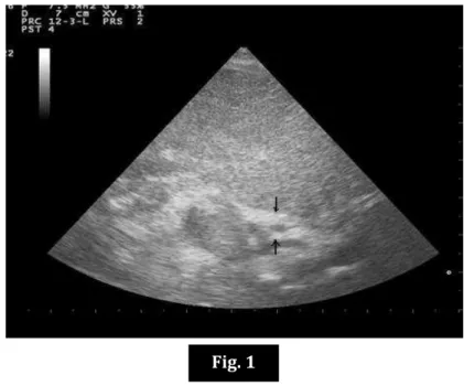
At longitudinal scanning, we measured thickness of the echogenic anterior wall of the right portal vein (EARPV) just proximal to the right portal vein bifurcation site. With US, the sole criterion for the TC sign was an EARPV that was thicker than 4 mm on longitudinal US images6 (Fig. 1).
The final diagnosis of EHBA was made on the basis of Per Operative Cholangiography. All confirmed cases of Biliary Atresia underwent kasai portoenterostomy and wedge biopsy. Fibrosis scoring in each case was noted. Other investigations like Tandem mass spectroscopy, Alpha feto protein, Gamma GT, Prothrombin time, Ferrtin, Eye examination, non-glucose reducing substance in urine, Lipid profile, liver biopsy were done to diagnose other causes of intrahepatic cholestasis.
Statistical analysis was performed using statistics software (SPSS package, version 11.5, SPSS). Mean and standard deviations were calculated for the relevant variables. Sensitivity, specificity, PPV, NPV were calculated for TC sign and HIDA scan.
RESULTS: A total of 65 infants were evaluated – 25 with biliary atresia and 40 with other causes of cholestasis. There were no stastistically significant differences in patient characterstics between the two groups i.e. age, male: female ration, weight in Kg and total bilirubin levels. (Table 1)
Charactristic BA Group (n=25) Non-BA Group (n=40)
Age in days 64±22.6 (46 to 184) 59±24.4(42 to 190)
Male: Female 15:10 26:14
Weight (kg) 4.2±0.81 (3.1 to 6.12) 3.62±0.72 (2.1 to 6.22) Total Bilirubin (mg/dl) 11±2.1 (5.8 to 22.1) 9.8±3.21 (5.3 to 21)
Table 1: Patient Characteristics
J of Evolution of Med and Dent Sci/ eISSN- 2278-4802, pISSN- 2278-4748/ Vol. 4/ Issue 07/Jan 22, 2015 Page 1162 septicemia presented with conjugated hyperbilirubinemia and after treatment with appropriated antibiotics, cholestasis subsided. Out of 2 babies with proven tyrosinemia (Positive urinary succinyl acetone) one baby died and the other baby was lost for follow up. 2 babies with galactosemia presented with cholestasis during study period. One baby had cataract, severe sepsis, ascites (Decompensated liver disease) at the time of presentation. Inspite of adequate supportive care baby could not be revived. Other baby with galactosemia is on lactose free diet and doing well.
Causes No. of cases (total 40)
Idiopathic neonatal hepatits 20
CMV hepatitis 06
PFIC 03
PILBD 03
Choledochal cyst 02
Galactosemia 02
Tyrosinemia 02
Sepsis 02
Table 2: Non biliary atresia causes of cholestasis
In all infants with biliary atresia (n=25), the diagnosis was confirmed at surgery and subsequent histologic examination. Out of the 25 operated cases, 9 babies had successful kasai and doing well in followup. Recurrent cholanigtis was the commonest complication noted in this group.
Triangular cord sign of > 4 mm was found in 20 babies in BA group (n=25) and 01 baby in non BA group. The baby with positive TC sign in non BA group was having choledochal cyst. TC sign was negative (<4mm) in 5 babies with biliary atresia (Table 3). The TC sign had a sensitivity, specificity, PPV, NPV of 80%, 97.5%, 95.23% and 88.63% respectively for the diagnosis of BA.
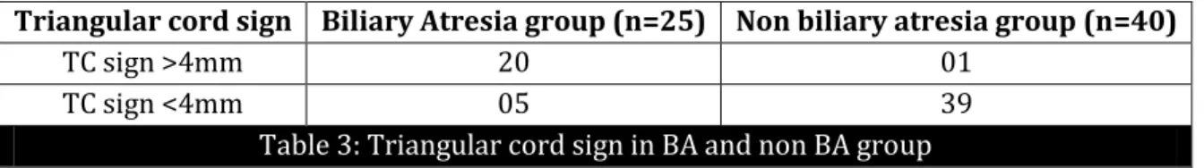
Triangular cord sign Biliary Atresia group (n=25) Non biliary atresia group (n=40)
TC sign >4mm 20 01
TC sign <4mm 05 39
Table 3: Triangular cord sign in BA and non BA group
Hepatoiminodiacetic acid (HIDA) scan was done in all 65 babies. In BA group, HIDA was positive in all 25 babies (Table 4). In nonBA group, HIDA was positive in 16 babies (n=40). The sensitivity, specificity, PPV and NPV of HIDA scanning for Biliary Atresia was found to be 100%, 60%, 60%, and 100%.
HIDA Scan Biliary atresia group (n=25) Non biliary atresia group (n=40)
Non-excretory (positive) 25 16
Excretory (negative) 0 24
Table 4: HIDA scan in BA and non BA group
J of Evolution of Med and Dent Sci/ eISSN- 2278-4802, pISSN- 2278-4748/ Vol. 4/ Issue 07/Jan 22, 2015 Page 1163
Fibrosis score TC sign >4 mm TC sign <4mm
Fibrosis score >3 18 01 Fibrosis score <3 02 04
Table 5: correlation of fibrosis score with TC sign
DISCUSSION: Neonatal cholestasis constitutes 19% of the total hepatobiliary diseases in children7.The two most common causes of neonatal cholestasis are biliary atresia and idiopathic neonatal hepatitis. Even with all available investigations, it is difficult to distinguish BA from INH, because of significant overlapping features between the two. BA should be picked up early, because survival, outcome and prognosis depends on timing of the surgery. There are several diagnostic tests which can be done to diagnose BA. Ultrasonography is one such valuable noninvasive test which can be used to diagnose BA. Among several US findings in BA, TC sign is simple, accurate, reliable sign.8
Biliary atresia is characterized by fibrous obliteration of extrahepatic bile duct with fibrous ductal remnant in the porta hepatis.9 The hepatic ducts that transform into a fibrous ductal remnant that is usually anterior and cranial to the hepatic artery and the portal vein. The original definition of TC sign was based on the idea that this fibrous ductal remnant could be seen as a thick tubular or triangular echogenic density along the anterior aspect of the portal vein.9
The TC sign has been reported to be highly specific for the diagnosis of BA in many studies, with values ranging from 96% to 100%.5,6,8,11
In our study also, TC sign was found to be highly specific but less sensitive marker of biliary atresia, with sensitivity, specificity, PPV and NPV of 80%, 97.5%, 95.25% and 88.63% respectively. Detection of TC sign requires a dedicated radiologist with adequate experience in hepatobiliary scans. In our study, the sensitivity, specificity, PPV and NPV of HIDA scan for the diagnosis of BA were found to be 100%, 60%, 60.97% and 100% respectively. These findings are comparable to the findings of previously published studies.12
In our study, we also found that fibrosis score in the histopathology varied proportionately with the TC sign. Fibrosis score of >3 was noticed in 18 out of 20 patients with positive TC sign (>4mm). To the best of our knowledge, no studies have compared TC sign with fibrosis score. We propose that TC sign also has a prognostic value in biliary atresia and indicates the extent of liver damage. Therefore TC sign can be used as one of the prognostic markers in biliary atresia along with other markers like prothrombin time, serum albumin, age etc. Further studies are required to confirm this finding.
CONCLUSION: The results of our study support those of previous studies that TC sign is a simple, time saving and specific diagnostic tool in the evaluation of infants with cholestasis. We propose that fibrosis score is proportional to the TC sign and TC sign should be considered as a prognostic markers along with other prognostic factors in biliary atresia.
REFERENCES:
1. Venigalla S, Gourley GR. Neonatal cholestasis.Semin Perinatol 2004; 28:348–355.
J of Evolution of Med and Dent Sci/ eISSN- 2278-4802, pISSN- 2278-4748/ Vol. 4/ Issue 07/Jan 22, 2015 Page 1164 3. Nicotra JJ, Kramer SS, Bellah RD, Redd DC. Congenital and acquired biliary disorders in children.
Semin Roentgenol 1997; 32: 215–227.
4. De Carvalho E, Ivantes CA, Bezerra JA. Extrahepatic biliary atresia: current concepts and future directions. J Pediatr (Rio J) 2007; 83:105–120.
5. Humphrey TM, Stringer MD. Biliary atresia: US diagnosis. Radiology 2007; 244:845–851.
6. Lee HJ, Lee SM, Park WH, Choi SO. Objective criteria of triangular cord sign in biliary atresia on US scans. Radiology 2003; 229: 395–400.
7. Yachha SK, Khanduri A, Kumar M, Sikora SS, Saxena R, Gupta RK, Kishore J. Neonatal cholestasis syndrome: an appraisal at tertiary center. Indian Pediatr 1996 Sep; 33(9): 729-34.
8. Kotb MA, Kotb A, Sheba MF, El Koofy NM, El-Karaksy HM, Abdel-Kahlik MK et al. Evaluation of the triangular cord sign in the diagnosis of biliary atresia. Pediatrics 2001; 108: 416-20.
9. Kasai M, Suzuki H, Ohashi E, Ohi R, Chiba P, Okamoto A. Techinque and results of operative management of biliary atrsia. World J Surg 1978; 1: 571-80.
10.Choi SO, Park WH, Lee HJ, Woo SK. Triangular cord : a sonographic finding applicable in the diagnosis of biliary atresia. J Pediatr Surg 1996; 31: 363-66.
11.Park WH, Choi SO, Lee HJ. The sonographic triangular cord coupled with gallbladder images in the diagnostic prediction of biliary atresia from infantile intrahepatic cholestasis. J Pediatr Surg 1999; 34: 1706-1710.
12.Poddar U, Bhattacharya A, Thapa BR, Mittal BR, Singh K. Ursodeoxycholic acid-augmented hepatobiliary scintigraphy in the evaluation of neonatal jaundice. J Nucl Med 2004; 45:1488-1492.
Fig. 1: Sagittal sonogram shows triangular cord sign (arrow) anterior to portal vein in a 12 weeks infant of biliary atresia.
J of Evolution of Med and Dent Sci/ eISSN- 2278-4802, pISSN- 2278-4748/ Vol. 4/ Issue 07/Jan 22, 2015 Page 1165
AUTHORS:
1. Ramesh R. L. 2. Vishnu Murthy G. S. 3. Vinay Jadhav
PARTICULARS OF CONTRIBUTORS:
1. Associate Professor, Department of Pediatric Radiology, IGICH.
2. Assistant Professor, Department of Pediatric Gastroenterology, IGICH. 3. Assistant Professor, Department of
Pediatric Surgery, IGICH.
NAME ADDRESS EMAIL ID OF THE CORRESPONDING AUTHOR:
Dr. Vishnu Murthy G. S,
No. 52/32, 37th Cross, 9th Block,
Jayanagar, Bangalore-560029. E-mail: [email protected]