ANAESTHESIA MANAGEMENT OF CSF RHINORRHEA REPAIR: A
CASE REPORT
Kirti Arvind Kundalwal1, Vinaya Rahul Kulkarni2, Kalpana Vinod Kelkar3
HOW TO CITE THIS ARTICLE:
Kirti Arvind Kundalwal, Vinaya Rahul Kulkarni, Kalpana Vinod Kelkar. “Anaesthesia Management of CSF Rhinorrhea Repair: A Case Report”. Journal of Evidence based Medicine and Healthcare; Volume 2,
Issue 37, September 14, 2015; Page: 5970-5975, DOI: 10.18410/jebmh/2015/822
ABSTRACT: Anaesthesiologist plays a major role in Cerebrospinal Fluid (CSF) rhinorrhea repair surgery as the prognosis of which is dependent on provision of clear bloodless surgical field and surgeons satisfaction. Anaesthesiologist also plays vital role in the management of CSF Lumbar drain. This case highlights the importance of hypotensive anaesthesia during endoscopic repair of a case of spontaneous CSF Rhinorrhea with successful perioperative management of Lumbar drainage of CSF
KEYWORDS: CSF Rhinorrhea, Endoscopic Repair, Lumbar Drainage, Hypotensive Anaesthesia.
INTRODUCTION: CSF rhinorrhea is the condition in which CSF leaks through nasal cavity due to dural fistula. Common causes are nontraumatic, traumatic, and iatrogenic. Nontraumatic CSF rhinorrhea may be associated with normal CSF pressure or CSF hypertension.1,2,3 Most of
traumatic type stop spontaneously within a week to 6 months. Anosmia and meningitis are two main dangers of it.2 In Nontraumatic type, there is greater flow of CSF. Side is not constant.
Headache is common than anosmia. It’s common in females and >30 years of age.2 Iatrogenic
type is commonly due to neurosurgical and otolaryngotic approaches to neoplastic diseases and functional endoscopic sinus surgery.
CASE REPORT:
PREOPERATIVE NOTES: 34yrs old female of 72kg weight and 160cm height came with chief complaints of sudden onset of watery discharge from nose since 15 days. Otherwise patient did not have history of any other significant systemic illness or any surgical history.
On Examination: General condition was fair, afebrile, pulse-96/min, BP-120/80mmHg, systemic examination-no abnormality noted.
Airway Examination: MPC grade 3, No missed or loose teeth, Neck/spine examination was normal.
Her routine laboratory examination, ECG, Chest X ray was within normal limits.
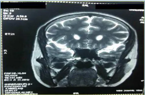
For diagnosis of CSF rhinorrhea CT PNS and MRI brain was done which showed a small defect in lateral wall of sphenoid sinus on right side with communication between sphenoid sinus and subarachnoid space. There was mild collection in sphenoid sinus. Rest of sinuses was normal. CT Cisternography- Was tried but there was block at T12-L1 level hence dye could not ascend.
On the day of surgery, patients NBM status and written informed consent for surgery and anaesthesia were confirmed. In operation theatre patients vitals checked, IV line secured in left hand with 18G intracath and 1 pint Ringers Lactate started. Multipara Monitor attached.
Before induction, as patient was obese we planned to put Lumbar drain in sitting position. Preloading with 1 pint of Ringers Lactate was done and patient was explained about the procedure. Patient was given sitting position. After painting and draping the spine of patient, Lumbar puncture was done with 18G epidural needle and immediately 18G catheter was put. 5-6cm length kept in subarachnoid space and fixed with Tegaderm. Filter was attached to outer end.
Now, patient was given supine position.
INTRAOPERATIVE NOTES After premedication patient was induced with inj pentothalsodium 500mg iv and Inj vecuronium 4mg. Patient was intubated with portex cuffed endotracheal tube no. 7.5. Throat packing was done. Patient was given 15% headup position. Maintenance of anaesthesia was done on O2 N2O-50 50% and Isoflurane. For the purpose of clear surgical field hypotensive anaesthesia with inj Dexmeditomedine was given. Bolus inj of 60microgram iv before induction followed by maintenance dose in the range of 30-60microgram/hour was given as per requirement after induction. Also 500mg Tranexamic acid IV was given for the same purpose. Then Patient was handed over to surgeon.
Surgeon cleaned face and nasal cavity with soap and betadine solution. Before introducing endoscope in nasal passage it was packed with cottonoids soaked in adrenaline 1 100000 solution and left inside for 3-5 min for hemostasis. After putting endoscope inside, VALSALVA maneuver was performed to confirm the leak site. The margin of defect was defined and made raw by removing any granulation tissue or bone chips. The defect was closed by cartilage by underlay technique, covered by two layers of fascia lata, each followed by fibrin glue by overlay technique, followed by abdominal fat. Throughout of the intraoperative period SystolicBlood Pressure was maintained around 80-90mmHg. After procedure was over after about 3 hours, Isoflurane and Dexmeditomedine was put off. After Posterior nasal packing patient was extubated in deep plane without coughing and observed in OT for 15-20min till she became wide awake. Before taking the patient out to recovery room, Lumbar drainage bag and filter was attached to epidural catheter and drainage of CSF flow was set with the help of regulator to 5-10 ml/hour and kept for 3 days in postoperative period.
DISCUSSION: Identification of CSF as the leaking fluid must precede demonstration of the cause as well as localization of the fistula itself.
Drops of CSF leak on absorbent filter paper show Double ring sign with central circle of blood and outer ring of CSF. Beta2 transferrin assay is more specific but unreliable with associated orbital injuries due to presence of beta2 transferrin in vitreous humour.4 Also we can
test for sugar level of leak fluid as it will be >30mg/dl in case of CSF but there are chances of false positive results with dextrstix reagent strip.5 CSF proteins can be differentiated from that of
Localisation of fistula can be done with dyes, fluorescent substance, radioactive tracer like 99mTc-DTPA, PET scan with EDTA, radiographic techniques like MRI, CT Cysternography.8
Pneumoencephalography has been used to show a dilated intrasellar subarachnoid pocket which by acting as a tense pulsating cyst may be responsible for a rupture in the sellar floor.9,10
There are several surgical options for routine repair of anterior cranial fossa CSF leaks. Intracranial repair was frequently done. Advantages of it being ability to inspect adjacent cerebral cortex, directly visualize the dural defect and seal a leak in presence of increased intracranial pressure with a large graft. But due to major disadvantages like increased morbidity, increased risk of permenant anosmia and trauma related to brain retraction including hematoma, cognitive dysfunction, seizures, oedema, hemorrhage, External approaches have taken place of intracranial approach. External approaches include external ethmoidectomy, transethmoidal sphenoidotomy, transseptal sphenoidotomy, transantral approach and endoscopic approach.
Endoscopic approach is a subset of the extracranial, extradural approach of CSF fistula. Since 1981 when Wigand first used endoscopic treatment to treat CSF rhinorrhea.11
Endoscopic approach has several advantages including better field visualization with enhanced illumination and magnified as well as angled visualization, ability to more accurately position the underlay or overlay grafts.12
Major role of anaesthesiologist in endoscopic surgery is to provide clear bloodless field for the surgeon to display his optimum expertise simultaneously providing the patient with the least hemodynamic changes and shorter recovery time. Dexmeditomedine was used by us for the same purpose.
Another crucial role of anaesthesiologist in CSF rhinorrhea surgeries is putting Lumbar drain and follow up in postoperative period. Drain can be put before induction or before extubation. We preferred to put it before induction as the patient was obese, sitting position was preferred. 18G epidural catheter was used for drainage purpose. Though putting 18G catheter is technically difficult and chances of blockage is more than larger diameter catheter, it will not allow CSF pressure to suddenly fall after lumbar puncture and over drainage in postoperative period.
Extubation in this scenario is crucial step. Aim of the anaesthesiologist is extubation without coughing in deeper plane as coughing can displace the graft. But risk factor in deeper plane of extubation is airway obstruction as patient should be conscious after extubation and should be able to breathe orally as nasal cavity is packed.
Postoperatively patient was instructed to avoid coughing, sneezing, walking and was given Head up position, stool softner for 4-5 days to avoid displacement and facilitate healing of graft. Lumbar drainage was performed at the rate of 5-10ml/hour for 3 days. Patient was observed for signs of meningitis and also complains of headache due to decreased CSF pressure. Patient was discharged on 7th day after check endoscopy.
So the surgery of CSF rhinorrhea is a success with pivotal role of anaesthesiologist in the form of:
Clear bloodless field.
Perfect placement of lumbar drain.
ACKNOWLEDGEMENT: Special Thanks to Dr. Rahul Telang, Associate Professor, Dept. of ENT, BJGMC, Pune.
Fig. 1: CT PNS Showing Collection in right sphenoid sinus and DNS with convexity towards right
Fig. 2: MRI brain Showing, 1. 2 mm sized defect in lateral wall of Sphenoid on Right side with communication between sphenoid and
REFERENCES:
1. Oberader S. Primary nontraumatic spontaneous CSF rhinorrhea with normal CSF pressure. Arch Neurol Neurochir Psychiatric 1972; 111 369-76.
2. Ommaya AK, Di Chiro et al. Hydrocephalus and leaks. In Wager H(ed) Principles of nuclear medicine, 2nd edition Phildelphia Saunder 1995.
3. Spetzler RF, Wilson CB. Dural fistula and their repair. In Youman JR(ed) Neurological surgery, 2nd edition. Philadelphia Saunder 1982PP 2209-27.
4. Ryall RG, Peacock MK, Simpson DA. Usefullness of beta2 transferrin assay in the detection of cerebrospinal fluid leaks following head injury. J Neurosurg 1992; 77 737-9.
5. Gadeholt H. The reaction of glucose oxidase test paper test in normal nasal secretion. Acta Otolaryngol (Stockh) 1964; 58 271-2.
6. Ricchetti A, Burkhard PR, Rodrigo N, et al. Skull base cerebrospinal fluid fistula. Neuroradiology 1972; 3 228-30.
7. Oberascher G, After E. First clinical experience with beta2 transferrin in cerebrospinal Fluid oto-andrhinoliquorrhea. HNO 1986; 34 151-5.
8. Allen MB Jr.El Gammal T, et al. Fistula detection in cerebrospinal fluid leakage. J Neurol Neurosurg Psychiat 1972; 35 664-8.
9. Busch W, Die. Morphologic der sella turcica und ihre Beziehungen Zur Hypophyse. Verchous Arch Pathol Anat 1951; 320 437-58.
10.Engels E P. Roentgenographic demonstration of a hypophyseal subarachnoid space. Am J Roentgenol 1958; 80 1001_4.
12.Kevin C Welch, Chief Editor Arlen D Meyers, et al, CSF Rhinorrhea Treatment and Management. (cited on 2015 Sept 04) Available from
http//emedicine.medscape.Com/article/861126-treatment.
NAME ADDRESS EMAIL ID OF THE CORRESPONDING AUTHOR:
Dr. Kirti Arvind Kundalwal, Assistant Professor,
Department of Anesthesiology, B. J. Government Medical College, Pune-411001, Maharashtra.
E-mail: [email protected]
Date of Submission: 07/09/2015. Date of Peer Review: 08/09/2015. Date of Acceptance: 10/09/2015. Date of Publishing: 14/09/2015.
AUTHORS:
1. Kirti Arvind Kundalwal 2. Vinaya Rahul Kulkarni 3. Kalpana Vinod Kelkar
PARTICULARS OF CONTRIBUTORS:
1. Assistant Professor, Department of Anesthesiology, B. J. Government Medical College, Pune, Maharashtra. 2. Associate Professor, Department of
Anesthesiology, B. J. Government Medical College, Pune, Maharashtra. 3. Professor & HOD, Department of