J. Evid. Based Med. Healthc., pISSN- 2349-2562, eISSN- 2349-2570/ Vol. 3/Issue 76/Sept. 22, 2016 Page 4114
STUDY OF 200 CASES OF PLEURAL FLUID
Ramakrishna R1, Venkata Kalyan Kumar P2
1Professor, Department of Pulmonary Medicine, Katuri Medical College, Guntur.
2Associate Professor, Department of Pulmonary Medicine, Katuri Medical College, Guntur.
ABSTRACT BACKGROUND
We have studied 200 patients of pleural fluid presenting to our tertiary care centre. Presence of cases of pleural fluid is a common presentation both in pulmonary and extrapulmonary diseases. We analysed the patients having both exudates and transudates and studied the results.
MATERIALS AND METHODS
We selected patients above 20 years of age and classified the patients with pleural fluid as having transudates and exudates. We studied the causes of transudates and exudates. A total of 200 patients are studied in this prospective study. Diagnosis of pleural exudates is made on the basis of Light’s criteria, chest x-ray, pleural fluid analysis, CT scan in selected patients, sputum examination, bronchoscopy and bronchial washings. Moribund and non-cooperative patients and HIV positives were excluded from the study.
RESULTS
Among the 200 patients, 91% have exudates. 9% have transudates by Light’s criteria. Tuberculosis is the commonest cause of effusions (64.83%) followed by malignancy (13.73%) and sympneumonic or parapneumonic effusions (9.89%). Pleural effusions occurred predominantly in males. Prevalence of diabetes Mellitus among cases of tuberculous pleural effusions is 13.56%. Tuberculous effusions are predominantly right-sided.
CONCLUSION
Predominant cases of pleural fluid are exudates. Commonest cause of pleural effusion is Tuberculosis followed by malignancy both pulmonary and extrapulmonary and sym. and parapneumonic effusions. Prevalence of Diabetes among Tuberculous pleural effusion cases is more or less same as in general population. Cough, expectoration fever, chest pain and breathlessness are the common symptoms occurring in three fourths of the patients of tuberculous pleural effusion. Most of the cases of Tuberculous effusion are above 30 years of age. In the diagnosis of tuberculous pleural effusion, Pleural fluid ADA is very important. Pleural fluid cytology, pleural biopsy, bronchoscopy, bronchial washings and sputum examination can aid in diagnosis of aetiology. Lung cancer is the commonest cause of malignant pleural effusions. Other causes of pleural effusion though rare should be considered in selective patients.
KEYWORDS
Tuberculous Pleural Effusion, Exudates, Cytology, Bronchoscopy, Malignant Effusions.
ABBREVIATIONS
HIV: Human immunodeficiency virus, ADA: Adenosine deaminase, LDH: Lactic dehydrogenase, FNAC: Fine needle aspiration cytology.
HOW TO CITE THIS ARTICLE: Ramakrishna R, Kumar VKP. Study of 200 cases of pleural fluid. J. Evid. Based Med. Healthc. 2016; 3(76), 4114-4118. DOI: 10.18410/jebmh/2016/879
INTRODUCTION: Pleural fluid arises from a number of Pulmonary and extrapulmonary problems. Pulmonary infections like Pneumonia, Tuberculosis, Malignancies of Lung and Pleura, Pancreatitis, Pulmonary thromboembolism, Oesophageal rupture, Hepatic and Splenic infections, Abdominal sepsis and Pancreatitis acute and chronic can be associated with pleural effusions.
Several malignancies like, carcinoma breast, osteogenic sarcoma, testicular and ovarian malignancies can be associated with pleural effusions. On the other hand, pleural fluid transudates are seen in congestive heart failure, cirrhosis of liver, hypoproteinaemia, sometimes in pulmonary thromboembolism secondary to atelectasis of lung. Pleural fluid aspiration and examination is a simple procedure and that can give enormous information regarding Pulmonary or extrapulmonary disease.
AIMS AND OBJECTIVES: To study the patients presenting with pleural fluid and identify the aetiology with the help of
clinical radiological biochemical histopathological
examination. Financial or Other, Competing Interest: None.
Submission 30-08-2016, Peer Review 12-09-2016, Acceptance 19-09-2016, Published 21-09-2016. Corresponding Author:
Dr. Ramakrishna R,
Sanjeevani Hospital, A1Q, Ram Kuteer Majestic, 3rd Line, 13th Cross Road, Brodipet, Guntur-522002.
J. Evid. Based Med. Healthc., pISSN- 2349-2562, eISSN- 2349-2570/ Vol. 3/Issue 76/Sept. 22, 2016 Page 4115
MATERIALS AND METHODS: We have done a
prospective study on the patients having pleural fluid attending Katuri Medical College Department of Pulmonary Medicine from 2013 April to 2015 June.
Inclusion Criteria: All the patients presenting with Pleural Fluid above 20 years of age.
Exclusion Criteria: Patients of Pleural fluid with severe comorbidities like HIV, unstable patients, moribund patients and terminal malignancies, severe hypoxia. Analysed pleural fluids using Light’s criteria along with thorough clinical and radiological, haematological and biochemical examination. Sputum microscopy for AFB was done for all the patients. Pleural biopsy, bronchoscopy, bronchial washings were done selectively. Other investigations were done based on the requirement of the patients.
RESULTS:
Total number of cases of Pleural Fluid Examined 200
No. of cases of Exudates 182
No. of cases of Transudates 18
Aetiology Total Male Female
18 14 4
COPD Cor pulmonale 8 07 1
Cardiac Disease with
CHF 4 03 01
Hypoproteinaemia 2 2 -
Associated with chronic
renal impairment 2 1 1
Associated with
generalised anasarca 2 1 1
Table 1: Analysis of 18 Cases of Pleural Transudates
Sl. No. Aetiology Total Males Females
1. Tuberculosis 118/182=64.83% 86/182=47.25% 32/182=17.58%
2. Primary or secondary malignancy 25=13.73% 18/182=9.89% 07=3.84%
3. Pleural effusion secondary to pulmonary
thromboembolism 03=1.65% 02=1.09% 01=0.55%
4. Pleural effusion secondary to chronic pancreatitis 03=1.65% 03=1.65% None
5. Pleural effusion secondary to
connective tissue disease 03=1.65% 03=1.65%
6. Sympneumonic and parapneumonic
effusions pleural effusions 18=9.89% 11=6.04% 07=3.85%
7. Pleural effusion secondary to amoebic hepatitis 02=1.09% 02=1.09% -
8. Pleural effusion secondary to chronic renal failure 04=2.19% 02=1.09% 02=1.09%
9. Pleural effusion after abdominal surgery 04=2.19% 03=1.65% 01=0.55%
10. Undiagnosed 02=1.09% 02=1.09% -
Table 2: Analysis of Exudates
Total No. of Exudates: 182.
Analysis of tuberculous pleural effusions among 118 cases of tuberculous pleural effusion, the following criteria were used for diagnosis. Patients met with multiple criteria. All the criteria together along with clinical and radiological examination.
Sl. No. Criteria No. of
Cases Percentage
1. Clinical Features 96/118 81.35%
2. Chest Radiology 44/118 37.29%
3. Pleural Fluid ADA
>40IU/L 101/118 85.59%
4.
Lymphocytic Predominance in
Pleural Fluid
84/118 74.20%
5. Sputum Positive
for AFB 26/118 22.3%
6.
Bronchial Washings Positive
for AFB
18/118 15.25%
7. Pleural Biopsy 6/118 5.8%
Table 3: Diagnosis of TB Pleural Effusion
Sl. No. Symptom No. of
Patients Percentage
1. Cough 82 69.49%
2. Expectoration 59 50%
3. Fever 80 67.79%
4. Chest Pain 76 64.46%
5. Haemoptysis 2 1.69%
6. Breathlessness 78 66.10%
7. Polyuria and
Nocturia 9 7.63%
8. Weight Loss 19 16.10%
9. Loss of Appetite 26 22.03%
J. Evid. Based Med. Healthc., pISSN- 2349-2562, eISSN- 2349-2570/ Vol. 3/Issue 76/Sept. 22, 2016 Page 4116
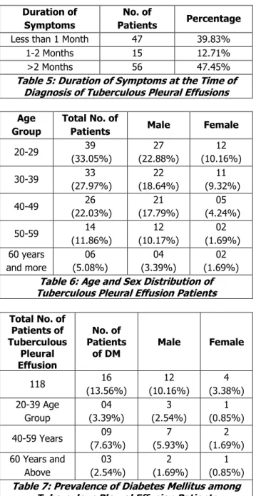
Duration of Symptoms
No. of
Patients Percentage
Less than 1 Month 47 39.83%
1-2 Months 15 12.71%
>2 Months 56 47.45%
Table 5: Duration of Symptoms at the Time of Diagnosis of Tuberculous Pleural Effusions
Age Group
Total No. of
Patients Male Female
20-29 39
(33.05%)
27 (22.88%)
12 (10.16%)
30-39 33
(27.97%)
22 (18.64%)
11 (9.32%)
40-49 26
(22.03%)
21 (17.79%)
05 (4.24%)
50-59 14
(11.86%) 12 (10.17%) 02 (1.69%) 60 years and more 06 (5.08%) 04 (3.39%) 02 (1.69%)
Table 6: Age and Sex Distribution of Tuberculous Pleural Effusion Patients
Total No. of Patients of Tuberculous Pleural Effusion No. of Patients of DM
Male Female
118 16
(13.56%) 12 (10.16%) 4 (3.38%) 20-39 Age Group 04 (3.39%) 3 (2.54%) 1 (0.85%)
40-59 Years 09
(7.63%)
7 (5.93%)
2 (1.69%) 60 Years and
Above 03 (2.54%) 2 (1.69%) 1 (0.85%)
Table 7: Prevalence of Diabetes Mellitus among Tuberculous Pleural Effusion Patients
Amount of Pleural Effusion as assessed by clinical, radiological and ultrasonographic examination among patients of Tuberculous Pleural Effusion. Total Number of Tuberculous Pleural Effusion: 118.
Amount of Pleural Effusion
No. of
Patients Percentage
Minimal (Obliteration of CP angle to 500
mL)
71 60.17%
Moderate (500 mL to
1000 mL) 33 27.97%
Large (>1000 mL) 14 11.9%
Table 8
Side of Effusion No. of Patients Percentage
Right sided 59 50%
Left sided 43 36.44%
Bilateral 16 13.56%
Table 9: Side of the Lesion in Tuberculous Pleural Effusion
Type of Pleural Effusion
No. of
Patients Male Female
Free Pleural Fluid 80 (67.79%) 52 (44.06%) 28 (23.72%) Encysted Pleural fluid 30 (25.42%) 28 (23.72%) 02 (1.69%) Multiple Encystments 08 (6.77%) 06 (95.08%) 02 (1.69%)
Table 10: Type of Tuberculous Pleural Effusion
Sl.
No. Aetiology
No. of
Cases Percentage
1.
Pleural effusion secondary to Primary Pulmonary Malignancy
21 84%
2. Secondary to ovarian
malignancy 1 04%
3. Secondary to
carcinoma breast 2 08%
4. Secondary to
osteogenic sarcoma 1 04%
Table 11: Analysis of Cases of Malignant Effusions: Total No. 25
DISCUSSION: Exudative pleural effusion poses a
diagnostic challenge as several pulmonary and
extrapulmonary causes lead to pleural exudates. Diagnosis exudates and transudates is routinely done on the basis of Light’s criteria,1 but multiple parameters have to be
considered in separating exudates and transudates.2 Other
parameters like pleural fluid albumin, cholesterol and comparison of pleural fluid and serum proteins and LDH can help in the differentiation. Some Indian authors have suggested limited criteria like cholesterol and LDH.3 In our
study of 200 patients, exudate constituted 91%. Total male patients exceeded female patients in several Indian studies also found predominant number of exudates and more number of male patients possibly because more male patients seek medical assistance.4 Manu Mohan K et al5 have
similar observations as in our study. Tuberculosis is the predominant cause of pleural effusion in our study occurring in 64% of patients. Other important causes included
sympneumonic and parapneumonic effusions and
malignancy.
J. Evid. Based Med. Healthc., pISSN- 2349-2562, eISSN- 2349-2570/ Vol. 3/Issue 76/Sept. 22, 2016 Page 4117 diagnosis of tuberculous pleural effusion. Our study showed
a male predominance, but predominant number of patients were in the above 30 years age group. Symptomatology varied in different studies. Cough, fever, chest pain and breathlessness are the predominant symptoms in our study occurring in predominant number of patients. Pleural effusion is predominantly right-sided in our study. 14% of the patients showed bilateral pleural effusions. Mohd Arif, Srivastava study6 had 60% of TB pleural effusions and they
were in 20-40 years age group. In our study, 51% of the patients are below 39 years age. About 13.56% of our Tuberculous pleural effusions have associated diabetes mellitus, which is probably similar to prevalence of diabetes
mellitus in South India.7 Predominant patients of
Tuberculous effusion patients had minimal pleural effusion of less than 500 mL as assessed by chest radiology and ultrasonographic examination.
This is because of better awareness of the patients and their seeking medical attention relatively early. 68% of our patients had free fluid and 32% had encysted effusion. This aspect is given attention because free fluid can be removed freely and with treatment patients have minimal pleural fibrosis and minimal restriction later on. C.H.S. Chan et al in their retrospective study found that only 6% of patients developed pleural thickening.8 In our study, we had 6.77%
of patients had multiply encysted pleural effusion, which can probably responsible for pleural thickening in subsequent years. Chan study from Hong Kong showed average age of Tuberculous pleural effusion was 44 years and that pleural biopsy is a better method of diagnosis and pleural fluid AFB was negative in all subjects. Pleural fluid ADA is more than 40 IU/L 85.6% of our tuberculous pleural effusion patients. This makes it an important tool in the diagnosis though there were a few borderline cases, which were decided on other diagnostic modalities. The importance of pleural fluid ADA was stressed in a number of studies. Sravan Kumar and Ritesh Agarwal study found poor utility, sensitivity as a
diagnostic tool in diagnosing tuberculous and
nontuberculous pleural effusion patients in chronic Kidney disease patients compared to DNA PCR.9 Sachin Kate and B.
K. Muthaa et al10 in their study found a sensitivity and
specificity of 93.3% and 90% respectively when ADA levels of 40 IU/L was taken as the criteria for the diagnosis of Tuberculous pleural effusion. Dr. Prabhakarra Rao et al11
also found Pleural fluid ADA as a very sensitive and specific marker of tuberculous pleural effusion and is simple, inexpensive and rapid.
Rama Saha et al12 in their study stressed the importance
of cytology of pleural fluid and histopathology of pleural biopsy specimen in the diagnosis of pleural effusions. We did pleural biopsy for only 8 patients and 6 of them were positive for tuberculosis. Among our Pleural effusions of other cause primary and secondary malignancies accounted for 13.65% and sympneumonic and parapneumonic effusions accounted for 9.89%. Other causes of pleural effusions were chronic pancreatitis, amoebic hepatitis, post abdominal surgery effusions, chronic renal impairment and pleural effusions secondary to connective tissue disease. Malignant effusions
were diagnosed by evidence of pulmonary and extrapulmonary malignant disease and analysis of pleural fluid and by pleural biopsy, bronchoscopy and bronchial washing, FNAC of lung lesions. Mohd Arif Siddiqui study6
showed parapneumonic effusions of 14.5% and malignant pleural effusions of 11.5% and the results in this regard are similar to our study. Arnab Maji et al13 in their study had
higher percentage of malignant pleural effusions of around 28% in their study of 568 patients of exudative Pleural effusions. Lung malignancies are the commonest cause of malignant pleural effusions in our study and also in Arnab Maji study,13 Basu A chakrbarty I et al14 concluded the
importance of pleural biopsy and ADA level and found that ADA levels of 70 IU/L are highly suggestive of tubercular aetiology. It requires extensive investigations when pleural effusions are found positive for secondary malignancy as a wide number of organs can cause secondary malignancy in the pleura.
Pleural effusions can also occur from obstruction of thoracic duct or pleural lymphatics secondary to malignant extension. In such cases, pleural fluid may not be positive for malignancy. Pleural effusions can occur in Chronic Kidney disease and it is necessary to differentiate Tuberculous pleural effusion from other causes. We had only small number of patients of pleural effusion associated with Chronic Kidney Disease. In a study among the CKD patients by Ray S, Mukherjee S. et al, they found a prevalence of 6.7% of pleural effusion among CKD patients. It is necessary to rule out Tuberculous pleural effusion in CKD patients.14
Massive recurrent pleural effusions can develop in asymptomatic pancreatic disease.15 Pleural effusions acute
respiratory distress syndrome associated with atelectasis and hypoxia can develop in acute pancreatitis.16 Chronic
massive effusions are reported with chronic pancreatitis.17
Pleural effusions of pancreatic origin are rich in pancreatic amylase.
Pleural effusion accompanying amoebic hepatitis and liver abscess is not uncommon in clinical practice. Though we have only two cases in our study group because of habit of alcoholism and high prevalence of amoebic hepatitis pleural effusion of this aetiology should be borne in mind in relevant cases. It can result because of extension of amoebic liver abscess into pleural cavity and lung18 or can be
reactionary effusion. Pleural effusions are also reported postoperatively after upper abdominal surgery can result from infection, atelectasis, sodium and water retention or age-related cardiac disease.19 They do not require any
specific treatment. In our series, we had four patients after abdominal surgery. Pleural effusions are also reported after CABG and cardiac valve surgery. They cause dyspnoea and chest pain and fever are uncommon, they disappear gradually over a few months.20
J. Evid. Based Med. Healthc., pISSN- 2349-2562, eISSN- 2349-2570/ Vol. 3/Issue 76/Sept. 22, 2016 Page 4118 failure. Predominant cause of exudates is Tuberculosis
occurring in 64.83% followed by primary or secondary malignancy (13.65%) and sym and parapneumonic effusions 9.89%. Other causes of pleural effusions included postoperative pleural effusions after abdominal surgeries, chronic pancreatitis, amoebic hepatitis, connective tissue disease, pulmonary thromboembolism and chronic renal failure.21 Tuberculous pleural effusions are diagnosed by
analysis of pleural fluid by microscopy, cytopathology, pleural fluid ADA, proteins, LDH, sputum microscopy and in selective cases by bronchoscopy and analysis of bronchial washings. Relevant history and extensive systemic examination is necessary in nontuberculous effusions.
REFERENCES
1. Light RW, Macgregor MI, Luchsinger PC,et al.Pleural effusions: the diagnostic separation of transudates and exudates. Ann Intern Med 1972;77(4):507-513.
2. Roth BJ, O'Meara TF, Cragun WH. The serum-effusion
albumin gradient in the evaluation of pleural effusions. Chest 1990;98(3):546-549.
3. Rungta R, Jha RK. Comparative analysis of pleural fluid biochemical parameters with cholesterol to differentiate transudates from exudates. J Assoc Chest Physicians 2013;1(2):54-57.
4. Patel R, Shah V, Kamdar D. Diagnostic approach to pleural effusion. GCSMC J Med Sci Vol 2015;4(1):57-61.
5. Mohan MK. Etiology and clinical profile of pleural effusion in a teaching hospital of south India: a descriptive study. Pulmon 2012;14(3):89-96. 6. Siddiqui MA, Srivastava VK. Clinical and etiological
profile of patients with pleural effusion: a retrospective cross-sectional study in North India. Medical Science 2016;6(5):285-287.
7. Mohan V, Sandeep S, Deepa R, et al. Epidemiology of
type 2 diabetes: Indian scenario. Indian J Med Res 2007;125(3):217-230.
8. Chan CH, Arnold M, Chan CY, et al. Clinical and pathological features of tuberculous pleural effusion and its long-term consequences. Respiration 1991;58(3-4):171-175.
9. Kumar S, Agarwal R, Bal A, et al. Utility of adenosine
deaminase (ADA), PCR & thoracoscopy in
differentiating tuberculous & non-tuberculous pleural effusion complicating chronic kidney disease. Indian J Med Res 2015;141(3):308-314.
10. Kate S, Mutha BK, Kulkarni G, et al. Study of diagnostic importance of adenosine deaminase levels in pleural effusions. MVP Journal of Medical Sciences 2015;2(2):104-109.
11. Rao PP, Rajakumari JH, Devi IV, et al. Adenosine deaminase as a diagnostic marker in tuberculous pleural effusion. International Journal of Innovative Research in Pharmaceutical and Medical Science 2015;3(1):1-5.
12. Saha R, Nayak P, Bhattacharya A, et al. The study of
Pleural diseases with special reference to
cytopathology and histopathology. International Journal of Biomedical and Advance Research 2015;6(8):599-610.
13. Maji A, Maikap MK, Jash D, et al. Role of common investigations in aetiological evaluation of exudative pleural effusions. Journal of Clinical and Diagnostic Research 2013;7(10):2223-2226.
14. Basu A, Chakrabarti I, Ghosh N, et al. A
clinicopathological study of tuberculous pleural effusion in a tertiary care hospital. Ann Trop Med Public Health 2012;5(3):168-172.
15. Ray S, Mukherjee S, Ganguly J, et al. A cross-sectional prospective study of pleural effusion among cases of chronic kidney disease, Indian J Chest Dis Allied Sci 2013;55(4):209-213.
16. Fernandes L, Mesquita A. Chronic massive recurrent
pleural effusion associated with asymptomatic pancreatic disease. Ind. J. Tub., 1996;43:31-34.
17. Browne GW, Pitchumoni CS. Pathophysiology of
pulmonary complications of acute pancreatitis. World J Gastroenterol 2006;12(44):7087-7096.
18. Dewan NA, Kinney WW, O'Donohue WJ. Chronic
massive pancreatic pleural effusion. Chest
1984;85(4):497-501.
19. Yokoyama T, Hirokawa M, Imamura Y. Respiratory failure caused by intrathoracic amoebiasis. Infect Drug Resist 2010;3:1-4.
20. Nielsen PH, Jepsen SB, Olsen AD. Postoperative pleural effusion following upper abdominal surgery. Chest 1989;96(5):1133-1135.