* Study carried out at the Hospital Israelita Albert Einstein, Hospital Infantil Darcy Vargas, Hospital Jaraguá and Hospital Iguatemi, São Paulo, Brazil. 1. Thoracic Surgeon. Hospital Israelita Albert Einstein, Hospital Jaraguá and Hospital Iguatemi, São Paulo, Brazil.
2. Attending Physician in the Department of Thoracic Surgery. Universidade de São Paulo – USP, University of São Paulo – School of Medicine, São Paulo, Brazil. 3. Coordinator of the Lung Transplant Program. Universidade Federal de São Paulo – UNIFESP, Federal University of São Paulo – Hospital São Paulo, São Paulo, Brazil. 4. Pediatric surgeon. Hospital Israelita Albert Einstein and Hospital Infantil Darcy Vargas, São Paulo, Brazil.
Correspondence to: Davi Wen Wei Kang. Avenida Rebouças, 3084, Cj. 14, Pinheiros, CEP 05402-600, São Paulo, SP, Brasil. Tel 55 11 3062-8295/3673-0765. E-mail [email protected]
Submitted: 29 January 2007. Accepted, after review: 21 July 2007.
Thoracoscopy in the treatment of pleural empyema in pediatric patients*
Toracoscopia no tratamento do empiema pleural em pacientes pediátricos
Davi Wen Wei Kang1, José Ribas Milanez de Campos2, Laert de Oliveira Andrade Filho1,
Fabiano Cataldi Engel1, Alexandre Martins Xavier3, Maurício Macedo4, Karine Meyer4
Abstract
Objective: To evaluate the results of thoracoscopy for the treatment of pleural empyema in pediatric patients. Methods: A retrospective study of 117 patients who underwent mediastinoscopy or video-assisted thoracoscopy for pleural empyema treatment. General anesthesia and single-lumen oral intubation were used. Surgery was indicated when there was pleural effusion and no clinical and radiological response to clinical treatment (antibiotics, physiotherapy and thoracocentesis) or severe sepsis, together with loculated pleural effusion (confirmed through ultrasound or computed tomography of the chest). Results: Between February of 1983 and July of 2006, 117 thoraco-scopies were performed in patients ranging in age from 5 months to 17 years (mean, 4 years). Mean time for thoracic drainage was 9 days (range, 2-33 days), and mean period of hospitalization was 16.4 days (range, 4 to 49 days). One patient (0.8%) died after surgery, and persistent fistula was observed in 33 patients (28%). In 7 cases (6%), open thoracotomy with pulmonary decortication was performed due to the disposition of the empyema. Conclusions: Management of pleural empyema in this age bracket is still controversial, and surgical indica-tion is often delayed, particularly when there are multiple loculaindica-tions or severe sepsis. Early thoracoscopy yields a better clinical outcome for pediatric patients with pleural empyema, with apparent decreased morbidity and mortality, earlier chest tube removal, earlier hospital discharge and improved response to antibiotic therapy.
Keywords: Empyema, pleural; Pediatrics/instrumentation; Thoracoscopy; Thoracic surgery, video-assisted.
Resumo
Objetivo: Apresentar resultados obtidos com a toracoscopia no tratamento do empiema pleural em pacientes pediátricos. Métodos: Foram avaliados 117 empiemas pleurais, utilizando-se o mediastinoscópio ou a videotoracoscopia, com anestesia geral e sonda de intubação simples. As indicações para a intervenção cirúrgica foram: derrame pleural com ausência de resposta clínica e radiológica ao tratamento clínico (antibióticos, fisioterapia e toracocentese) ou sepse grave, e derrame pleural loculado (documentado por ultrassonografia ou tomografia computadorizada do tórax). Resultados: De fevereiro de 1983 a julho de 2006, 117 toracoscopias foram realizadas em pacientes com idade entre 5 meses e 17 anos (média, 4 anos). O tempo médio de permanência do dreno torácico foi de 9 dias (2 a 33), e o tempo de internação hospitalar foi de 16,44 dias (4 a 49). Houve apenas um óbito (0,8%), e 33 pacientes (28%) tiveram como complicação fístula aérea prolongada. Em 7 pacientes (6%), houve necessidade de conversão para toracotomia com decorticação pulmonar em decorrência da organização do empiema. Conclusão: Não existe consenso para o tratamento do empiema pleural nesta faixa etária. A terapêutica cirúrgica é geralmente requisitada tardiamente no curso da doença, particularmente quando já existem múltiplas loculações ou quadro séptico grave. A toracoscopia indicada mais precocemente no tratamento do empiema pleural em pacientes pediátricos proporcionou uma melhor resposta à terapêutica clínica, aparentemente reduzindo o índice de morbi-mortalidade, o tempo de permanência do dreno torácico, o tempo de internação hospitalar e o tempo de antibioticoterapia.
Descritores: Empiema pleural; Pediatria/instrumentação; Toracoscopia; Cirurgia torácica vídeo-assistida.
Introduction
Pleural empyema is defined as pleural effusion in which bacteria have invaded the pleural cavity. Parapneumonic pleural effusion (pleural effusion associated with pneu-monia) is one of the most common causes of pleural empyema. Pleural empyema results from the progression
of the inflammation or pulmonary infection toward the pleural space. This progression can be divided into three stages: exudative, fibrinopurulent and organizational.
Hospital Israelita Albert Einstein, Hospital Jaraguá and Hospital Iguatemi, all located in São Paulo, Brazil. Of these 117 patients, 69 (59%) were male and 48 (41%) were female. They all underwent surgery under general anesthesia and with simple intubation: 54 (46%) underwent pleuroscopy (or mediastinoscopy); and 63 (54%) underwent video-assisted thoracoscopy.
Three different lengths of pleuroscope were used (11, 13 and 17 cm) depending on the age of the patient and on the size of the chest cavity. This instrument was introduced through the 4th or 5th intercostal space on the mid-axillary line, on the anterior axillary line or near the area of greatest interest. In addition, a second trocar port was typi-cally placed at the base of the operated hemithorax in order to introduce endoscopic instruments such as the suction pump, biopsy forceps, electric scalpel, etc. At the end of the surgical procedure, this opening accommodated the chest tube.
Depending on the age of the patient and on the size of the chest cavity, a 5 mm or 10 mm endo-scope was used in the video-assisted thoracoscopy. Endoscopes with a 30° angle, or with no angle, were used in order to facilitate the visualization of the pleural cavity. After the introduction in the chest cavity, similar to pleuroscopy, the second trocar was inserted, under direct viewing, at the base (mid-ax-illary line) of the operated hemithorax. This port was the principal site for the manipulation of the endo-scope, providing a wide general view of the pleural cavity, as well as providing the subsequent access for the chest tube. The first port, therefore, became the principal working channel. A third trocar was used at times in the triangle of auscultation to aid pulmonary decortication.
Material for culture and pathological anatomy was collected during all surgical procedures. In the post-operative period, all patients received common analgesics (dipyrone and paracetamol), with or without opioids. All patients received empirical antibiotic therapy prior to the surgical treatment of the empyema, and changes were made based on the clinical evolution and on the results of the culture of the material collected during the intra-operative period.
Chest tube diameter varied (20, 22, 24, 26 or 28 F) depending on the age of the patient and the size of the chest cavity, and all chest tubes were water-sealed. Criteria for removal of chest tubes draining the effusion and re-expanding the lung.(2)
Therefore, since empyema is a progressive disease, treatment methods differ in each phase. The use of broad spectrum antibiotics, with adequate pleural penetration, is mandatory at any stage of the disease. According to the stage of the disease, the drainage of the pleural effusion and lung re-expansion can be performed using the following techniques: thoracocentesis; pleural drainage with a chest tube; intrapleural fibrinolytic therapy; or surgical treatment (thoracoscopy/pleuroscopy, mini-thoracotomy, or even pulmonary decortication).(3)
The development of pleural empyema is deter-mined by three factors: resistance of the host; virulence of the bacteria; and time since disease onset.(4) We have noticed an increased incidence
of empyema in pediatric patients, as reported in the literature, regardless of early diagnosis and treatment, together with an increase in anti-biotic-resistant microorganisms.(5,6) Therefore,
there is a greater need for surgical treatments in the management of pleural empyema in this age bracket, principally through thoracoscopy, in order to perform pleural fluid drainage, since there are many cases of more rapid evolution to complicated empyema of greater severity.
To date, the management of empyema in pedi-atric patients is a controversial issue. There is as yet no consensus among pediatricians, thoracic surgeons and pediatric surgeons as to if and when surgical treatment is indicated. Data from controlled studies in adult patients cannot be extrapolated to pediatric patients, since the microorganisms responsible for the disease, the pulmonary patholo-gies and the comorbidities differ between the two populations. This study presents results of the use of thoracoscopy, with video-assisted thoracoscopy or mediastinoscopy, in the treatment of pleural empyema in pediatric patients. The surgical findings and peculiarities of the two methods and of this age bracket will also be discussed and compared to data in the literature.
Methods
without pleural fistula. This difference was statisti-cally significant (p = 0.003).
The microorganism that caused pleural empyema was identified in only 14.5% of the patients, and, in these patients, 64.7% of the microorganisms identified were Streptococcus spp. or Streptococcus pneumoniae. The other agents identified were as follows, in decreasing order: Staphylococcus aureus (17.6%), coagulase-negative Staphylococcus (5.9%), Staphylococcus warneri (5.9%) and Mycobacterium tuberculosis (5.9%).
Discussion
Pleural inflammation results in increased capil-lary permeability to proteins, fluid and leukocytes, forming sterile pleural effusion. This is the exudative stage, in which parapneumonic effusion is simple, or free in the cavity (it flows on lateral decubitus chest X-rays), and occurs within the first 24-72 h. The biochemical characteristics of this fluid are normal pH, normal glucose and low leukocyte count.
After this period, if the response to treatment in inadequate, there will be an accumulation of poly-morphonuclear leukocytes, bacterial invasion and fibrin deposition, which can result in the forma-tion of loculaforma-tions (complicated pleural effusion) or extremely purulent pleural fluid. The biochemical characteristics of the fluid are decreased pH and decreased glucose, as well as increased lactate dehy-drogenase. This stage can last from 7 to 10 days.
At two to four weeks after the onset of pleural empyema, fibroblast growth begins on the parietal and visceral surfaces, forming a membrane that is not elastic and entraps the lung, creating a fixed pleural cavity that has a propensity to maintain the infection.(1)
The treatment of pleural empyema has the following objectives: cleaning the pleural cavity; draining the pleural fluid; and re-expanding the lung. Drainage of the empyematous cavity can be performed through thoracocentesis, pleural drainage with a chest tube, or debridement, through thora-coscopy or more aggressive surgical techniques (such as thoracotomy) when there is organization of the pleural space.(3,11)
The first use of thoracoscopy in the treatment of pleural diseases was described by Jacobaeus, in 1910, when he performed the lysis of intrapleural adhesions using a modified cystoscope.(7) With
were as follows: output rate of < 50 mL within the preceding 24 h; absence of fistula; and X-ray showing an expanded lung. When there was persistent fistula (for more than 7 days), we changed the water seal using a Heimlich valve in order to decrease resist-ance of the drainage system.
For the statistical analysis, data were compiled in a Microsoft Excel spreadsheet, and analyses were carried out using the Statistical Package for the Social Sciences program, version 13.0 (SPSS Inc., Chicago, IL, USA). The Student’s t-test was used, together with the chi-square test or Fisher’s exact test, as indicated. The statistical significance was set at p < 0.05.
Results
Of the 117 patients submitted to thoracoscopy, 97 (82.9%) presented fibrinopurulent empyema (stage II), and 20 (17.1%) presented organized empyema (stage III). The mean drainage time was 8.98 days, ranging from 2 to 33 days.
Surgery was indicated in the following cases: pleural effusion with lack of clinical and radiological response to clinical treatment (antibiotics, physical therapy and thoracocentesis), with severe sepsis, in 43 patients (36.8%); and loculated pleural effu-sion (confirmed through ultrasound or computed tomography of the chest) in 74 patients (63.2%).
The mean total hospital stay, including the period of conservative treatment, was 16 days (range, 4 to 49 days). There was only 1 death (0.8%) during the peri-operative period (within the first 30 post-operative days). This 17-year-old patient presented diabetes mellitus and chronic renal insufficiency, and death occurred due to complications associated with the underlying disease.
The principal surgical complication was fistula (with or without residual pneumothorax on chest X-ray), which occurred in 33 patients (28.2%). Necrosis and lung abscesses were identified in 26 patients (22.2%) during surgery. In 7 patients (6%), thoracotomy with pulmonary decortica-tion was performed due to the disposidecortica-tion of the empyema.
pleural drainage and longer hospital stays. These findings reveal the severity of our patients, since a great number of these patients presented necrosis and lung abscesses, which were identified during debridement of the visceral pleura. In addition, early surgical intervention was necessary to prevent fistula, since surgical procedures performed during a more organized stage make debridement of visceral pleura more difficult, with a risk of pulmonary laceration. In the present study, these two factors were considered the principal factors responsible for longer hospital stays and longer use of pleural drainage, in contrast with data in the literature.
Thoracoscopy is an important instrument in the treatment of empyema, in adult and pedi-atric patients alike, principally because it has the following objectives: to obtain material for micro-biological and histopathological diagnosis; to promote the treatment of the pleural cavity with less surgical trauma; and to decrease hospital stays, promoting the early resumption of normal physical activities.(6,8,25) The smaller surgical scar, in
compar-ison with that resulting from conventional surgical procedures (thoracotomies, thoracoplasties and open thoracostomies),(24) is also an important factor,
not only for the patients but for the parents as well, since it minimizes the social stigma and helps physicians persuade the family of the real need to perform the surgical procedure.
Since endoscopes and microcameras have become more widely available, video-assisted thoracoscopy has been employed more frequently in the treatment of empyemas.(18-20) In this type of approach, the use
of a third trocar has become important, since one of the trocars is used for the introduction of the endoscope (4, 5 or 10 mm, depending on the age of the patient and size of the chest cavity). At the end of the surgery, the chest tube was inserted into one of the ports (typically the lower port).
In pediatric patients, we find some peculi-arities inherent to this age bracket regarding the endoscopic instrument used and the ventilation technique in the intra-operative period, principally in patients under the age of 12. The fact that there are no appropriate tubes for selective orotracheal intubation in pediatric patients prevents blockage and isolation of the affected hemithorax. In order to avoid contamination of the contralateral lung, constant and careful bronchial aspiration should be performed during the intra-operative period, so that the advent of antituberculosis drugs, the use of
thoracoscopy for surgical treatment (principally of tuberculosis, through artificial pneumothorax) was practically abandoned, and the value of the method was restricted to investigating pleural diseases and pleural effusion of unknown etiology.
However, in the 1970s, the development of endoscopic instruments, fiber optics and more accu-rate anesthetic methods allowed thoracoscopy to be used to greater advantage, in the diagnosis and in the treatment of pleural diseases. The description of the use of the mediastinoscope for the diagnosis of intrathoracic diseases, by Carlens, dates back to 1976,(9,10) and this instrument is currently used with
great efficacy in the investigation, treatment and staging of pleuropulmonary diseases.
In the 1990s, these technological improve-ments reached an apex, with the appearance of endoscopes with microcameras and adequate endo-scopic instruments that minimized surgical trauma, post-operative pain, hospital stay and hospital costs without decreasing the accuracy of the diagnosis or the effectiveness of the treatment. Since then, there has been a considerable increase in the use of thora-coscopy, which has expanded to include surgical procedures that were formerly performed as ‘open’ procedures (for example, thoracic sympathectomy and correction of pectus excavatum), and providing easier treatment of the conditions in which surgery is currently indicated (for example, treatment of multiloculated empyemas).(8)
The decision to perform a surgical interven-tion is controversial, since there is no consensus regarding the role of conservative treatment versus surgical treatment. Many studies indicate that surgical treatment is rarely necessary,(11-13) whereas
others show the benefits of early decortication or debridement of the infected pleura.(14-17) A
system-atic review of 67 studies, between 1981 and 2004, comparing conservative treatment (antibiotics and chest tube drainage of the pleural effusion) with surgical treatment, revealed that 76% of all patients evolved satisfactorily when receiving the former. However, comparing patients receiving conservative treatment to those undergoing surgery, mortality rates were higher (3.3 vs. 0%) and the duration of antibiotic therapy was longer (21.3 vs. 12.8 days), as was that of thoracic drainage (10.6 vs. 4.4 days) and hospitalization (20 vs. 10.8 days).(21) Pleural
lower lobe. The need for conversion to thoracotomy to allow pulmonary resection could have been avoided, in this last case, had there been pediatric endoscopic staplers available. Based on our experi-ence, as well as on the rapid clinical and radiological improvement of the patients, in whom we could visually diagnose lung abscesses during surgery, we realized that there was no need to resect areas presenting pulmonary gangrene. Lung parenchyma with necrosis and abscesses often suffers remodeling, appearing absolutely normal on chest X-rays and tomography scans of the chest during outpatient follow-up evaluations. This always occurred natu-rally if the pleural cavity had been properly drained during the intra-operative period, allowing enough yield to the alveolopleural or bronchopleural fistula and providing the necessary lung expansion to avoid the accumulation of fluid or air in the pleura.
The mean duration of drainage in patients with pleural fistula was 17.5 days, compared to 5.9 days in patients without. Abscess or necrosis of the lung parenchyma was observed in 26 patients (22.2%) diagnosed during thoracoscopy. Of those 26 patients, only 5 (4%) presented persistent fistula and were discharged, and thoracic drainage was maintained with the chest tube connected to the unidirectional Heimlich valve. All drains were removed one week after hospital discharge.
The fact that we found a low incidence of persistent fistula in patients with abscesses and pulmonary necrosis called our attention, and we concluded that there was no need for pulmonary resection in these cases, thus avoiding the morbidity and surgical trauma of a thoracotomy.
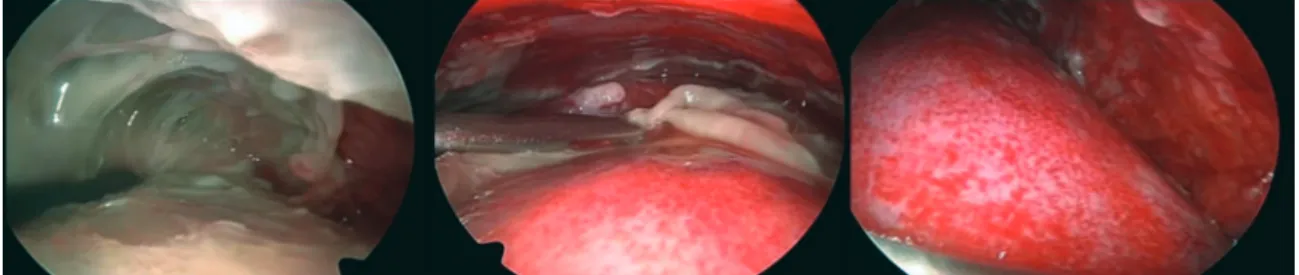
All patients submitted to thoracoscopy presented rapid clinical improvement (Figure 1), despite some residual pleural alterations seen on chest X-rays in some cases. The alterations were minor and did not represent any type of complication or symptom, which is in agreement with data in the literature.(11,27)
There was only one case (0.8%) in which the patient was submitted to an additional pleuroscopy, which was performed 5 days after the first proce-dure, due to the absence of lung expansion and the worsening of the sepsis. The drain was removed 4 days later, and there was clinical improvement.
Although indications for thoracoscopy in pedi-atric patients are practically the same as those applied in adult patients, it is rare for thoracoscopy secretions from the affected lung do not drain into
the contralateral lung. Bronchial blockage of the operated lung with balloon devices is an alternative that can prevent such secretions from contami-nating the contralateral lung.
In many cases, we opted for the use of the medi-astinoscope (at least to begin the procedure) for the following reasons: 1) it allows access to the pleural cavity through a small incision, under direct viewing, thereby avoiding false trajectories and pulmonary lesions, which are likely, since this is a region rich in neovascularization and inflammatory pleuropulmo-nary adhesions; 2) since younger patients maintain ventilation in the hemithorax that is being oper-ated, the pleuroscope itself maintains an artificial pneumothorax, displacing and lowering the lung, allowing visualization and debridement of the pleural cavity; 3) it allows very young children to be operated upon by a single surgeon, which is impor-tant, since the surgical field is extremely limited; 4) it allows the initial inventory of the cavity and the choice of the best sites for the introduction of the trocars.
However, the use of video-assisted thoracos-copy has other advantages: 1) it enables a wide and magnified view of the pleural cavity; 2) it allows the procedure, as well as the pathological condi-tions of the lung and pleura, to be monitored by the entire team that treats the patient, particularly the pediatrician; 3) the pleural cavity can be insufflated with carbon dioxide in order to create an artificial pneumothorax for better visualization of the pleural cavity (a technique that we typically avoid due to the possibility that it will lead to worsening of the ventilatory or hemodynamic status of the patient). Whenever necessary, we introduced a third trocar in order to insert a lung spreader.
surgeon is typically called in late in the course of pleural empyema, after the patient has developed multiple loculations or severe sepsis. In the present study, early thoracoscopy in the treatment of pleural empyema in pediatric patients provided a better response to clinical therapy, apparently resulting in lower rates of morbidity and mortality, as well as reducing the duration of thoracic drainage, hospital stays and antibiotic therapy.
References
1. Rodgers BM, McGahren ED. Mediastinum and pleura. In: Oldham KT, Colombani PM, Foglia RP, Skinner MA, editors. Principles and Practice of Pediatric Surgery. Philadelphia: Lippincott Williams & Wilkins; 2005. p. 929.
2. Jaffé A, Balfour-Lynn IM. Management of empyema in children. Pediatr Pulmonol. 2005;40(2):148-56.
3. Moran JF. Surgical management of pleural space infections. Semin Respir Infect. 1988;3(4):383-94.
4. Tuomanen EI, Austrian R, Masure HR. Pathogenesis of pneumococcal infection. N Engl J Med. 1995;332 (19):1280-4.
5. Byington CL, Spencer LY, Johnson TA, Pavia AT, Allen D, Mason EO, et al. An epidemiological investigation of a sustained high rate of pediatric parapneumonic empyema: risk factors and microbiological associations. Clin Infect Dis. 2002;34(4):434-40.
6. Schultz KD, Fan LL, Pinsky J, Ochoa L, Smith EO, Kaplan SL, et al. The changing face of pleural empyemas in children: epidemiology and management. Pediatrics. 2004;113(6):1735-40.
7. Hatzinger M, Kwon ST, Langbein S, Kamp S, Häcker A, Alken P. Hans Christian Jacobaeus: Inventor of human laparoscopy and thoracoscopy. J Endourol. 2006;20(11):848-50. 8. de Campos JR, Andrade Filho LO, Werebe EC, Minamoto H,
Quim AO, Filomeno LT, et al. Thoracoscopy in children and adolescents. Chest. 1997;111(2):494-7.
9. Carlens E. Mediastinoscopy: a method for inspection and tissue biopsy in the superior mediastinum. Dis Chest. 1959;36:343-52.
10. Rodgers BM, Talbert JL. Thoracoscopy for diagnosis of intrathoracic lesions in children. J Pediatr Surg. 1976;11(5):703-8.
to be performed in a pediatric patient based on the etiological diagnosis of the pleural effusion.(8) At our
facility, we have seen only one case of pleural tuber-culosis diagnosed through thoracoscopic biopsy of the pleura in a pediatric patient (an 11-year-old boy).
For the comfort of the parents, the pediatric patient and the pediatrician, as well as of the surgeon, simple thoracic drainage is typically performed under general anesthesia. Combining thoracoscopy with simple thoracic drainage allows the cleaning of the pleural cavity and the lung expansion, as well as the pleural drainage, to be performed under direct viewing, facilitating the surgical treatment and making it more efficacious.(18,22,23) Thoracoscopy
should be scheduled for as early as possible in order to avoid organization within the pleural space, and subsequent loss of pulmonary function, due to lung entrapment or chronic empyema. This would result in the need for pulmonary decortication, which involves a posterior lateral thoracotomy for the excision of the rigid, thick pleura, as well as evacua-tion of the purulent material.(24,26) The participation
of the thoracic surgeon from the beginning of the treatment of such patients ensures that the neces-sary surgical intervention will be performed at an earlier and more appropriate time.(24)
Most pediatric patients with empyema improve after antibiotic therapy and simple pleural drainage.(21)
However, early, aggressive therapy, with the partici-pation of the thoracic surgeon, principally using thoracoscopy, can result in shorter disease duration and a shorter hospital stay. Bronchopleural fistulas and hypertensive pneumothorax are rare compli-cations in pediatric patients. However, when they occur, they increase the time to convalescence.
Thoracoscopy is the method of choice in the treatment of stage II pleural empyema. The thoracic
in the era of video-assisted thoracoscopy. Am Surg. 2005;71(6):512-4.
20. Kalfa N, Allal H, Montes-Tapia F, Lopez M, Forgues D, Guibal MP, et al. Ideal timing of thoracoscopic decortication and drainage for empyema in children. Surg Endosc. 2004;18(3):472-7.
21. Avansino JR, Goldman B, Sawin RS, Flum DR. Primary operative versus nonoperative therapy for pediatric empyema: a meta-analysis. Pediatrics. 2005;115(6):1652-9.
22. Klena JW, Cameron BH, Langer JC, Winthrop AL, Perez CR. Timing of video-assisted thoracoscopic debridement for pediatric empyema. J Am Coll Surg. 1998;187(4):404-8. 23. Merry CM, Bufo AJ, Shah RS, Schropp KP, Lobe TE. Early
definitive intervention by thoracoscopy in pediatric empyema. J Pediatr Surg. 1999;34(1):178-80; discussion 180-1. 24. Balfour-Lynn IM, Abrahamson E, Cohen G, Hartley J, King S,
Parikh D, et al. BTS guidelines for the management of pleural infection in children. Thorax. 2005;60(Suppl 1):i1-21. 25. Gates RL, Caniano DA, Hayes JR, Arca MJ. Does VATS provide
optimal treatment of empyema in children? A systematic review. J Pediatr Surg. 2004;39(3):381-6.
26. Fraga JC, Kim P. Surgical treatment of parapneumonic pleural effusion and its complications. J Pediatr (Rio J.). 2002;78(Supl 2):161-70.
27. Murphy D, Lockhart CH, Todd JK. Pneumococcal empyema: outcome of medical management. Am J Dis Child. 1980;134(7):659-62.
11. McLaughlin FJ, Goldmann DA, Rosenbaum DM, Harris GB, Schuster SR, Strieder DJ. Empyema in children: clinical course and long-term follow-up. Pediatrics. 1984;73(5):587-93. 12. Berger HA, Morganroth ML. Immediate drainage is not
required for all patients with complicated parapneumonic effusions. Chest. 1990;97(3):731-5.
13. Kennedy AS, Agness M, Bailey L, White JJ. Decortication for childhood empyema. The primary provider’s peccadillo. Arch Surg. 1991;126(10):1287-91.
14. Gustafson RA, Murray GF, Warden HE, Hill RC. Role of lung decortication in symptomatic empyemas in children. Ann Thorac Surg. 1990;49(6):940-6; discussion 946-7. 15. Kercher KW, Attorri RJ, Hoover JD, Morton D Jr.
Thoracoscopic decortication as first-line therapy for pediatric parapneumonic empyema. A case series. Chest. 2000;118(1):24-7.
16. Kern JA, Rodgers BM. Thoracoscopy in the management of empyema in children. J Pediatr Surg. 1993;28(9):1128-32. 17. Silen ML, Weber TR. Thoracoscopic debridement of
loculated empyema thoracis in children. Ann Thorac Surg. 1995;59(5):1166-8.
18. Cohen G, Hjortdal V, Ricci M, Jaffe A, Wallis C, Dinwiddie R, et al. Primary thoracoscopic treatment of empyema in children. J Thorac Cardiovasc Surg. 2003;125(1):79-83; discussion 83-4.