CLINICAL SCIENCE
1 Service of Cytology of Division of the Central Laboratory- Hospital das
Clínicas - University of São Paulo Medical School, São Paulo, Brazil.
2 Pneumology, Hospital das Clínicas Heart Institute-Incor University of São
Paulo Medical School, São Paulo, Brazil. Email: [email protected] Received for publication on June 20, 2007 Accepted for publication on July 07, 2007
CLINICAL AND LABORATORY PARAMETERS IN THE
DIFFERENTIAL DIAGNOSIS OF PLEURAL EFFUSION
SECONDARY TO TUBERCULOSIS OR CANCER
Leila Antonangelo1, Francisco Suso Vargas2, Marcia Seiscento2, Sidney Bombarda2, Lisete Teixera2, Roberta Karla Barbosa de Sales2
Antonangelo L, Vargas FS, Seiscento M, Bombarda S, Teixeira L, Sales RKB. Clinical and Laboratory Parameters in the Differential Diagnosis of Pleural Effusion Secondary to Tuberculosis or Cancer. Clinics. 2007;62(5):585-90.
PURPOSE: To evaluate the clinical and laboratory characteristics of pleural effusions secondary to tuberculosis (TB) or cancer (CA).
METHODS: A total of 326 patients with pleural effusion due to TB (n=182) or CA (n=144) were studied. The following parameters were analyzed: patient gender, age and pleural effusion characteristics (size, location, macroscopic fluid aspect, protein concentration, lactate dehydrogenase (DHL) and adenosine deaminase activity (ADA) and nucleated cell counts).
RESULTS: Young male patients predominated in the tuberculosis group. The effusions were generally moderate in size and unilateral in both groups. Yellow-citrine fluid with higher protein (p < 0.001) levels predominated in effusions from the tuberculosis group (5.3 + 0.8 g/dL) when compared to the CA group (4.2 ± 1.0 g/dL), whereas DHL levels were more elevated in CA (1,177 ± 675 x 1,030 ± 788 IU; p = 0.003) than in TB. As expected, ADA activity was higher in the TB group (107.6 ± 44.2 x 30.6 ± 57.5 U/L; p < 0.001). Both types of effusions presented with high nucleated cell counts, which were more pronounced in the malignant group (p < 0.001). TB effusion was characterized by a larger percentage of leukocytes and lymphocytes (p < 0.001) and a smaller number of mesothelial cells (p = 0.005). Lymphocytes and macrophages were the predominant nucleated cell in neoplastic effusions. CONCLUSION: Our results demonstrate that in lymphocytic pleural exudate obtained from patients with clinical and radiological evidence of tuberculosis, protein and ADA were the parameters that better characterize these effusions. In the same way, when the clinical suspicion is malignancy, serous-hemorrhagic lymphocytic fluid should be submitted to oncotic cytology once this easy and inexpensive exam reaches a high diagnostic performance (≅ 80%). In this context, we suggest thoracocentesis with fluid
biochemical and cytological examination as the first diagnostic approach for these patients.
KEY-WORDS: Pleural effusion. Neoplasm. Tuberculosis. Adenosine deaminase. Exudates.
INTRODUCTION
The first step in the etiological investigation of a pleu-ral effusion is to determine whether the effusion is a tran-sudate or an exudate. Trantran-sudates reflect the presence of systemic disease with repercussions on the mechanisms of
pleural fluid production and resorption1. In contrast, exu-dates reflect the presence of primary pleural disease and require etiological investigation1. However, considering worldwide epidemiological aspects, most cases of pleural effusion in Brazil result as a consequence of tuberculosis or cancer2,3. In this respect, although differential diagnosis must be a priority, it is often difficult due to the similar biochemical profiles and the predominance of lymphocytes in both conditions.
rate of the bacillus (less than 30% in pleural fluid and ap-proximately 50% in the pleura) and the slow growth of mycobacterium in culture (about 60 days)4. Therefore, in regions with a high prevalence of tuberculosis, pleural bi-opsy demonstrating a granulomatous inflammatory proc-ess is used for diagnosis since other granulomatous diseases such as sarcoidosis, mycosis and rheumatoid arthritis ac-count for less than 10% of granulomatous findings in the pleura5.
On the other hand, the diagnosis of neoplastic pleural effusion is made based on the presence of malignant cells in the pleural fluid or tissue. The positivity rate of the cy-tological exam ranges from 40 to 87%, higher than that obtained with a needle biopsy which ranges from 35 to 65%1,6.
In this context, the objective of the present study was to describe the characteristics and laboratory performance of pleural fluid biochemical and cytological parameters from patients with tuberculosis or cancer.
METHODS
A total of 326 consecutive patients from the Pleural Outpatient Clinic with a diagnosis of pleural effusion due to tuberculosis (n=182) or cancer (n=144) were included. The criteria for establishment of the diagnosis of tubercu-losis were: a) pleural biopsy demonstrating a granuloma-tous process, b) detection of M. tuberculosis in pleural fluid or tissue, and c) a compatible clinical history and radio-logic exams, in patients with a lymphocytic exudate and adenosine deaminase (ADA) levels higher than 40 U/L as well as favorable clinical evolution after specific treatment. The diagnosis of cancer was based on the finding of neoplastic cells in pleural fluid or tissue obtained by nee-dle biopsy. In inconclusive cases, diagnosis was established by thoracoscopy-guided biopsy, transbronchial biopsy or surgery.
The following variables were analyzed: gender, age and characteristics of the pleural effusion including size (evalu-ated by chest X-ray - postero-anterior and lateral views) which was classified according to involvement of hemitho-rax as: discrete (<25%); moderate (25 to 75%) or intense (>75%). Location (uni- or bilateral) and macroscopic as-pects of the pleural fluid (yellow-citrine, serohemorrhagic or chylous) were also evaluated. Total protein concentra-tion (colorimetric biuret reacconcentra-tion, Cobas Integra 700) and lactate dehydrogenase (LDH) levels (kinetic ultraviolet method, Cobas Integra 700) were assayed in pleural fluid samples collected without the addition of anticoagulant. Samples collected with EDTA were used for oncotic cy-tology and cell count and for the quantification of ADA
(colorimetric method of Giusti et al.7).
The study was approved by the local Ethics Commit-tee and all patients signed a free informed consent form.
Statistical Analysis. The Mann-Whitney test and Z test were used for comparison between groups, with differences being considered significant when p < 0.05. Statistical analysis was performed using the SigmaStat program (V2.03; Jandel Scientific, San Rafael, CA).
RESULTS
The demographic data of the patients and the charac-teristics of the pleural effusions are shown in table 1. Pa-tients with tuberculosis were significantly (p < 0.001) younger (36.7 ± 13.6 years) than those with cancer (58.5 ± 14.5 years). Among patients with tuberculosis, there was a predominance (p < 0.001) of males (132/182; 72.5%) over females (50/182; 27.5%), while the opposite was observed in the cancer group (females: 95/144, 66% versus males: 49/144, 34%; p < 0.001). This finding reflects the charac-teristics of our outpatient clinic, where mainly breast can-cer patients are seen.
With respect to fluid volume, we observed a predomi-nance of moderate effusion in both groups (tuberculosis: 142/182; 78%, and cancer: 103/144; 71.5%), with no sig-nificant difference between them (p = 0.187). Although most effusions were moderate, the lowest volumes (<25%) were observed in the tuberculosis group (32/182; 17.6%), whereas more voluminous effusions (>75%) were most fre-quent in the cancer group (40/144; 27.8%). Unilateral ef-Table 1 - Demographic data and characteristics of the pleural effusions.
Tuberculosis Cancer p
N 182 144 NS
Age (years) 36.7 ± 13.6 58.5 ± 14.5 < 0.001
Gender (M/F) 132/50 49/95 < 0.001
(72.5/27.5%) (34/66%) Size (% of the hemithorax)
< 25 32 (17.6%) 1 (0.7%) < 0.001
25 – 75 142 (78.0%) 103 (71.5%) 0.187
> 75 8 (4.4%) 40 (27.8%) < 0.001
Location
Unilateral 174 (95.6%) 133 (92.4%) 0.381
Bilateral 8 (4.4%) 11 (7.6%) 0.342
Aspect
Yellow-citrine 161 (88.5%) 69 (47.9%) < 0.001 Serohemorrhagic 21 (11.5%) 73 (50.7%) < 0.001
fusions predominated in both tuberculosis (174/182; 95.6%) and cancer (133/144; 92.4%) groups.
Macroscopically, most pleural effusions presented a yel-low-citrine aspect (230/326; 70.5%), demonstrating a sig-nificant difference (p < 0.001) between the tuberculosis (161/182; 88.5%) and cancer (69/144; 47.9%) groups. Al-though half of these effusions were yellow-citrine, serohemorrhagic effusions predominated (p< 0.001) in the cancer group (73/144; 50.7%) when compared to the tu-berculosis group (21/182; 11.5%).
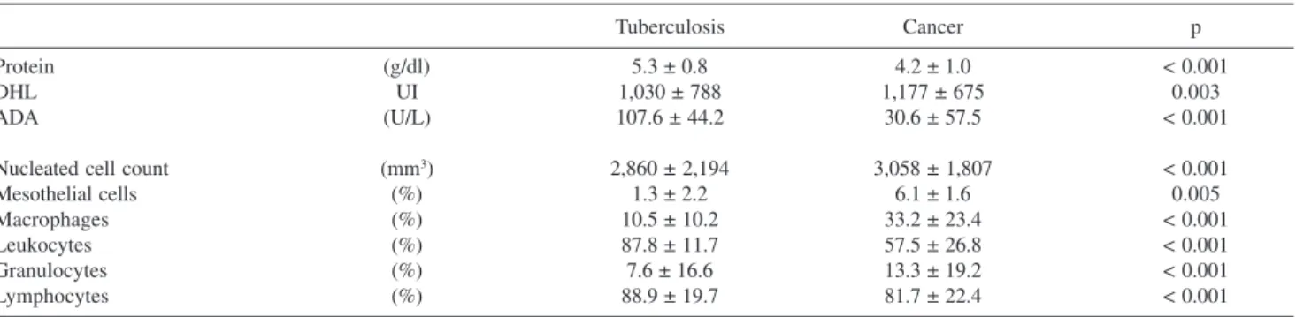
Table 2 shows the biochemical and cytological charac-teristics of the pleural fluids. Protein concentrations were significantly higher (p < 0.001) in the tuberculosis group (5.3 ± 0.8 g/dL) compared to the cancer group (4.2 ± 1.0 g/dL). In contrast, LDH levels were higher (p = 0.003) in the cancer group (1,177 ± 675 IU) than in the tuberculosis (1,030 ± 788 IU) group. Finally, as expected, ADA levels were significantly higher in the tuberculosis group (107.6 ± 44.2 U/L versus 30.6 ± 57.5 U/L; p < 0.001).
Although the pleural effusions were characterized by high cellularity, neoplastic effusions were more richly cel-lular than tuberculous effusions (3,058 ± 1,807 versus 2,860 ± 2,194 cells/mm3; p < 0.001). Analysis of the proportion of nucleated cells revealed a significantly lower percent-age of mesothelial cells in the tuberculosis group (1.3 ± 2.2% versus 6.1 ± 1.6%; p = 0.005), whereas the percent-age of macrophpercent-ages was significantly higher (p < 0.001) in the cancer group (33.2 ± 23.4% versus10.5 ± 10.2%). A higher proportion of leukocytes (p < 0.001) was observed in the tuberculosis group (87.8 ± 11.7%) when compared to the cancer group (57.5 ± 26.8%). Although both effu-sions were lymphocytic, the number of lymphocytes was significantly higher (p < 0.001) in the tuberculosis (88.9 ± 19.7%) compared to the cancer group (81.7 ± 22.4%).
In cancer effusions, oncotic cytology was positive in 70% (101/144) of cases and highly suggestive in 19.2% (28/ 144), permitting suspicion or confirmation of diagnosis in approximately 90% of the patients. A second pleural ap-proach (thoracocentesis) confirmed the diagnosis of
can-cer in four suspected cases and in additional two whose cytology was negative. In the remaining 24 patients, cy-tology was inconclusive. The diagnosis of cancer was con-firmed by immunophenotyping of pleural fluid in four cases (lymphoma), by closed pleural biopsy in 13 (carcinoma), and by thoracoscopy-guided pleural biopsy in seven (car-cinoma).
Closed pleural biopsy was conclusive in 68/78 (87.1%) and 21/37 (56%) of tuberculosis and cancer patients, re-spectively. A second pleural biopsy increased the cancer rate to 23/37 (62.1%).
DISCUSSION
A systematic approach to the classification of pleural effusion permits the diagnosis of a large number of pleu-ral diseases, especially when considering the high incidence of tuberculosis and cancer in Brazil. Diagnostic explora-tion is based on the analysis of clinical variables (gender, age and symptoms), imaging characteristics (volume and location of fluid), and laboratory (biochemical and cyto-logical) data.
A detailed clinical history of previous neoplasms or complaints related to consumptive states, as well as the presence of fever, night sweats, cough and contact with tu-berculous patients are fundamental. Since thoracocentesis is the first approach proposed in cases of pleural effusion, analysis of the removed fluid is the easiest and fastest way of assessment8. Although the cause of pleural effusion re-mains indeterminate in about 20% of cases, cytological, biochemical and microbiological analysis of pleural fluid is fundamental for adequate screening, avoiding more ex-pensive exams that would increase the morbidity and/or mortality of the patient9.
Tuberculosis is a ubiquitous disease with a high preva-lence in Brazil3. This disease usually predominates among individuals younger than those affected by cancer, a fact confirmed in the present study in which only 4.2% of the cases of tuberculosis occurred in patients older than 35
Table 2 - Biochemical and cytological characteristics of the pleural fluids.
Tuberculosis Cancer p
Protein (g/dl) 5.3 ± 0.8 4.2 ± 1.0 < 0.001
DHL UI 1,030 ± 788 1,177 ± 675 0.003
ADA (U/L) 107.6 ± 44.2 30.6 ± 57.5 < 0.001
Nucleated cell count (mm3) 2,860 ± 2,194 3,058 ± 1,807 < 0.001
Mesothelial cells (%) 1.3 ± 2.2 6.1 ± 1.6 0.005
Macrophages (%) 10.5 ± 10.2 33.2 ± 23.4 < 0.001
Leukocytes (%) 87.8 ± 11.7 57.5 ± 26.8 < 0.001
Granulocytes (%) 7.6 ± 16.6 13.3 ± 19.2 < 0.001
(only one patient older than 60) years. However, in the state of São Paulo (1998 to 2000), notifications of pleural effu-sion due to tuberculosis indicate that 40% of cases occurred in male individuals older than 40 years.2 Thus, although important, age should only be considered as a complemen-tary variable in the diagnosis of tuberculosis.
With respect to gender, it is known that men are more predisposed to both tuberculosis10 and lung cancer,11,12 al-though the incidence of cancer has been increasing among women over the last few decades. The predominance of fe-males among patients with cancer observed in the present study is in contrast to international epidemiological data. This bias is easily explained by the tertiary character of our hospital, which is a referral center for cancer with pleu-ral involvement, particularly that secondary to breast can-cer.
The pleural effusions were moderate in most patients with tuberculosis or cancer, with unilateral effusions pre-dominating in more than 90% of cases. This finding agrees with literature showing that pleural effusions are preferen-tially unilateral and moderate.1,12 Despite this similarity, a higher incidence of voluminous collections is found in ma-lignant pleural effusions, a fact also observed in the present study1.
After evaluation of demographic variables and chest images, the pleural space should be submitted to invasive investigation. The first step is thoracocentesis, i.e., thoracic puncture for pleural fluid collection, with the objective, in addition to the determination of macroscopic aspects, of material collection for adequate laboratory examination.
Yellow-citrine fluid (sometimes slightly turbid) dominates in tuberculosis while serohemorrhagic fluid pre-dominates in cancer. Our results confirmed this observa-tion8,13 but also showed overlaps between the groups con-cerning the classification of pleural effusion based only on this variable.
As a rule, thoracocentesis should always be considered for collection of pleural fluid for diagnosis. Only in excep-tional cases, when diagnosis has already been established or during an emergency (respiratory insufficiency), will it be performed when no laboratory infrastructure is available. Otherwise, thoracocentesis should include fluid collection and subsequent biochemical and cytological exams.
The first parameters analyzed (LDH and protein) should permit the differentiation between a transudate and an exu-date (Light’s criteria).14 Clinically, this discrimination is extremely valuable, since transudative effusions generally reflect systemic diseases and therefore do not require a complementary diagnostic approach. On the other hand, exudates indicate pleural involvement resulting from sys-temic disorders or primary pleural diseases. In these cases,
diagnostic exploration should be complemented by other pleural fluid exams or histological evaluation of the pleura.1,8
Since our objective was to differentiate between tuber-culosis and cancer, the first step was to recognize the pres-ence of an exudate. All pleural effusions included in the study were exudates and, although not specific, protein lev-els were significantly higher in the tuberculosis group, in agreement with the findings of Liam et al.15 Since the pro-tein concentration in pleural tuberculosis is frequently higher than 4.5 g/dL, Melo et al.16 proposed this value as a cutoff for diagnostic presumption. As confirmation of this proposal, this was the minimum protein level detected in the present study.
Lactic dehydrogenase, a nonspecific inflammatory marker,8 is in general discretely elevated in pleural tuber-culosis. Neoplasms present higher levels, suggesting a greater extent of pleural disease or the presence of blood in the pleural cavity.17 Despite this difference and, as is the case for the macroscopic evaluation of effusion, these bio-chemical parameters do not permit differentiation between the two diseases because of overlapping values.
The next step is the quantification of ADA, an enzyme
produced by macrophages and activated T lymphocytes,18
which is usually elevated in tuberculosis. In this situation, the activity of the enzyme is generally higher than 40 U/ L, although similar levels are observed in lymphocytic pleu-ral effusions secondary to rheumatoid arthritis and certain lymphoproliferative diseases. Other lymphocytic exudates, such as those secondary to pulmonary edema or metastatic
tumors, generally show levels below 40 U/L.19-22 In the
present study, using a cutoff value of 40 U/L as a presump-tive diagnosis, all cases of tuberculosis were correctly sified. This cutoff value shows high efficiency in the clas-sification of lymphocytic effusions (sensitivity: 87 to 100% and specificity: 81 to 97%).19-25 Despite the high diagnos-tic sensitivity of ADA, its specificity is influenced by other clinical conditions and by the regional prevalence of tu-berculosis.26,27
Considering the cytological profile of the pleural flu-ids in these two diseases, no cellular finding is character-istic, with exception to the presence of neoplastic cells in cancer effusions.
Oncotic cytology of the pleural fluid is considered to be the most sensitive method for the diagnosis of malig-nant pleural effusion.1,8 However, its performance varies according to the histological type and exfoliative capacity of the tumor, the techniques used to process the pleural samples, the number of slides analyzed per case and, fi-nally, the cytologist expertise. These factors explain the variation in the efficacy of the exam (40 to 87%) reported in the literature.28 In this study, the first fluid examination led to the diagnosis of cancer in 70.1% (101) of cases. In 19.4% (28), oncotic cytology was highly suspicious of ma-lignancy and in 10.5% (15) it was negative. A second method performed in the 28 suspicious patients (thoraco-centesis: 4; immunophenotyping: 4; closed pleural biopsy: 13 and VATS: 7) defined the diagnosis of neoplasia. In all patients (15) with negative cytology, the diagnosis of can-cer was confirmed.
This study demonstrates that although the pleural effu-sion exudates secondary to tuberculosis or cancer were
pre-dominantly lymphocitic, some clinical and laboratory as-pects could aid in their differentiation. In general, tuber-culosis occurred in younger persons, and effusions were unilateral and of a median size. The pleural fluid was fre-quently yellow (citrine or turbid) with high protein and ADA levels and with a low number of mesothelial cells. In contrast, patients with malignant effusions were gener-ally older and, although the effusions were predominantly unilateral, the presence of a bilateral effusion was more as-sociated with malignancy. In addition, the fluid was pre-dominantly turbid or serohemorrhagic. The level of pro-tein was lower than in tuberculosis, while ADA was nor-mal and LDH was frequently higher. Cytological exami-nation revealed a predominance of lymphocytes and macrophages. In patients with clinical and radiological evi-dence of tuberculosis presenting an exudative effusion rich in lymphocytes, protein and ADA levels are important to the etiological diagnosis. Similarly, when the clinical sus-picion is malignancy, serous hemorrhagic limphocytic fluid should be submitted to oncotic cytology, once this easy and inexpensive exam is an efficient diagnostic tool (≅ 80%).
As a result, we suggest thoracocentesis with pleural fluid examination as the first diagnostic approach.
RESUMO
Antonangelo L, Vargas FS, Seiscento M, Bombarda S, Teixeira L, Sales RKB. Parâmetros clínicos e laboratoriais no diagnóstico diferencial de efusões pleurais secundárias à tuberculose ou ao cancer. Clinics.2007;62(5):585-90.
OBJETIVO: Avaliar as características clínicas e
labora-toriais de derrames pleurais secundários à tuberculose ou câncer.
MÉTODOS: Um total de 326 pacientes com derrame pleural
por tuberculose (n=182) ou câncer (n=144) foi avaliado. Os seguintes parâmetros foram analisados: sexo e idade dos pa-cientes e características do líquido pleural (tamanho, locali-zação, aspecto macroscópico, concentração de proteínas, ati-vidade da desidrogenase lática (DHL) e da adenosina deaminase (ADA) e contagem de células nucleadas).
RESULTADOS: A tuberculose pleural predominou nos
pa-cientes mais jovens e do sexo masculino. Em ambos os gru-pos, os derrames pleurais foram de tamanho moderado e uni-laterais. Derrames com aspecto amarelo-citrino com níveis mais elevados de proteínas predominaram na tuberculose (5,3 ± 0,8 g/dL), quando comparados aos neoplásicos (4,2 ± 1,0 g/dL), enquanto que níveis mais elevados de DHL foram observados nos derrames neoplásicos (1.177 ± 675 x 1.030 ± 788 UI; p = 0,003). Conforme esperado, a atividade da
ADA foi maior na tuberculose que no câncer (107,6 ± 44,2 x 30,6 ± 57,5 U/L; p < 0,001). Ambos os derrames apresen-taram alta celularidade, embora mais pronunciada no grupo neoplásico (p < 0,001). Os derrames de etiologia tuberculosa se caracterizaram por apresentar uma maior percentagem de leucócitos e de linfócitos (p < 0,001) e um pequeno número de células mesoteliais (p = 0,005). Linfócitos e macrófagos foram as células nucleadas que predominaram nos derrames pleurais malignos.
CONCLUSÃO: Nossos resultados demonstram que em
exsudatos pleurais linfocíticos de pacientes com evidênci-as clínicevidênci-as e radiológicevidênci-as de tuberculose, os níveis de pro-teína e de ADA foram os parâmetros que melhor caracte-rizaram esses derrames. Da mesma maneira, quando a sus-peita clínica é câncer, um líquido serohemorrágico e linfocítico deve ser submetido à citologia oncótica, uma vez que este exame laboratorial de fácil realização e
bai-xo custo apresenta alto desempenho diagnóstico (≅ 80%).
Neste contexto, sugerimos que a toracocentese, com exa-mes bioquímicos e citológicos do líquido pleural, seja a primeira abordagem diagnóstica do paciente.
UNITERMOS: Derrame pleural. Neoplasia. Tuberculose
REFERENCES
1. Maskell NA, Butland RJA. BTS Guidelines for the Investigation of a Unilateral Pleural Effusion in Adults. Thorax 2003;58:8-17. 2. Centro de Vigilancia Epidemiológica Alexandre Vranjac. Secretaria da
Saúde de São Paulo. URL:http//www.cve.saude.sp.gov.br/tuberculose (20/08/2006).
3. Instituto Nacional de Câncer - Ministério da Saúde. URL:http:// www.inca.gov.br (20/08/2006).
4. Gopi A, Madhavan SM, Sharma SK and Sahn SA. Diagnosis and Treatment of Tuberculous Pleural Effusion in 2006. Chest 2007;131:880-89.
5. Valdés L, Pose A, San José E and Martínez VázquezJM.Tuberculous Pleural Effusions.EurJ Intern Med 2003;14:77-88.
6. Di Bonito I, Falconieri G, Colautti I, Bonifacio D and Dudine S. The Positive Pleural Effusions. A Retrospective Study of Cytopathologic Diagnosis with Autopsy Confirmation. Acta Cytol 1992;36:329-32. 7. Giust G. Adenosine Deaminase In: Bergmeyer HU. Methods of
Enzymatic Analysis. New York Academic Press 1974;1092-99. 8. Light RW. Clinical Manifestations and Useful Tests. In: Pleural Diseases.
4thed, Philadelphia, Lippincott-Williams & Wilkins, 2001. p. 42-86. 9. Villena V, López Encuentraz A, Echave –Sustaeta J, Alvarez Martinez
C, Martin Escribano P. Prospective study of 1,000 consecutive patients with pleural effusion. Etiology the effusion and characteristics of the patients. Arch. Bronconeumol. 2002;38:21-6.
10. World Health Organization URL:www.who.int/gtb/policyrd/ Gender&tb.htm (20/08/2006)
11. Parkin DM. Bray F, Ferlay J and Pisani P. Global Cancer Statistics, 2002. CA Cancer J Clin 2005;55:74-108.
12. Antunes G, Neville E, Duffy J, Ali N. BTS Guidelines for the Management of Malignant Pleural Effusions. Thorax 2003;58:29-38. 13. Porcel-Perez JM, Vives Soto M, Esquerda Serrano A, Jover Saenz A.
Cutoff Values of Biochemical Tests on Pleural Fluid: Their Usefulness in Differential Diagnosis of 1040 Patients with Pleural Effusion. Ann Med Intern 2004;21:113-17.
14. Light RW, MacGregor MI, Luchsinger PC et al. Pleural Effusions: The Diagnostic Separation of Transudates and Exudates. Ann Intern Med 1972;77:507-13
15. Liam CK, Lim KH, Wong CM. Differences in Pleural Fluid Characteristics, white Cell Count a Biochemistry of Tuberculous and Malignant Pleural Effusions. Med J Malaysia 2000;55:21-28
16. Melo FAF, AJB Santos ML et al. Diagnóstico da Tuberculose Pleural pela ADA, Isolada ou Combinada a Outras Variáveis, Inclusive em HIV-Positivos. Folha Med 2000;119:19-21.
17. Vergnon JM, Guidollet J, Gateau O, Ripoll JP, Collet P, Louisot P et al. Lactic dehydrogenase isoenzyme electrophoretic patterns in the diagnosis of pleural effusion. Cancer 1984;54:507-11.
18. Gaga M, Papamichalis G, Bakakos P, Latsi P, Samara I, Koulouris NG, et al. Tuberculous Effusion: ADA Activity Correlates with CD4+ Cell Numbers in the Fluid the Pleura. Respiration 2005;72:160-65. 19. Pettersson T, Ojala K and Weber TH. Adenosine Deaminase in the
Diagnosis of Pleural Effusions. Acta Med Scand 1984;215:299-304. 20. Ocanã I, Martinez-Vasquez JM, Ribera E, Segura RM and Pascual C.
Adenosine Deaminase Activity in the Diagnosis of Lymphocytic Pleural Effusions of Tuberculous, Neoplastic and Lymphomatous Origin. Tubercle 1986;67:141-45.
21. Lee YC, Rogers JT, Rodriguez RM, Miller KD and Light RW. Adenosine Deaminase Levels in Nontuberculous Lymphocytic Pleural Effusions. Chest 2001;120:356-61.
22. Jimenez Castro D, Diaz Nuevo G, Perez Rodríguez E and Light RW. Diagnostic Value of Adenosine Deaminase in Nontuberculous Lymphocytic Pleural Effusions. Eur Respir J 2003;21:220-24. 23. Goto M, Noguchi Y, Koyama H, Hira K, Shimbo T and Fukui T.
Diagnostic Value of Adenosine Deaminase in Tuberculous Pleural Effusion: A meta-analysis. Ann Clin Biochem 2003;40:374-81. 24. Chen ML, Yu WC, Lam CW, Au KM, Kong FY and Chan AY. Diagnostic
Value of Pleural Fluid Adenosine Deaminase in Tuberculous Pleurisy. Clin Chim Acta 2004;341:101-107.
25. Smach MA, Garouch A, Charfeddine B, Ben Abdelaziz A, Dridi H, Krayem B, et al. Diagnostic Value of Serum and Pleural Fluid Adenosine Deaminase Activity in Tuberculous Pleurisy. Ann Biol Clin (Paris) 2006;64:265-70.
26. Valdés L, Alvarez D, San Jose E, Juanatey JR, Pose A, Valle JM et al. Value of Adenosine Deaminase in the Diagnosis of Tuberculous Pleural Effusion in Young Patients in a Region of High Prevalence of Tuberculosis. Thorax 1995;50:600-603.
27. Diacon AH, Van de Wal BW, Wyser C ,Smedema JP, Bezuidenhout J and Bolliger CT et al. Diagnostic Tools in Tuberculous pleurisy: A Direct Comparative Study. Eur Respir 2003;22:589-91.