DOI: 10.1590/0004-282X20160108
ARTICLE
Propentofylline reduces glial scar development
following gliotoxic damage in the rat brainstem
Propentofilina reduz o desenvolvimento da cicatriz glial após dano gliotóxico no tronco
encefálico de ratos
Eduardo Fernandes Bondan1,2, Maria de Fátima Monteiro Martins1,2, Pietro Domingues Dossa1,
Lígia Bocamino Viebig1, Carolina Vieira Cardoso1, João Lopes Martins Júnior1, Maria Martha Bernardi1
It is widely described that ethidium bromide (EB) injec-tion in the white matter of the central nervous system (CNS) acts like a gliotoxin causing local oligodendroglial and astro-cytic death, with consequent demyelination (although the naked axons remained preserved), blood-brain barrier dis-ruption and Schwann cell invasion due to the glia limitans breakdown1,2,3. Surviving astrocytes presented a vigorous
re-action around the injury site with increased
immunoreac-tivity to the speciic cell marker glial ibrillary acidic protein
(GFAP) and reexpression of vimentin (VIM)2.
Propentofylline [PPF, 3-methyl-1-(5´-oxohexyl)- 7-propylxanthine]
is a xanthine derivative with pharmacological efects distinct
from those of the classical methylxanthines theophylline and
cafeine4. In vitro and in vivo studies have demonstrated
ex-tensive neuroprotective, antiproliferative and anti-inlamma
-tory efects of PPF in several experimental models in animals4.
It was sucessfully used in degenerative vascular dementia and as a potential adjuvant treatment to Alzheimer’s disease, schizophrenia and multiple sclerosis4. Propentofylline
de-creases activation of microglial cells and astrocytes, whose
1Universidade Paulista, Departamento de Patologia Ambiental e Experimental, São Paulo SP, Brasil; 2Universidade Cruzeiro do Sul, Departamento de Medicina Veterinária, São Paulo SP, Brasil.
Correspondence: Eduardo Fernandes Bondan; Rua Caconde, 125/51; 01425-011 São Paulo SP, Brasil; E-mail: [email protected] Conflict of interest: There is no conlict of interest to declare.
Received 29 February 2016; Received in inal form 24 May 2016; Accepted 08 June 2016.
ABSTRACT
Propentofylline is a xanthine derivative that depresses activation of glial cells, whose responses contribute to neural tissue damage during inlammation. Ethidium bromide injection into the central nervous system induces local oligodendroglial and astrocytic loss, resulting in primary demyelination, neuroinlammation and blood-brain barrier disruption. Surviving astrocytes present a vigorous reaction around the injury site with increased immunoreactivity to glial ibrillary acidic protein (GFAP). Objective: This study aimed to evaluate the effect of propentofylline administration on astrocytic response following gliotoxic injury. Method: Wistar rats were injected with ethidium bromide into the cisterna pontis and treated or not with propentofylline (12.5mg/kg/day, intraperitoneal) during the experimental period. Brainstem sections were collected from 15 to 31 days after gliotoxic injection and processed for GFAP immunohistochemistry. Results and Conclusion: Results demonstrate that propentofylline decreased astrocytic activation until the 21st day, suggesting that this drug may have
a role in reducing glial scar development following injury.
Key words: astrocytes; ethidium; gliosis; gliotoxin; xanthine.
RESUMO
A propentoilina é uma xantina que deprime a ativação das células gliais, cujas respostas contribuem para o dano neural durante inlamação. A injeção de brometo de etídio no sistema nervoso central induz a perda oligodendroglial e astrocitária, resultando em desmielinização, neuroinlamação e ruptura da barreira hematoencefálica. Os astrócitos sobreviventes apresentam vigorosa reação ao redor da lesão com aumento da imunorreatividade à proteína glial ibrilar ácida (GFAP). Objetivo: Este estudo objetivou avaliar o efeito da propentoilina sobre a resposta astrocitária após injúria gliotóxica. Método: Ratos Wistar foram injetados com brometo de etídio na cisterna basal e tratados ou não com propentoilina (12.5mg/kg/dia, intraperitoneal). Amostras do tronco encefálico foram coletadas dos 15 aos 31 dias pós-injeção do gliotóxico e processadas para estudo ultraestrutural e imuno-histoquímico para GFAP. Resultados e Conclusão: Os resultados demonstram que a propentoilina reduziu a ativação astrocitária até o 21o dia, sugerindo que essa droga pode
atuar na redução da cicatriz glial após injúria.
responses are associated with neuronal damage during inflammation and hypoxia, and PPF consequently de-creases glial production and release of damaging proin-flammatory factors5,6.
In the EB-demyelinating model, PPF administration has
been shown to signiicantly increase both oligodendroglial and
Schwann cell remyelination following gliotoxic damage7 and
even reverse the impairment in remyelination found in diabetic rats8. Despite the beneicial efects of PPF observed on oligoden
-drocyte remyelinating activity in these investigations, astrocyte
behavior has not been properly evaluated. hus, the aim of this study was to evaluate whether PPF had the capacity to afect
astrocyte responses during the process of demyelination and re-myelination following gliotoxic injury induced by EB.
METHOD
he animal procedures were performed in accordance
with the guidelines of the Committee on Care and Use of Laboratory Animal Resources and Brazilian Institutional Ethics Committee, Universidade Paulista (protocol number 182/13, CEUA/ICS/UNIP). Seventy-two adult (4–5-month-old) male Wistar rats were submitted to a local injection of 10 microlitres of 0.1% EB into the cisterna pontis, an en-larged subarachnoid space below the ventral surface of the pons. All rats were anaesthetized with ketamine and xylazine (5:1; 0.1 ml/100g) and 2.5% thiopental (40 mg/ml) by intraper-itoneal route and a burr-hole was made on the right side of the skull, 8 mm behind the fronto-parietal suture. Injections
were performed freehand using a Hamilton syringe, itted with
a 35° angled polished 26 gauge needle into the cisterna pontis. Rats were then distributed into two groups – untreated rats (group I, n = 36) and rats treated with 12.5 mg/kg/day of PPF (Agener União Química, São Paulo, SP, 20 mg/ml solution) by intraperitoneal route during the experimental period (group II,
n = 36). he animals were kept under controlled light condi -tions (12 h light-dark cycle) and water and food were given ad libitum during the experimental period.
For ultrastructural investigation, four rats from each group were anaesthetized and were submitted to intracardiac per-fusion with 4% glutaraldehyde in 0.1 M Sorensen phosphate
bufer (pH 7.4) at each of the following periods – 15, 21 and 31 days post-injection (p.i.). hin slices of the brainstem (pons and mesencephalon) were collected and post-ixed in 0.1%
osmium tetroxide, dehydrated with graded acetones and em-bedded in Araldite 502 resin, following transitional stages in
acetone. hick sections were stained with 0.25% alkaline to -luidine blue. Selected areas were trimmed and thin sections were stained with 2% uranyl and lead acetate and viewed in a JEM -1200 EX2 JEOL transmission electron microscope.
For immunohistochemical study of the expression of the astrocytic marker GFAP, eight rats were anaesthetized
and submitted to intracardiac perfusion with bufered 10%
formaldehyde solution at each of the same periods. heir
brains were then removed and kept for three days in the
same ixative. Coronal sections from the brainstem were
mounted on silanized slides and submitted to GFAP immu-nostaining using the avidin-biotin peroxidase complex (ABC)
method. Briely, the sections were deparainized in xylene
and rehydrated in a crescent graded series of ethanol solu-tions. Antigen retrieval was done by transferring the slides to
10 mM sodium citrate bufer (pH 6.0) at 95° C for 20 minutes.
Endogenous peroxidase was blocked by 3% hydrogen perox-ide for 10 minutes at room temperature. Two washes with
Tris/HCl bufer pH 6.0 (Wash bufer 10x, S3006, Dako, Glostrup,
Danmark) were done between incubations. Polyclonal rabbit anti-GFAP immunoglobulin (Z0334, Dako), at a dilution of 1:1000, was used as primary antibody, for 16 hours, followed by the application of biotinylated secondary antibody (Dako Universal LSABTM 2 System – HRP, K0690), according to the
manufacturer`s instructions. Immunoreactivity was visual-ized by incubating the sections in a solution containing 0.1% diaminobenzidine (DAB, K3467, Dako). Sections were then
counterstained by Harris’ modiied hematoxylin solution, de -hydrated and mounted in Entellan (Merck, Germany).
Astrocytic evaluation was done in the brainstem of animals from both groups using a computerized image analysis system (Image-Pro-Plus 4.5, Media Cybernetics, Silver Spring, USA), measuring, by colorimetry, the area stained brown in a total area
of 302,952.5 µm2. Negative controls for immunostaining (sections
lacking primary antibody application) were done. Data were
ana-lyzed by t test and statistical signiicance was set at p < 0.05.
RESULTS
he general aspect of the EB-induced lesions found in this
investigation in both groups at 15, 21 and 31 days was sim-ilar to that previously described in other studies using this gliotoxin in the rat brainstem1,2 (Figure 1). Briely, they pre
-sented extensive demyelinated areas in the ventral surface of the mesencephalon and pons and contained, in the cen-tral region, phagocytic cells, myelin debris and naked axons. At the periphery, oligodendrocytes and Schwann cells were observed, the latter occurring in areas of enlarged extracel-lular spaces devoid of astrocytic extensions. Astrocyte pro-cesses were invariably seen near the incipient, but prepon-derant, oligodendroglial remyelination at the periphery, and Schwann cells also appeared to contribute to myelin repair. Ultrastructural analysis apparently showed that astrocytic processes among oligodendrocyte remyelinated axons were slightly thinner in PPF-treated animals (Figure 2B) compared to those that had not received the xanthine (Figure 2A). Although oligodendroglia prevailed in the brainstem my-elin repair from the 15th to the 31st day, sheaths formed by
As described earlier in a former investigation7, PPF-treated
rats presented an increased remyelination from the 15th to
the 31st day following EB injection. Some lymphocytes and
iniltrating pial cells were occasionally seen, the irst contact -ing phagocytic cells and myelin debris.
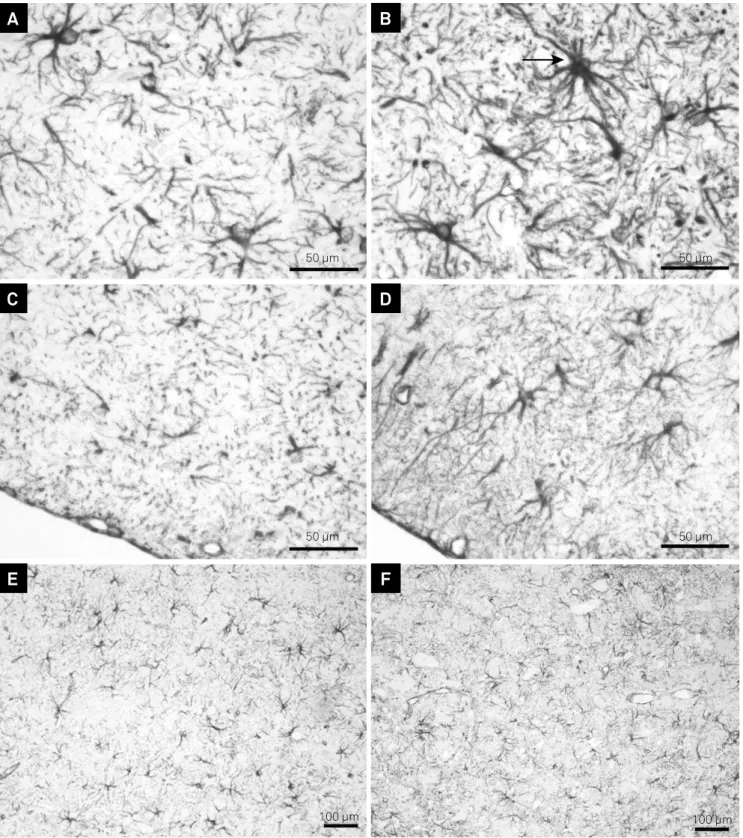
By GFAP immunohistochemical staining, it was observed that the EB-induced lesions from group II (PPF-treated rats) apparently presented a decreased astrocytic reaction close to
the edges of the injury site, with the observation of fewer and thinner GFAP-stained processes at the periphery at both 15 days (Figure 3A,B) and 21 days (Figure 3C,D). No astrocytes were observed in the central areas of the lesions from both groups even at 31 days after EB injection.
he Table presents the mean areas with GFAP staining in µm2 from both groups at all analyzed periods (15, 21 and
31 days). hese results showed that, at 15 days, the mean brown-stained area was signiicantly smaller in rats treated with PPF (group II – 41,653 ± 7,306.61 µm2) compared to untreated rats
(group I – 55,391.38 ± 5,819.91 µm2). A similar inding was
seen at 21 days (44,829.38 ± 6,164.66 µm2 in group II versus
55,381.75 ± 5,785.65 µm2 in group I), but no statistical diference
was seen at 31 days (mean areas of 50,227.38 ± 7,612.02 µm2 and
50,020.37 ± 6,308.2 µm2, respectively, in groups I and II).
DISCUSSION
Astrocytes respond to all forms of CNS insults through a
process referred to as reactive astrogliosis, which is a inely
gradated continuum of progressive changes in gene expres-sion and cell morphology9,10. Intermediate ilaments of as
-trocytes are composed mainly of GFAP and this protein has become the best-known astrocytic marker11. In mild reactive
astrogliosis there is variable upregulation of expression of GFAP and other genes as well as hypertrophy of the cell body and processes, but this occurs within the domains of
indi-vidual astrocytes without signiicant overlap of processes of
neighboring astrocytes or loss of individual domains12. In this
discrete reaction there is little or no astrocyte proliferation,
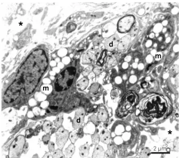
Figure 1. Electronmicrograph from a central area at 15 days following ethidium bromide (EB) injection in rats not treated with propentofylline (PPF). Demyelinated axons (d) and macrophages (m) in different stages of myelin degradation are seen in a distended extracellular space (asterisk). Bar = 2 µm.
m
d
d
m
*
*
2 µm
Figure 2. Electronmicrographs from peripheral areas of the ethidium bromide (EB)-induced lesions in untreated (A) and propentofylline (PPF)-treated (B) rats in the ventral surface of the pons at 21 days. Arrows indicate astrocytic processes. Note the thicker astrocyte processes among oligodendrocyte remyelinated axons in A (no treatment) and the greater amount of remyelinated axons in B (PPF treatment). A) Bar = 3 µm; B) Bar = 3 µm.
3 µm 3 µm
but the increased expression of GFAP can lead to the staining of more cells, giving the false impression of proliferation12,13.
On the other hand, severe astrogliosis leads to a more pro-nounced upregulation of GFAP, among other genes, with blurring and disruption of individual astrocyte domains, as usually found in areas surrounding severe focal lesions12.
Astrocyte precursors and immature astrocytes present prin-cipally nestin and vimentin (VIM) and, during development, as astrocytes mature, nestin expression disappears, GFAP be-comes increasingly expressed and VIM decreases to undetect-able levels11. In both mild or severe astrogliosis, astrocytes also
reexpress VIM and nestin11. In the EB demyelinating model,
Figure 3. Peripheral glial ibrillary acidic protein (GFAP) expression by immunohistochemistry in the ventral surface of the pons at 15 days (A, B), 21 days (C, D) and 31 days (E, F) in ethidium bromide (EB)-induced lesions from untreated (B, D, F) and propentofylline (PFF)-treated (A, C, E) rats. Observe a strongly stained GFAP-positive astrocyte (arrow) in B. (A, B, C, D) Bar = 50 µm; E, F) Bar = 100 µm.
50 µm 50 µm
50 µm 50 µm
100 µm 100 µm
A
B
C
D
reexpression of VIM and strong astrocytic immunoreactivity to GFAP were described by Bondan et al.2 in the rat brainstem
from the 3rd to the 31st day following gliotoxic injection. his in
-creased GFAP expression around the EB-induced lesions was
also conirmed in the present study, in a pattern suggestive of
mild astrogliosis.
Many diferent types of signaling molecules are able to trig -ger and/or regulate astrogliosis and can be released by all cell types of the CNS tissue, including neurons, microglia, oligo-dendrocyte lineage cells, pericytes, endothelia and other
as-trocytes, as well as by invasive inlammatory/immune cells12,13.
While it was initially thought that astrocyte proliferation was a major component of glial scar, it has been repeatedly demonstrated that there are actually few astrocytes undergo-ing cell division durundergo-ing glial scar formation9. his observation
is conirmed by the fact that no astrocytes in mitotic activity
were seen in this study and also in previous investigations focusing on astrocytic response following gliotoxic lesions2.
Concerning the mechanisms of PPF action, it has been shown that (i) inhibition of cyclic AMP and GMP-phosphodiesterases (PDE), (ii) inhibition of membrane adenosine transporters and (iii) reinforcement of adenosine A2 receptor-mediated efects
in a synergistic manner are potent pathways responsible for the protective adenosine-mediated actions of this xanthine4,14.
here is also evidence that PPF is a weak adenosine autorecep -tor A1 antagonist, which can additionally inhibit its reuptake and the activity of the 5´-nucleotidase14.
hus, PPF leads to increased intracellular cAMP levels
and greater extracellular concentrations of adenosine, stimu-lating adenosinergic neurotransmission and adenosine 2 (A2) receptor-mediated cAMP synthesis5,15.
Intracellular levels of the second messenger cAMP can be elevated by activation of the adenylate cyclase or by inhibi-tion of cAMP-degrading phosphodiesterases (PDE). Eleven
PDE families have been identiied with diferent speciicity
towards cAMP and cGMP16.
Regulation of cytokine production includes the adenylate cyclase – cAMP – protein kinase pathway17. Yoshiwawa et al.18
reported that PPF, a type III-IV speciic PDE inhibitor, although
decreasing in a dose-dependent manner the production of the
inlammatory cytokines TNF-α, IL-1 and IL-6 by mouse mi-croglia stimulated by LPS in vitro, increased up to two or three
times the production of the inhibitory cytokine IL-10. In turn, IL-10 acts by suppressing cytokine release by microglia and macrophages and attenuating astroglial reactivity in vivo19.
he GFAP is regulated in part by the secretion of factors into the extracellular space. he common pathway for GFAP expres -sion in astrocytes is triggered by the binding of cytokines from
the IL-6 family to their receptors. hese receptors subsequent -ly activate the JAK/STAT intracellular pathway, leading to the expression of GFAP in astrocytes. Most of the other pathways known to participate in GFAP expression are connected at some point to this pathway. For example, some members of the TGF-β
superfamily of cytokines have little or no efect on GFAP synthe -sis by themselves, but they strongly potentiate GFAP induced by the IL-6 family of cytokines20. he PDE inhibitor pentoxifylline is
also known to decrease the synthesis of TNF-α, IL-1β and IL-6 through the inhibition of nuclear factor-κB and stimulation of IL-10 expression in the CNS21,22. In the EB demyelinating
mod-el, PPF has already been shown to decrease the production of TNF-α and IL-1β in the rat brainstem23.
In the CNS, PPF acts as a glial modulator, with direct ac-tions on microglia, decreasing microglial proliferation and
expression of inlammatory cytokines, such as TNF-α and IL-1β, in vitro and in vivo6,14,18,24.
In the present study, PPF was shown to decrease the astro-cytic reaction to the gliotoxic injury as seen through the expres-sion of GFAP and by ultrastructural observation. Morphometric
analysis conirmed, at 15 and 21 days the initial impression sug -gested by the observation of semithin and ultrathin sections, that PPF treatment decreased the astrocytic reaction to the
glio-toxic injection as peripheral GFAP stained areas were signii -cantly greater in EB injected rats that were not treated with PPF compared to rats treated with the xanthine.
Decreased activation of astrocytes and microglia in rats treat-ed with PPF, as shown by rtreat-eductreat-ed GFAP and OX-42 expression,
Table. Areas with glial ibrillary acidic protein (GFAP) staining in µm2 in a total area of 302,952.5 µm2 in rats injected with ethidium bromide (EB), treated (group II) or not (group I) with propentofylline (PPF).
SD: standard deviation; distinct letters indicate signiicant differences between groups I and II at each period (p < 0.05).
Animal Group I – EB injection Group II – EB injection + PPF
15 days 21 days 31 days 15 days 21 days 31 days
1 50,231 45,924 39,523 47,292 45,435 46,417
2 60,812 50,125 58,126 39,548 36,021 58,352
3 48,154 54,531 45,132 51,63 43,19 55,325
4 53,824 56,642 44,243 40,226 47,611 45,131
5 57,122 56,134 55,232 34,135 47,163 47,361
6 61,326 57,288 56,785 36,177 56,236 44,372
7 62,451 58,125 44,457 50,723 44,634 44,527
8 49,211 64,915 58,321 33,453 38,345 58,678
Mean 55,391.38A 55,381.75A 50,227.38C 41,653B 44,829.38B 50,020.37C
respectively, was also observed in vivo by Young et al.25 after
spi-nal cord injury. he PPF also inhibited injury-induced GFAP ex -pression along with enhancement of glutamate transporters GLT-1 and GLAST in the dorsal horn upper laminae in mice sub-mitted to L5-spinal nerve transection26.
Activated astrocytes may lose their homeostatic functions upon exposure to stressors, decreasing glutamate uptake and
increasing the expression of deleterious proinlammatory mol -ecules such as cytokines, nitric oxide, prostaglandins, among others, as an injury response13. hus, reactive astrocytes dis
-play decreased glutamate transporters and as a result synaptic glutamate clearance is impaired. In vitro PPF was capable of
diferentiating astrocytes back to a homeostatic, mature phe -notype, competent for glutamate clearance26.
Both oligodendrocyte and astrocyte loss are hallmarks within the epicenter of an EB lesion while axons remain
unaf-fected. he mechanism of selective glial death has been sug -gested to occur through EB’s action as a minor-groove DNA intercalator3. However, other evidences suggest that while EB
does intercalate both chromosomal and mitochondrial DNA,
it only afects transcription of mtDNA27. So, it is likely that EB
injection into the white matter compromises mtDNA in all cells in the lesion site although neurons and endothelial cells appear to be less sensitive than glia in rat models3.
After trauma, blood-brain barrier dysfunction is
im-mediately observed as well as activation of inlamma -tory cells including microglia, astrocytes and invading monocytes/macrophages1,2,3. Activation and recruitment
of inlammatory cells into the injured CNS generate pro
-inlammatory cytokines, free radicals and other damaging molecules. he two most important cytokines found in the
CNS after trauma are TNF-α and IL-1β, which are highly cy-totoxic and regulated by cAMP signaling16. he beneits of
PDE4 inhibition in reducing inlammation have been well
studied in rodent models of ischemia14 and traumatic
inju-ry16. PDE4 inhibitors have been found to improve neuronal
survival, reduce infarct size, and attenuate inlammation
and blood-brain barrier breakdown28. In experimental
auto-immune encephalomyelitis, rolipram, a PDE4 inhibitor, pre-vents the progression of neurodegeneration and demyelin-ation by increasing cAMP levels29,30.
It is possible that macrophage and lymphocyte products
during the inlammatory response triggered by the EB injec
-tion may provide a greater harmful inluence to the nervous tissue than the early gliotoxin injection itself. herefore the anti-inlammatory efects performed by PPF may possibly be beneicial to remyelination.
A Ca++-dependent and excessive activation of glial cells
is usually found in neuroinlammation and, in this con -text, increased levels of adenosine induced by PPF admin-istration may perform a regulatory role on these Ca++- and
cAMP-dependent molecular signaling pathways that deter-mine many cell-related functions, such as cellular proliferation
rate, diferentiation state, cytokine production, among others5.
A strengthening of the cAMP signaling, which can be achieved by adenosine agonists and by PPF, stimulates the production of neurotrophic factors in astrocytes, apparently preventing a deleterious and secondary astrocytic activation caused by previous microglial upregulation15.
Although not entirely understood, it has been accepted that drugs that elevate extracellular adenosine and/or block the deg-radation of cyclic nucleotides, like PPF, may be used to counter-act glia-related damage in CNS pathological processes15.
hus, ultrastructural observation along with morphomet -ric analysis in the present study unequivocally demonstrated that PPF decreased astrocytic activation until the 21st day
af-ter gliotoxic lesion, probably by simultaneously suppressing
the release of proinlammatory molecules, such as the above
mentioned TNF-α and IL-1β, as well as IL-6, which may trig-ger and promote astrogliosis following CNS injury, and by
in-creasing secretion of the anti-inlammatory cytokine IL-10.
In conclusion, our results clearly indicate that PPF may have a role in preventing or reducing glial scar development following injury.
References
1. Bondan EF, Lallo MA, Sinhorini IL, Pereira LAVD, Graça DL. The effect of cyclophosphamide on brainstem remyelination following local ethidium bromide injection in Wistar rats. J Submicrosc Cytol Pathol. 2000;32(4):603-12.
2. Bondan EF, Lallo MA, Dagli MLZ, Sanchez M, Graça DL. [Investigation into the astrocytic immunoreactivity to GFAP and vimentin in the brainstem of Wistar rats submitted to the ethidium bromide gliotoxic model]. Arq Neuropsiquiatr. 2003;61(3A):642-9. Portuguese. doi:10.1590/S0004-282X2003000400022
3. Kuypers NJ, James KT, Enzmann GU, Magnuson DSK, Whittemore SR. Functional consequences of ethidium bromide demyelination of the mouse ventral spinal cord. Exp Neurol. 2013;247:615-22. doi:10.1016/j.expneurol.2013.02.014
4. Sweitzer S, De Leo J. Propentofylline: glial modulation, neuroprotection, and alleviation of chronic pain. Handb Exp Pharmacol. 2011;200:235-50. doi:10.1007/978-3-642-13443-2_8
5. Schubert P, Ogata T, Rudolphi K, Marchini C, McRae A, Ferroni, S. Support of homeostatic glial cell signaling: a novel therapeutic approach by propentofylline. Ann NY Acad. Sci. 1997;826:337-47. doi:10.1111/j.1749-6632.1997.tb48484.x
6. Si, Q, Nakamura Y, Ogata T, Kataoka K, Schubert P. Differential regulation of microglial activation by propentofylline via cAMP signaling. Brain Res. 1998;812(1-2):97-104. doi:10.1016/S0006-8993(98)00954-8
7. Bondan EF, Martins MFM, Baliellas DEM, Gimenez CFM, Poppe SC, Bernard MM. Effects of propentofylline on CNS remyelination in the rat brainstem. Microsc Res Tech. 2014;77(1):23-30. doi:10.1002/jemt.22308
9. Fitch MT, Silver J. Astrocytes are dynamic participants in central nervous system development and injury responses. In: Jessen KR, Richardson WD. Glial cell development. Oxford: Oxford University Press; 2001. p. 263-77.
10. Cregg JC, DePaul MA, Filous AR, Lang GBT, Tran A, Silver J. Functional regeneration beyond glial scar. Exp Neurol. 2014;253:197-207. doi:10.1016/j.expneurol.2013.12.024
11. Pekny M, Pekna A. Astrocyte intermediate ilaments in CNS pathologies and regeneration. J Pathol. 2004;204(4):428-37. doi:10.1002/path.1645
12. Sofroniew MV, Vinters HV. Astrocytes: biology and pathology. Acta Neuropathol. 2010;119(1):7-35. doi:10.1007/s00401-009-0619-8
13. Sofroniew MV. Molecular dissection of reactive astrogliosis and glial scar formation. Trends Neurosci. 2009;32(12):638-47. doi:10.1016/j.tins.2009.08.002
14. Plaschke K, Grant M, Weigand MA, Züchner J, Martin E, Bardenheuer HJ. Neuromodulatory effect of propentofylline on rat brain under acute and long-term hypoperfusion. Br J Pharmacol. 2001;133(1):107-16. doi:10.1038/sj.bjp.0704061
15. Schubert P, Morino T, Miyazaki H, Ogata T, Nakamura Y, Marchini C et al. Cascading glia reactions: a common pathomechanism and its differentiated control by cyclic nucleotide signaling. Ann N Y Acad Sci. 2000;903:24-33. doi:10.1111/j.1749-6632.2000.tb06346.x
16. Titus DJ, Oliva AA, Wilson NM, Atkins CM. Phosphodiesterase inhibitors as therapeutics for traumatic brain injury. Curr Pharm Des. 2014;21(3):332-42. doi:10.2174/1381612820666140826113731
17. Kammer GA. The adenylate cyclase-cAMP-protein kinase A pathway and regulation of the immune response. Immunol Today. 1988;9(7-8):222-9. doi:10.1016/0167-5699(88)91220-0
18. Yoshikawa M, Suzumura A, Tamaru T, Takayanagi T, Sawada M. Effects of phosphodiesterase inhibitors on cytokine production by microglia. Mult Scler. 1999;5(2):126-33. doi:10.1177/135245859900500210
19. Balasingam V, Yong VW. Attenuation of astroglial reactivity by interleukin-10. J Neurosci. 1996;16(9):2945-55.
20. Herrera F, Chen Q, Schubert D. Synergistic effect of retinoic acid and cytokines in the regulation of glial fibrillary acidic protein expression. J Biol Chem. 2010;285(50):38915-22. doi:10.1074/jbc.M110.170274
21. Neuner P, Klosner G, Schauer E, Pourmojib M, Macheiner W, Grünwald C et al. Pentoxifylline in vivo down-regulates the release of IL-1β, IL-6, IL-8 and tumour necrosis factor-α by human peripheral blood mononuclear cells. Immunol. 1994;83(2):262-7.
22. Lundblad R, Ekstrøm P, Giercksky KE. Pentoxifylline improves survival and reduces tumor necrosis factor, interleukin-6, and endothelin-1 in fulminant intra-abdominal sepsis in rats. Shock. 1995;3(3):210-5. doi:10.1097/00024382-199503000-00009
23. Bondan EF. Propentofylline decreases the production of TNF-alpha and IL-1 in the rat brainstem after a gliotoxic injury induced by ethidium bromide. J Neuroimmunol. 2014;275:139. doi:10.1016/j.jneuroim.2014.08.373
24. Jung S, Donhauser T, Toyka KV, Hartung HP. Propentofylline and iloprost suppress the production of TNF-α by macrophages but fail to ameliorate experimental autoimmune encephalomyelitis in Lewis rats. J Autoimmun. 1997;10(6):519-29. doi:10.1006/jaut.1997.0159
25. Gwak YS, Crown ED, Unabia GC, Hulsebosch CE. Propentofylline attenuates allodynia, glial activation and modulates GABAergic tone after spinal cord injury in the rat. Pain. 2008;138(2):410-22. doi:10.1016/j.pain.2008.01.021
26. Tawik VL, Regan MR, Haenggeli C, LaCroix-Fralish ML, Nutile-McMenemy N, Perez N et al. Propentofylline-induced astrocyte modulation leads to alterations in glial glutamate promoter activation following spinal nerve transection. Neuroscience. 2014;152(4):1086-92. doi:10.1016/j.neuroscience.2008.01.065
27. Hayakawa T, Noda M, Yasuda K, Yorifuji H, Taniguchi S, Miwa I et al. Ethidium bromide-induced inhibition of mitochondrial gene transcription suppresses glucose-stimulated insulin in the mouse pancreatic beta-cell line betaHC9. J Biol Chem. 1998;273(32):20300-7. doi:10.1074/jbc.273.32.20300
28. Li, LX, Cheng YF, Lin HB, Wang C, Xu JP, Zhang HT. Prevention of cerebral ischemia-induced memory deicits by inhibition of phosphodiesterase-4 in rats. Metab Brain Dis. 2011;26(1):37-47. doi:10.1007/s11011-011-9235-0
29. Sommer N, Löschmann PA, Northoff GH, Weller M, Steinbrecher A, Steinbach JP et al. The antidepressant rolipram suppresses cytokine production and prevents autoimmune encephalomyelitis. Nat Med. 1995;1(3):244-8. doi:10.1038/nm0395-244