Deficiency in Wistar Rats
Mariele Castilho Pansani1, Paula S. Azevedo1, Bruna Paola Murino Rafacho1, Marcos F. Minicucci1, Fernanda Chiuso-Minicucci2 , Sofia Gonc¸alves Zorzella-Pezavento2 , Julio Sergio Marchini4, Gilberto Joa˜o Padovan4, Ana Angelica Henrique Fernandes3, Beatriz B. Matsubara1, Luiz S. Matsubara1, Leonardo A. M. Zornoff1, Sergio A. R. Paiva1*
1Internal Medicine Department, Faculdade de Medicina de Botucatu, Universidade Estadual Paulista (UNESP), Botucatu, Sa˜o Paulo, Brazil,2Department of Microbiology and Immunology, Instituto de Biocieˆncias de Botucatu, Universidade Estadual Paulista (UNESP), Botucatu, Sa˜o Paulo, Brazil,3Chemistry and Biochemistry Department, Instituto de Biocieˆncias de Botucatu, Universidade Estadual Paulista (UNESP), Botucatu, Sa˜o Paulo, Brazil,4Internal Medicine Department, Faculdade de Medicina de Ribeira˜o Preto, Universidade de Sa˜o Paulo (FMRP – USP), Ribeira˜o Preto, Sa˜o Paulo, Brazil
Abstract
Introduction:Micronutrient deficiency is observed in heart failure patients. Taurine, for example, represents 50% of total free amino acids in the heart, andin vivostudies have linked taurine deficiency with cardiomyopathy.
Methods:Thirty-four male Wistar rats (body weight = 100 g) were weighed and randomly assigned to one of two groups: Control (C) or taurine-deficient (T (-)). Beta-alanine at a concentration of 3% was added to the animals’ water to induce taurine deficiency in the T (-) group. On day 30, the rats were individually submitted to echocardiography; morphometrical and histopathological evaluation and metalloproteinase activity, oxidative stress and inflammation evaluation were performed. Tissue samples were collected to determine the taurine concentration in the heart.
Results:Taurine deficiency led to decreases in: ventricular wall thickness, left ventricle dry weight, myocyte sectional area, left ventricle posterior wall thickness and ventricular geometry. With regard to heart function, the velocity of the A wave, the ratio between the E and A wave, the ejection fraction, fractional shortening and cardiac output values were decreased in T (-) rats, suggesting abnormal diastolic and systolic function. Increased fibrosis, inflammation and increased activation of metalloproteinases were not observed. Oxidative stress was increased in deficient animals.
Conclusions:These data suggest that taurine deficiency promotes structural and functional cardiac alterations with unique characteristics.
Citation:Pansani MC, Azevedo PS, Rafacho BPM, Minicucci MF, Chiuso-Minicucci F, et al. (2012) Atrophic Cardiac Remodeling Induced by Taurine Deficiency in Wistar Rats. PLoS ONE 7(7): e41439. doi:10.1371/journal.pone.0041439
Editor:Lynette Kay Rogers, The Ohio State Unversity, United States of America ReceivedMarch 20, 2012;AcceptedJune 21, 2012;PublishedJuly 23, 2012
Copyright:ß2012 Pansani et al. This is an open-access article distributed under the terms of the Creative Commons Attribution License, which permits unrestricted use, distribution, and reproduction in any medium, provided the original author and source are credited.
Funding:This work was supported by the CAPES (MCP scholarship) and Faculdade de Medicina de Botucatu - UNESP (FC-M, SGZ-P, JSM, GJP, AAHF, PSA, BBM, LSM, BPMR, MFM, LAMZ and SARP salaries). The funders had no role in study design, data collection and analysis, decision to publish, or preparation of the manuscript.
Competing Interests:The authors have declared that no competing interests exist. * E-mail: [email protected]
Introduction
Patients with heart failure (HF) may have different nutritional needs than those with a normal physiological state [1]. There is evidence that patients with HF are deficient in many micronutri-ents that play important roles in maintaining calcium homeostasis, controlling oxidative stress and regulating energy and protein metabolism [2]. Among these nutrients, taurine is very important. It is involved in biological processes such as bile salt formation, reduction of the levels of pro-inflammatory cytokines in various organs, insulin activity modulation, anti-hypertension, anti-ath-erogenic action, hepatoprotection and neurotransmission [3,4,5,6,7,8].
Taurine accounts for 50% of total free amino acids in the heart [9]. Allard et al. reported that taurine deficiency contributes to HF in cats and dogs [1]. There are many factors involved in cardiac
remodeling and progression of HF, including oxidative stress and inflammation [1,10,11,12]. Taurine is described as a nutrient with the following functions in the heart: osmoregulation, indirect regulator of oxidative stress, anti-inflammatory action, stabilizing membranes through direct interactions with phospholipids, maintenance of normal contractile function, modulation of cellular calcium levels, modulator of protein kinases and phosphatases, inhibiting apoptosis, [8,13].
Materials and Methods
Animals and treatment: Male Wistar rats, 21 days old and weighting 100 g, were used in the study; the animals were housed and cared for in accordance with the National Institute of Health’s Guide for the Care and Use of Laboratory Animals. The experimental protocol was approved by the Animal Ethics Committee of the Botucatu School of Medicine, UNESP, Sa˜o Paulo, Brazil. The animals were randomly allocated into two groups: the control group (C; n = 17) and the taurine-deficient group (T (-); n = 17). The animals were housed in individual cages; their feeding was monitored daily, and water was administeredad libitum. In group T (-), 3% b-alanine (diluted in water), a well
known antagonist of taurine transport, was administered [14,15]. The consumption of water or solution was measured every other day. The animals were weighed weekly. After 30 days, the animals underwent an echocardiographic study. Then, euthanasia was performed via a large dose of pentobarbital, and biological materials were collected for biochemical and morphometric evaluation.
Doppler-echocardiography: After the observation period, all animals were weighed and evaluated by a transthoracic echocar-diographic exam [16]. The exams were performed using a commercially available echocardiographic machine (Philips model TDI 5500) equipped with a 12 MHz phased array transducer. Imaging was performed using a 60u sector angle and a 3 cm imaging depth. Left ventricle (LV) end-diastolic dimension (LVDD) and posterior wall thickness (LVWT) were measured at a maximal diastolic dimension, and the end-systolic dimension (LVSD) was taken at the maximal anterior motion of the posterior wall. The left atrium (LA) was measured at its maximal diameter, and the aorta was measured at the end of diastole. The LV systolic function was assessed by calculating the ejection fraction [(LVDD3– LVSD3)/LVDD3], fractional shortening index [(LVDD – LVSD)/LVDD] x 100, cardiac output (CO) (LVDD32LVSD3) x heart rate), flow velocity through the aorta (VAO), and cardiac index (CI) (CO divided by body weight). The velocities of transmitral diastolic flow (E and A velocities) were obtained from the apical four-chamber view. The E/A ratio, the
isovolumetric relaxation time (IRT), and the isovolumetric relaxation time normalized to heart rate (IRT/RR0.5) were used as indices of LV diastolic function.
Collection of biological material: Hearts were collected from the experimental animals. The right and left ventricles (including the interventricular septum) were dissected, separated and weighed. The water content and mass of the tissues was calculated by measuring the weight of the fragment after dissection, called the wet weight (WW), and the weight of the same fragment after it was dried for 48 hours in an oven at 65uC, called the dry weight (DW). Thus, the equation [(WW-DW)/WW]6100 provides information
on the water content in the tissue. The LV dry weight was determined by subtracting the water content from the original tissue weight.
Histopathologic study: A morphometric analysis of the myo-cardium was performed as described previously [17]. Transverse sections of the LV were fixed in 10% buffered formalin and embedded in paraffin, and stained with hematoxylin and eosin (HE) or the collagen-specific stainPicrosiriusred (Sirius red F3BA in
aqueous saturated picric acid). The measurements were obtained from digital images (406magnification) that were collected with a
video camera attached to a Leica microscope; the images were analyzed using Image-Pro Plus 3.0 software (Media Cybernetics; Silver Spring, MD). The myocyte cross-sectional area (CSA) was measured with a digital pad, and the selected cells were transversely cut so that the nucleus was in the center of the myocyte [18]. The interstitial collagen volume fraction was determined for the entire cardiac section that was stained with
Picrosiriusred by analyzing digital images that were captured under
polarized light (206 magnification). Perivascular collagen was
excluded from this analysis [18].
Determination of concentrations of taurine in the myocardium: Tissue concentrations of taurine were measured by high-perfor-mance liquid chromatography, using a Schimadzu H LC10AD and a Shimadzu RF535 fluorescence detector. Phase A was a 25 mmol/L sodium phosphate solution, pH 6.9, containing
Table 1.Echocardiographic morphological data.
C n = 17
T(-)
n = 17 P
LVDD(mm) 6.76 (6.69–7.34) 6.90 (6.76–7.36) 0.654 LVDD/BW(mm/
kg)
26.462.45 26.462.17 0.962
LVWT (mm) 1.3760.11 1.2460.16* 0.013 LVWT/LVDD 0.2060.02 0.1760.02* 0.016 LA (mm) 4.060.61 3.8360.39 0.356 AD (mm) 3.1660.30 3.1360.18 0.755 LA/AD 1.2660.17 1.2260.15 0.489 LA/BW (mm/kg) 15.262.38 14.462.31 0.298 LVM (g) 0.34 (0.33–0.43) 0.36 (0.33–0.43) 0.654 LVMI (g/kg) 1.9460.49 1.8760.32 0.708
Group C: control animals, Group T (-): taurine-deficient animals; LVDD: left ventricular end-diastolic diameter; BW: body weight; LVWT: left ventricular posterior wall thickness; LA: left atrium diameter; AD: aortic diameter; LVM: left ventricle mass; LVMI: left ventricular mass index. Data are expressed as means6
standard deviations or medians (quartile 1– quartile 3). Bold values indicate statistical significance (P#0.05).
doi:10.1371/journal.pone.0041439.t001
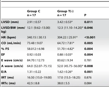
Table 2.Echocardiographic functional data.
Group C n = 17
Group T(-)
n = 17 P
LVSD (mm) 2.9160.57 3.4260.53* 0.011 LVSD/BW (mm/
kg)
12.1 (9.62–13.00) 12.3 (11.10–14.20)*0.046
HR (bpm) 340.15630.13 304.22625.91* ,0.001 CO (mL/min) 73.4869.07 64.7267.81* 0.005 % FS 58.01266.98 51.7064.82* 0.004
EF 0.9260.03 0.8860.03* 0.004
E wave (cm/s) 84.70612.73 83.6269.34 0.781 A wave (cm/s) 64.0 (52.07–75.15) 52.0 (45.75–56.00)*0.008
E/A 1.3160.22 1.6260.26* 0.001
IRT (ms) 16.50 (15.0–19.00) 17.0 (15.5–18.25) 0.876 IRTc (ms) 42.568.8 38.065.5 0.084
Group C: control animals, Group T (-): taurine-deficient animals; LVSD: left ventricular systolic diameter; BW: body weight; HR: heart rate; CO: cardiac output; % FS: fractional shortening; EF: ejection fraction; E/A: relationship between the E and A waves; IRT: isovolumetric relaxation time; IRTc: isovolumetric relaxation time normalized by the HR. Data are expressed as means6standard deviations or medians (quartile 1 - quartile 3). Bold values indicate statistical significance (P#0.05).
20 mL/L methanol, 20 ml/L acetonitrile, and 20 ml/L tetrahy-drofuran, and phase B was a solution of 65% chromatographic-grade methanol, as described by Lo¨ser et al. (1988) e Sussman (1988) [19,20]. The flow of solvent used was 0.8 ml/minute. An adsorbosphere OPA HR C18 column was used for chromato-graphic separation. For analysis, 10 mL of sample was used, and 200 mL of MeOH was added. This solution was centrifuged, and the supernatant was transferred to another tube, followed by MeOH evaporation. The solution was resuspended in 100 mL of 0.05 N HCl and then stirred and filtered through a 25mm membrane; it was then injected into the equipment [21].
Metalloproteinase-2 and -9 activity: Metalloproteinase-2 and -9 activity was determined as previously reported by Tyagi et al [22]. Briefly, samples for analysis were prepared by dilution in an extraction sample buffer consisting of 50 mM Tris, pH 7.4, 0.2 M NaCl, 0.1% Triton-X and 10 mM CaCl2. The samples
were then diluted in application sample buffer consisting of 0.5 M Tris, pH 6.8, 100% glycerol, and 0.05% bromophenol blue. The samples were loaded into wells of 8% SDS-polyacrylamide containing 1% gelatin. Electrophoresis was performed in a Bio-Rad apparatus at 80 V for 2 h, until the bromophenol blue reached the bottom of the gel. The gel was then incubated at 37uC overnight in activation solution consisting of 50 mM Tris, pH 8.4, 5 mM CaCl2 and ZnCl2. Staining with 0.5% Coomassie blue,
30% MeOH, and 10% acetic acid was performed for 2 h until clear bands over a dark background were observed. The gels were photographed, and the intensity of gelatinolytic action (clear bands) was analyzed in a UVP, UV, White Darkhon image analyzer.
Evaluation of cytokine production and adhesion molecules: The production of tumor necrosis factor alpha (TNF-a), interferon gamma (IFN-c) and interleukin 10 (IL-10) were evaluated. Briefly, 60 mg of cardiac tissue sample was homogenized and solubilized in a solution containing 50 mM potassium phosphate buffer, pH 7.4, 0.3 M sucrose, 0.5 mM DTT, 1 mM EDTA, pH 8.0, 0.3 mM PMSF, 10 mM NaF, and 1:100 protease inhibitor. Cytokine levels in cardiac homogenate were evaluated by ELISA, according to the manufacturer’s instructions (R & D Systems, Minneapolis, MN, USA) [17].
Cardiac lipid hydroperoxide and antioxidant enzyme analysis: lipid hydroperoxide (LH) based on the hydro peroxide-mediated oxidation of Fe2+; and antioxidant enzyme activities [23].
Glutathione peroxidase (GSHPx, E.C.1.11.1.9), superoxide dis-mutase (SOD, E.C.1.15.1.1) and catalase (CAT, E.C.1.11.1.6) activities were assessed as previously described [23,24,25]. Enzyme activity assays were performed at 25uC using a micro-plate reader (lQuant-MQX 200 with Kcjunior software connected to computer system control, Bio-Tec Instruments, Winooski, Vermont, USA). The absorbance was measured using a Pharmacia Biotech spectrophotometer (UV/visible Ultrospec 5000 with Swift II Applications software connected to computer system control, 974213, Cambridge, England, UK) at 560 nm. All reagents were purchased from Sigma (St. Louis, Missouri, USA) [23].
Statistical analysis: Comparisons between groups were made using Student’s t test for parameters with normal distribution. Otherwise, groups were compared using the Mann-Whitney U test. Data were expressed as means6SD or medians (including the lower quartile and upper quartile). Data analysis was carried out using SigmaStat for Windows v2.03 (SPSS Inc, Chicago, IL). The significance level was 5%. This study was designed to have 80% power of detecting a difference of several parameters between the mean for the control animals and the mean for taurine-deficient animals: left ventricular end-diastolic diameter adjusted by body weight (2 mm/kg); and cross sectional area
(30mm2), left ventricular systolic diameter adjusted by body
weight (0.2 mm/kg).
Results
The groups did not differ with regard to final body weight (C = 257 (254–277) g, T(-) = 262 (252–288) g; p = 0.558). Group T(-) animals had lower levels of taurine in the left ventricle (C = 1.860.8, T(-) = 0.460.1mmol/mg of tissue; p = 0.007).
The morphological data assessed by echocardiogram are shown in Table 1. Taurine deficiency resulted in lower values of LV posterior wall thickness and the ratio of LV wall thickness (LVWT)/LV end-diastolic diameter (LVDD). There were no differences in the other morphological echocardiographic variables between the groups.
The functional data assessed by echocardiogram are shown in Table 2. LV systolic diameter, heart rate, ejection fraction and fractional shortening were decreased in the T(-) group, compared to the C group. In contrast, the T(-) group had increased E/A ratios.
Histological analysis data and metalloproteinases values are shown in Table 3. The CSA was decreased in the T(-) group compared to the C group. There were no differences in the collagen volume fraction or metalloproteinase values between the groups.
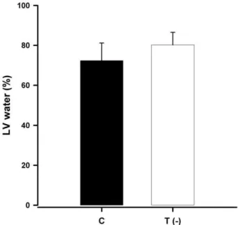
The LV water content was higher (Figure 1) and the LV dry weight was lower in the T(-) group compared to the C group (Figure 2).
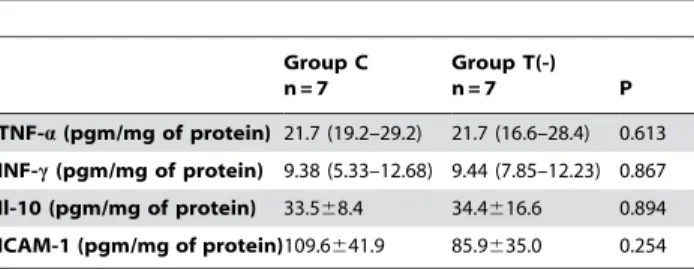
The data showing the cardiac lipid hydro peroxide and antioxidant enzyme activities are presented in Table 4. The T(-) group showed a higher concentration of lipid hydro peroxide and a lower catalase and glutathione peroxide activity than the C group. No differences were observed for cytokine and adhesion molecule levels (Table 5).
Discussion
The rat taurine deficiency model used in this study consisted of treatment with a solution of 3% ß-alanine. The treatment caused a
Figure 1. Bar graph representing the mean and standard deviation of water content (%) in the left ventricles of control and taurine-deficient animals (P = 0.03).
77% decrease in the concentration of taurine in the LV. This decrease in taurine concentration is higher than those observed by Parildar et al. [14] and Dawson Jr. et al. [15] (20% and 50% respectively). The difference can be explained by the age of the animals used in the experimental protocols. Parildar et al. [14] used rats at 22 months, and Dawson, Jr. et al. [15] used rats weighing 180 to 200 g. The animals studied by Dawson Jr. et al. [15] were 2 to 3 weeks older than the rats used in the present study. In a review article, Schuller-Lewis & Park [26] demon-strated that young animals show a lower capacity for taurine synthesis when compared to older animals. Thus, the observed differences between studies may be due to animal age differences. In humans, ß-alanine is used as an ergogenic supplement. The principal role is acting as a substrate of carnosine synthesis in skeletal muscle (a major contributor to H+
buffering during high-intensity exercise) [27]. ß-alanine supplementation-induce increase in muscle carnosine and the result is the opposite effect of the muscle taurine depletion [28]. It is not known if ß-alanine/ carnosine has compensatory changes in the heart. However, animals treated with ß-alanine and decreased taurine levels shows a lipid peroxidation potential in the heart [14].
The noteworthy finding in the present study was that taurine deficiency resulted in cardiac atrophy, as confirmed by thinning of the ventricular wall, reduced left ventricular dry weight, decreased myocyte cross sectional area, and increased oxidative stress. Regarding diastolic function, these data are consistent with decreased diastolic function in animals that are deficient in taurine. Indeed, the taurine-deficient group showed a lower velocity A and E, a higher E/A ratio, and a tendency for lower IRT/HR. With regard to systolic function, the echocardiographic data are consistent with systolic dysfunction. Decreased ejection fraction, fractional shortening and cardiac output were observed in the taurine-deficient group, compared to the control group. Moreover, in the present study, increased fibrosis and greater activation of metalloproteinases were not observed upon optical
microscopy. These findings are similar to those described by Ito et al. [29] in taut2/2 transgenic mice. The mice displayed small areas of atrophied cardiac myocytes, an absence of fibrosis, eccentric remodeling and systolic dysfunction [29].
Baskin & Taegtmeyer (2011) stated that these atrophic remodeling characteristics are cellular consequences of metabolic and hemodynamic unloading of a stressed heart [30]. Therefore, the atrophic cardiac remodeling observed in our study may result from two possible sources: 1) metabolic unloading (food restriction, protein-energy malnutrition) [31], abnormal protein metabolism [32] and inflammation [33] or 2) hemodynamic unloading [34,35].
Food restriction and protein-energy malnutrition induce changes in the heart such as atrophy and cardiac dysfunction [36]. Animal studies with food restriction and cachectic rats showed lower body weight, lower LV weight, a higher collagen concentration, and no change in cardiac performance [31,37]. Other common findings in malnourished animals include brady-cardia, hypotension and reduced cardiac output [38]. Thus, our data are similar to those found in malnourished rats. However, our animals suffered no food restrictions, and no differences in body weight or collagen percentage were observed.
Another mechanism that could explain our study findings is a decrease in protein synthesis and/or increase in protein catabo-lism. Protein synthesis is dependent on amino acids, and decreased protein synthesis may occur when amino acids are limiting, such as in the case of deficient diet or amino acid overuse (catabolic stress) [32]. For example, sulfur-containing amino acids such as methionine and cysteine can be limiting. However, this is not
Figure 2. Bar graph representing the mean and standard deviation of the left ventricular (LV) dry weight and normal-ized to body weight (BW) of control animals and taurine-deficient animals (P = 0.05).
doi:10.1371/journal.pone.0041439.g002
Table 3.Histopathology by light microscopy, myocyte cross-sectional area, collagen value fraction and determination of metalloproteinases -2 and -9 activation.
Group C n = 7
Group T(-)
n = 7 P
CSA (mm2) 238.3624.2 196.3629.7* 0.013 CVF (%) 1.3760.8 1.3461.3 0.955 MMP2 A/I 0.5160.1 0.5160.1 0.870 MMP9 A/I 0.6560.7 0.6060.4 0.898
Group C: control animals, Group T (-): taurine-deficient animals; CSA: myocyte cross sectional area; CVF: collagen value fraction; MPP2 A/I: ratio of the active and inactive forms of metalloproteinase 2; MPP9 A/I: ratio of the active and inactive forms of metalloproteinase 9. Data are expressed as means6standard deviations or medians (quartile 1 - quartile 3). Bold values indicate statistical significance (P#0.05).
doi:10.1371/journal.pone.0041439.t003
Table 4.Oxidative stress evaluation.
Group C n = 7
Group T(-)
n = 7 P
LH (nmol/g of tissue) 158.16614.36 197.75 33.29 0.023 GPx (mmol/g of tissue) 55.9567.83 35.7367.42 ,0.001 SOD (nmol/mg of protein) 20.0362.41 20.5761.80 0.673 Catalase (mmol/g of tissue) 112.58624.64 77.40616.42 0.016
Group C: control animals, Group T (-): taurine-deficient animals; LH: Lipid hydro peroxide; GPx: glutathione peroxidase; SOD: superoxide dismutase. Data are expressed as means6standard deviations. Bold values indicate statistical significance (P#0.05).
observed in taurine deficiency. Taurine is not incorporated into proteins; rather, it is the end product of the metabolic pathway of methionine [32]. Another possible mechanism of taurine is to regulate intracellular signaling pathways that are involved in protein synthesis. An example of this mechanism is the action of inhibitors of the renin angiotensin converting enzyme in the protein synthesis and catabolism promoting a reduction of left ventricular mass [39]. However, unlike other sulfur-containing amino acids, taurine does not participate in signaling pathways that control protein turnover [32].
Thus, our results suggest that the mechanisms that led to cardiac atrophy and ventricular dysfunction in taurine-deficient animals are not the same mechanisms involved in food restriction, protein-energy malnutrition or protein metabolism alteration.
Another potential mechanism involved in cardiac atrophy is load alterations. Indeed, changes in the hemodynamic mechanism of myocardium "load" may result in structural remodeling. In situations in which the heart undergoes pressure overload, concentric hypertrophy is observed, whereas volume overload leads to eccentric hypertrophy [34]. In the latter situation, dilatation of the cavity, changes in geometry, ventricular dysfunction and increased cardiac mass can be observed [34]. In contrast, atrophy occurs in situations in which there is a reduction in afterload [34]. Left ventricular mass regression is also observed
when blood pressure is reduced [40]. However, in this study, no blood pressure measurements were made, although a worsening of systolic and diastolic function was observed. In studies in which animals underwent the same model of taurine deficiency, no differences in blood pressure were observed when compared with non-deficient animals [41,42]. Thus, it is unlikely that the changes observed in this study are due to a reduction in blood pressure.
Inflammation was not responsible to the cardiac dysfunction observed in our study. Since the findings atrophy with decreased cardiac myocyte area, absence of fibrosis, no differences in metalloproteinase activity and no cytokine profile differences are not compatible with changes due to inflammation.
So, considering the critical role of oxidative stress in cardiac remodeling, we suggest that oxidative stress is associated with the cardiac dysfunction observed in our study. Oxidative stress has direct effects on cellular structure and function, and it can activate signaling molecules that are involved in cardiac remodeling, including apoptotic cascade [43,44]. In previous studies, taurine has been shown to promote antioxidant activity, regulating the rate of ROS generation by the mitochondria [8]. Gokce et al. reported that taurine significantly inhibited glutathione depletion and DNA damage caused by buthionine sulfoximine, an effective GSH-depleting compound [45]. Additionally, Ghosh et al. showed that taurine prevents arsenic-induced cardiac oxidative stress in cardiomyocytes [46]. Thus, the heart could be more susceptible to oxidative stress in taurine-deficient animals [14]. However, although the association between oxidative stress and remodeling, this study provides no evidence that there is causal relationship between oxidative stress and cardiac function and further study is needed to test this hypothesis.
In conclusion, taurine deficiency promoted structural and functional cardiac alterations, particularly with regard to the left ventricle.
Author Contributions
Analyzed the data: BPMR MFM SARP. Contributed reagents/materials/ analysis tools: LAMZ SARP. Wrote the paper: BPMR LAMZ SARP. Designed research: MCP LAMZ SARP. Conducted research: MCP FC-M SGZ-P JSM GJP AAHF BBM LSM PSA.
References
1. Allard ML, Jeejeebhoy KN, Sole MJ (2006) The management of conditioned nutritional requirements in heart failure. Heart Fail Rev 11: 75–82. 2. Witte KK, Clark AL, Cleland JG (2001) Chronic heart failure and
micronutrients. J Am Coll Cardiol 37: 1765–1774.
3. Spaeth DG, Schneider DL, Sarett HP (1974) Taurine synthesis, concentration, and bile salt conjugation in rat, guinea pig, and rabbit. Proc Soc Exp Biol Med 147: 855–858.
4. Birdsall TC (1998) Therapeutic applications of taurine. Altern Med Rev 3: 128– 136.
5. Bouckenooghe T, Remacle C, Reusens B (2006) Is taurine a functional nutrient? Curr Opin Clin Nutr Metab Care 9: 728–733.
6. Xu YJ, Arneja AS, Tappia PS, Dhalla NS (2008) The potential health benefits of taurine in cardiovascular disease. Exp Clin Cardiol 13: 57–65.
7. Wojcik OP, Koenig KL, Zeleniuch-Jacquotte A, Costa M, Chen Y (2010) The potential protective effects of taurine on coronary heart disease. Atherosclerosis 208: 19–25.
8. Schaffer SW, Jong CJ, Ramila KC, Azuma J (2010) Physiological roles of taurine in heart and muscle. J Biomed Sci 17 Suppl 1: S2.
9. Jacobsen JG, Smith LH (1968) Biochemistry and physiology of taurine and taurine derivatives. Physiol Rev 48: 424–511.
10. Hori M, Nishida K (2009) Oxidative stress and left ventricular remodelling after myocardial infarction. Cardiovasc Res 81: 457–464.
11. Denipote F, Ardisson LP, Azevedo PS, Minicucci MF, Lima-Leopoldo AP, et al. (2011) Influence of taurine on cardiac remodeling induced by tobacco smoke exposure. Cell Physiol Biochem 27: 291–298.
12. Rafacho BP, Azevedo PS, Polegato BF, Fernandes AA, Bertoline MA, et al. (2011) Tobacco smoke induces ventricular remodeling associated with an increase in NADPH oxidase activity. Cell Physiol Biochem 27: 305–312.
13. Das J, Vasan V, Sil P (2012) Taurine exerts hypoglycemic effect in alloxan-induced diabetic rats, improves insulin-mediated glucose transport signaling pathway in heart and ameliorates cardiac oxidative stress and apoptosis. Toxicology and Applied Pharmacology 258: 296–308.
14. Parildar H, Dogru-Abbasoglu S, Mehmetcik G, Ozdemirler G, Kocak-Toker N, et al. (2008) Lipid peroxidation potential and antioxidants in the heart tissue of beta-alanine- or taurine-treated old rats. J Nutr Sci Vitaminol (Tokyo) 54: 61– 65.
15. Dawson R, Jr., Biasetti M, Messina S, Dominy J (2002) The cytoprotective role of taurine in exercise-induced muscle injury. Amino Acids 22: 309–324. 16. Lang RM, Bierig M, Devereux RB, Flachskampf FA, Foster E, et al. (2005)
Recommendations for chamber quantification: a report from the American Society of Echocardiography’s Guidelines and Standards Committee and the Chamber Quantification Writing Group, developed in conjunction with the European Association of Echocardiography, a branch of the European Society of Cardiology. J Am Soc Echocardiogr 18: 1440–1463.
17. Minicucci MF, Azevedo PS, Oliveira SA, Jr., Martinez PF, Chiuso-Minicucci F, et al. (2010) Tissue vitamin A insufficiency results in adverse ventricular remodeling after experimental myocardial infarction. Cell Physiol Biochem 26: 523–530.
18. de Paiva SA, Zornoff LA, Okoshi MP, Okoshi K, Matsubara LS, et al. (2003) Ventricular remodeling induced by retinoic acid supplementation in adult rats. Am J Physiol Heart Circ Physiol 284: H2242–2246.
19. Lo¨ser C, Wunderlich U, Fo¨lsch UR (1988) Reversed-phase liquid chromato-graphic separation and simultaneous fluorimetric detection of polyamines and theirs monoacetyl derivatives in human and animal urine, serum and tissue samples: an improved, rapid and sensitive method for routine application. J Chromatogra 430: 249–262.
Table 5.Cytokine production and adhesion molecules.
Group C n = 7
Group T(-)
n = 7 P
TNF-a(pgm/mg of protein) 21.7 (19.2–29.2) 21.7 (16.6–28.4) 0.613
INF-c(pgm/mg of protein) 9.38 (5.33–12.68) 9.44 (7.85–12.23) 0.867 Il-10 (pgm/mg of protein) 33.568.4 34.4616.6 0.894 ICAM-1 (pgm/mg of protein)109.6641.9 85.9635.0 0.254
Group C: control animals, Group T (-): taurine-deficient animals, TNF-a: tumor necrosis factor alpha, INF-c: interferon-gamma, Il-10: interleukin 10; ICAM-1: intercellular adhesion molecule 1. Data are expressed as means6standard deviations or medians (quartile 1 - quartile 3).
20. Sussman MR (1988) Purification of integral plasma membrane protein by reversed phase high performance liquid chromatography. Anal Bioch 169: 395– 399.
21. Deminice R, Portari GV, Marchini JS, Vannucchi H, Jordao AA (2009) Effects of a low-protein diet on plasma amino acid and homocysteine levels and oxidative status in rats. Ann Nutr Metab 54: 202–207.
22. Tyagi SC, Matsubara L, Weber KT (1993) Direct extraction and estimation of collagenase(s) activity by zymography in microquantities of rat myocardium and uterus. Clin Biochem 26: 191–198.
23. Nakamura W, Hosoda S, Hayashi K (1974) Purification and properties of rat liver glutathione peroxidase. Biochem Biophys Acta 358: 251–261.
24. Ewing JF, Janero DR (1995) Microplate superoxide dismutase assay employing a nonenzymatic superoxide generator. Anal Biochem 232: 243–248.
25. Burneiko RC, Diniz YS, Galhardi CM, Rodrigues HG, Ebaid GM, et al. (2006) Interaction of hypercaloric diet and physical exercise on lipid profile, oxidative stress and antioxidant defenses. Food Chem Toxicol 44: 1167–1172. 26. Schuller-Levis GB, Park E (2003) Taurine: new implications for an old amino
acid. FEMS Microbiol Lett 226: 195–202.
27. Hobson RM, Saunders B, Ball G, Harris RC, Sale C (2012) Effects ofb-alanine supplementation on exercise performance: a meta-analysis. Amino Acids DOI 10.1007/s00726-011-1200-z.
28. Stellingwerff T, Decombaz J, Harris R, Boesch C (2012) Optimizing human in vivo dosing and delivery of b-alanine supplements for muscle carnosine synthesis. Amino Acids DOI 10.1007/s00726-012-1245-7.
29. Ito T, Kimura Y, Uozumi Y, Takai M, Muraoka S, et al. (2008) Taurine depletion caused by knocking out the taurine transporter gene leads to cardiomyopathy with cardiac atrophy. J Mol Cell Cardiol 44: 927–937. 30. Baskin KK, Taegtmeyer H (2011) Taking pressure off the heart: the ins and outs
of atrophic remodelling. Cardiovasc Res 90: 243–250.
31. Fioretto JR, Querioz SS, Padovani CR, Matsubara LS, Okoshi K, et al. (2002) Ventricular remodeling and diastolic myocardial dysfunction in rats submitted to protein-calorie malnutrition. Am J Physiol Heart Circ Physiol 282: H1327– 1333.
32. Metayer S, Seiliez I, Collin A, Duchene S, Mercier Y, et al. (2008) Mechanisms through which sulfur amino acids control protein metabolism and oxidative status. J Nutr Biochem 19: 207–215.
33. Yndestad A, Damas JK, Oie E, Ueland T, Gullestad L, et al. (2007) Role of inflammation in the progression of heart failure. Curr Cardiol Rep 9: 236–241.
34. Campbell SE, Korecky B, Rakusan K (1991) Remodeling of myocyte dimensions in hypertrophic and atrophic rat hearts. Circ Res 68: 984–996. 35. Welsh DC, Dipla K, McNulty PH, Mu A, Ojamaa KM, et al. (2001) Preserved
contractile function despite atrophic remodeling in unloaded rat hearts. Am J Physiol Heart Circ Physiol 281: H1131–1136.
36. Pissaia O, Rossi MA, Oliveira JS (1980) The heart in protein-calorie malnutrition in rats: morphological, electrophysiological and biochemical changes. J Nutr 110: 2035–2044.
37. Okoshi MP, Okoshi K, Pai VD, Pai-Silva MD, Matsubara LS, et al. (2001) Mechanical, biochemical, and morphological changes in the heart from chronic food-restricted rats. Can J Physiol Pharmacol 79: 754–760.
38. Nutter DO, Murray TG, Heymsfield SB, Fuller EO (1979) The effect of chronic protein-calorie undernutrition in the rat on myocardial function and cardiac function. Circ Res 45: 144–152.
39. Siddiq T, Richardson PJ, Trotter SE, Preedy VR (1996) Protein synthesis during regression of left ventricular hypertrophy with lisinopril in abdominal aortic constriction model of hypertension. Biochem Mol Med 57: 19–24.
40. Pauletto P, Vescovo G, Scannapieco G, Angelini A, Piccolo D, et al. (1986) Progression and regression of cardiac hypertrophy in hypertensive rats: biochemical and molecular changes in ventricular myosin. J Hypertens Suppl 4: S135–137.
41. Mozaffari MS, Abebe W (2000) Cardiovascular responses of the taurine-depleted rat to vasoactive agents. Amino Acids 19: 625–634.
42. Roysommuti S, Suwanich A, Jirakulsomchok D, Wyss JM (2009) Perinatal taurine depletion increases susceptibility to adult sugar-induced hypertension in rats. Adv Exp Med Biol 643: 123–133.
43. Ricci C, Pastukh V, Leonard J, Turrens J, Wilson G, et al. (2008) Mitochondrial DNA damage triggers mitochondrial-superoxide generation and apoptosis. Am J Physiol Cell Physiol 294: C413–422.
44. Tsutsui H, Kinugawa S, Matsushima S (2009) Mitochondrial oxidative stress and dysfunction in myocardial remodelling. Cardiovasc Res 81: 449–456. 45. Gokce G, Ozsarlak-Sozer G, Oktay G, Kirkali G, Jaruga P, et al. (2009)
Glutathione depletion by buthionine sulfoximine induces oxidative damage to DNA in organs of rabbits in vivo. Biochemistry 48: 4980–4987.