Mailing Address: Arturo Jaramillo •
Departamento de Neurología y Neurocirugía, Hospital Clínico de la Univer-sidad of Chile. Santos Dumont 999. Independencia, Santiago, Chile. Email: [email protected]
Manuscript received September 11, 2008; revised manuscript received December 11, 2008; accepted february 02, 2009.
Ischemic Stroke as the First Manifestation of Severe Ventricular
Hypertrabeculation/Non-compaction
Arturo Jaramillo
1, Alfredo Ramírez
2, Lorna Galleguillos
1, José Vallejos
1, Sergio Illanes
1Department of Neurology and Neurosurgery, Clinical Hospital and School of Medicine, University of Chile1; Department of Cardiology, Clinical
Hospital and School of Medicine, University of Chile2, Chile
Introduction
Left Ventricular Hypertrabeculation/non-compaction (LVHT) is a rare congenital cardiomyopathy characterized by a multiple, prominent myocardial trabeculations and deep intertrabecular recesses communicating with the left ventricular cavity. Its prevalence is about 0.05%/y to 0.24%/y in echocardiographic series1. Some necropsy and
echocardiographic studies have shown that clots can be formed in LVHT with a potential secondary embolism risk2,3.
We report a case of ischemic cerebral infarction associated with severe LVHT associated to ventricular dysfunction as a possible synergistic cardioembolic mechanism.
Clinical case
A 56-year-old man with an isolated history of hypertension presented a sudden and severe weakness of the left side. Physical examination found a S3 without murmurs. The neurological examination showed that the patient was spatially-disoriented and gave a wrong answer about his
age; he also presented right hemianopsia, mild dysarthria, mild weakness and sensory loss on the left side and sensory extinction on the left side (basal NIHSS=7). The initial CT scan showed an extensive area of hypodensity in the right superficial and deep middle cerebral artery (MCA) territory. The MRI showed an ischemic lesion in the same artery territory with a small hemorrhagic transformation in the basal ganglia (Figure 1). The electrocardiogram suggested left ventricular hypertrophy. The chest radiography showed severe cardiomegaly without pleural effusion. A two-dimensional Doppler color-flow echocardiography (TTE) and transesophageal (TEE) echocardiography (Vivid 7 Dimension Echocardiograph, General Electric), showed severe dilated myocardiopathy with a diffuse hypokinesia of the left ventricle, ejection fraction < 20% and severe pulmonary hypertension. No cardiac thrombus was detected (Figure 2A). Subsequently, a cardiac MRI was performed to evaluate the damage to the ventricular wall, which showed a characteristic ventricular image of severe non-compacted left ventricle (LVHT) in the lateral, septal and apical walls (Figure 2B). The ratio between the non-compacted/ compacted (N/C) endomyocardium layers was 3.0, according with the LVHT TTE diagnostic criteria. An associated global hypokinesia in the affected regions was observed, especially in the anteroseptal wall, with 18% of ejection fraction. Not myocardial delayed enhancement was demonstrated. A carotid Doppler study failed to show any abnormality. No other associated blood abnormalities (including cardiac enzymes) were found as potential etiologies for the ischemic stroke. Finally, a regimen of warfarin (INR 2-3), carvedilol, spironolactone and furosemide was started and maintained until hospital discharge.
Discussion
During the development of the embryonic myocardium, trabeculations emerge from the apical region of the primitive ventricles at about day 32 and then involutes through a process of resorption and remodeling. Afterwards, the myocardium gradually condenses and the large spaces within the trabecular meshwork flatten or disappear4. A failure in this process
results in multiple trabeculations and intertrabecular recesses in the left ventricular myocardium, a phenomenon known as LVHT5. Apparently, both sexes are similarly affected. The
LVHT may manifest from infancy to young adulthood, with a high mortality rate in most cases1. In children, it can be
associated with different neurological abnormalities, affecting nerves and/or striated muscle6. However, controversies still
remain about its embolic role as an isolated myocardial defect, when compared with an associated ventricular dysfunction,
:
A rare congenital myocardial defect, known as left ventricular hypertrabeculation/non-compaction (LVHT), has been occasionally described associated with thrombus formation with a potential systemic embolic risk, but its association with ischemic strokes remains controversial.
:
We report a case of ischemic stroke in a patient with severe LVHT and ventricular dysfunction as a possible etiologic synergistic association.
:
In absence of other embolic sources, a severe LVTH associated with ventricular dysfunction could constitute a potential source of brain embolism, especially in patients with high suspicion of an embolic mechanism of ischemic stroke.
Key Words
Heart defects, congenital; ventricular dysfunction, left; intracranial embolism; stroke.
e28
Figure 1 - (A) TTE - Blood low among trabecular recesses (white arrow). (B) Cardiac Magnetic Resonance using gadolinium shows a LVHT on the lateral, septal and
apical myocardial walls (See details in text).
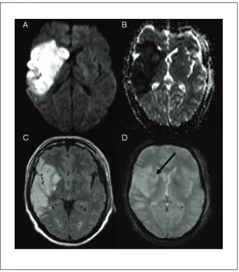
Figure 2 - MRI Stroke-protocol.(A) DWI, (B) ADC map, (C) FLAIR, (D), T2
weighted. The indings are compatible with a large ischemic lesion in the right basal ganglia and right frontal, parietal and temporal lobes, territories of the deep and supericial branches of the right middle cerebral artery. In (D) there are signs of hemorrhagic transformation in the basal ganglia (black arrow)
considering that a relevant myocardial hypokinesia constitutes in itself a recognized embolic source from young boys7 to
adult women8.
Our patient did not have a family history of cardiopathy or genetic disorders and ischemic stroke was the first clinical
manifestation. The current literature shows that the clinical spectrum of LVHT has yet to be established. From the first publication in 19904, LVHT has been associated with systemic
thromboembolism, cardiac failure, cardiac arrhythmia, depressed left ventricular systolic function and Ischemic stroke or peripheral embolism9. Cerebral embolism has been reported
at 38% of LVHT in different series2,4. In a necropsy retrospective
study2 that included 6 children, 3 had systemic embolism,
with 2 of them involving the brain. In the study by Chin4, 3
of 10 included patients had cerebral embolism, which was the direct cause of death in one of them2. The intertrabecular
recesses in the cardiac apex and/or the inferior and lateral left ventricular walls, which have been considered the more frequently affected segments3, are prone to thrombus formation
with a potential risk of secondary systemic embolism18. Thus,
in the study by Chin4, left ventricular mural thrombi were
identified in two patients, one of them by echocardiography and the other one at necropsy, with clots being found within the intertrabecular recesses in the latter one. We failed to find any intertrabecular clots using echocardiography or cardiac MR studies, but blood flow among the trabecular recesses was detected on TTE (Figure 1).
Hemorrhagic cerebral infarction, in contrast to the pale form, results from reperfusion of the vascular bed of the infarction after relief of the occlusion, with fragmentation and distal migration of an embolus10. This hypothesis has
been supported by the higher frequency of hemorrhagic infarctions secondary to cardioembolic mechanisms. Based on accumulated experience, it is probably to find a cardioembolic source when a hemorrhagic brain infarction has occurred, as it was suspected in our patient who had a small hemorrhagic transformation (Figure 2). Classic cardioembolic sources such as atrial fibrillation, acute myocardial infarction, ventricular aneurysm, rheumatic heart disease or prosthetic cardiac valve, among others, were excluded in our patient.
Arq Bras Cardiol 2009; 94(3) : e28-e30
Jaramillo et al Myocardial hypertrabeculation and embolic stroke
e29
The diagnosis of LVHT is usually based on two-dimensional echocardiography4,9,11. The TTE diagnostic criterion consists
in calculating the ratio of non-compacted/compacted (N/C) endomyocardium layers, which has been defined as N/C > 211. In our patient, this criterion was met, with an N/C ratio =
3.0, suggesting LVTH. A contrast left ventricular angiography using an iodine contrast agent (gadolinium) demonstrated a dysfunctional inferior wall and an apical region with multiple trabecular recesses. Following our institutional algorithm, we decided to perform a new echocardiography using a sonic contrast (Air–filled serum albumin microcapsules, Quantison™) which demonstrated numerous, excessively prominent trabeculations and deep intertrabecular recesses, concordant with LVHT, in the same dysfunctional regions demonstrated at left ventricular angiography, with absence of co-existing cardiac structural abnormalities. A cardiac MR study was performed, which helped us confirm a diagnosis of LVHT.
In conclusion, based on the echocardiographic and cardiac MRI findings, we considered that our patient probably presented a cardioembolic ischemic stroke, where
a severe and localized LVHT associated to ventricular dysfunction were the only relevant cardiac findings observed. We propose that both conditions may act as a synergistic cardioembolic mechanism, but this proposition needs to be confirmed in future studies. The combined use of Doppler color-flow echocardiography and cardiac MRI in patients without coronary disease could help confirm this unusual cardiac disorder.
Potential Conflict of Interest
No potential conflict of interest relevant to this article was reported.
Sources of Funding
There were no external funding sources for this study.
Study Association
This study is not associated with any post-graduation program.
References
1. Stöllberg C, Finsterer J. Left ventricular hypertrabeculation/noncompactation. J Am Soc Echocardiogr. 2004; 17: 91-100.
2. Stollberger C, Finsterer J. Left ventricular hypertrabeculation/noncompaction and stroke or embolism. Cardiology. 2005; 103: 68-72.
3. Domnitskaia TM, Sidorenko BA, Erokhina MG, Saidova MA. [Non-compact left ventricular myocardium: Current state of the problem]. Kardiologiia. 2006; 46: 63-8.
4. Chin TK, Perloff JK, Williams RG, Jue K, Mohrmann R. Isolated noncompaction of left ventricular myocardium: a study of eight cases. Circulation. 1990; 82: 507-13.
5. Breckenridge RA, Anderson RH, Elliott PM. Isolated left ventricular non-compaction: the case for abnormal myocardial development. Cardiol Young. 2007; 17 (2): 124-9.
6. Stollberger C, Finsterer J, Blazek G. Left ventricular hypertrabeculation/ noncompaction and association with additional cardiac abnormalities and neuromuscular disorders. Am J Cardiol. 2002; 90: 899-902.
7. Vijayvergiya R, Jha A, Panda SN, Grover A. Biventricular non-compactation: the rare cause of stroke in a young boy. Int J Cardiol. 2008; 129: E84-E85.
8. Ker J, Van der Merwe C. Isolated left ventricular non-compactation as a cause of thrombo-embolic stroke: a case report and review. Cardiovasc J S Afr. 2006; 17: 146-7.
9. Stollberger C, Winkler-Dworak M, Blazek G, Finsterer J. Cardiologic and neurologic findings in left ventricular hypertrabeculation/noncompaction relating to echocardiographic indication. Int J Cardiol. 2007; 119 (1): 28-32.
10. Fisher M, Adams RD. Observations on brain embolism with special reference to the mechanism of hemorrhagic infarction. J Neuropathol Exp Neurol. 1951; 10: 92-4.
11. Murphy RT, Thaman R, Blanes JG, Ward D, Sevdalis E, Papra E, et al. Natural history and familial characteristics of isolated left ventricular non-compaction. Eur Heart J. 2005; 26: 187-92.
Arq Bras Cardiol 2009; 94(3) : e28-e30
Jaramillo et al
Myocardial hypertrabeculation and embolic stroke