PRISCILLA LARA FARIA
SCREENING FUNGAL ISOLATES FOR BIOLOGICAL CONTROL OF CULICID VECTORS BASED ON FORMATION OF APPRESSORIA
Dissertação apresentada à Universidade Federal de Viçosa, como parte das exigências do Programa de Pós-Graduação em Entomologia, para obtenção do título de Magister Scientiae.
VIÇOSA
Ficha catalográfica preparada pela Seção de Catalogação e Classificação da Biblioteca Central da UFV
T
Faria, Priscila Lara, 1984-
F224s Screening fungal isolates for biological control of culicid 2011 vectores based on formation of appressoria / Priscila Lara
Faria. – Viçosa, MG, 2011.
x, 53f. : il. (algumas col.) ; 29cm.
Inclui apêndices.
Orientador: Simon Luke Elliot.
Dissertação (mestrado) - Universidade Federal de Viçosa. Referências bibliográficas: f. 37-53.
1. Mosqueiro - Controle biológico. 2. Aedes aegypti. 3. Anopheles aquasalis. 4. Fungos entomopatogênicos. 5. Metarhizium anisopliae. 6. Beauveria bassicena. 7. Inseto
nocivo - Controle biológico. 8. Fungos - Seleção. I. Universidade Federal de Viçosa. II. Título.
PRISCILLA LARA FARIA
SCREENING FUNGAL ISOLATES FOR BIOLOGICAL CONTROL OF CULICID VECTORS BASED ON FORMATION OF APPRESSORIA
Dissertação apresentada à Universidade Federal de Viçosa, como parte das exigências do Programa de Pós-Graduação em Entomologia, para obtenção do título de Magister Scientiae.
APROVADA: 27 de julho de 2011.
_____________________________ _____________________________ Prof. Gustavo Ferreira Martins Dra. Alessandra Aparecida Guarneri (Coorientador)
_____________________________ Prof. Simon Luke Elliot
ii
À minha avó Vitória e ao meu avô
Messias (in memoriam) pela dedicação e amor incondicionais.
À minha mãe Luiza, ao meu pai
Aluísio, ao meu padrasto Paulo e
aos meus irmãos Diego e
Jéssica, por serem sempre o
motivo da minha busca por ser
alguém melhor,
iii AGRADECIMENTOS
Gostaria de agradecer à Universidade Federal de Viçosa e ao programa
de Pós-graduação em Entomologia, pela oportunidade e estrutura oferecidas;
À Coordenação de Aperfeiçoamento de Pessoal de Nível Superior
(Capes) pelo financiamento deste estudo;
Ao meu orientador Dr. Simon Luke Elliot pelos seus valiosos conselhos
científicos, críticas, paciência, apoio e incentivo;
Ao meu coorientador Dr. Gustavo Ferreira Martins, que se empenhou
muito em me ajudar e pela atenção sempre a mim cedida e ao Dr. Angelo
Pallini por ter aceitado tão prontamente ser meu coorientador;
À Dra. Alessandra Aparecida Guarneri, ao Dr. Arne Janssen e Dr. Eraldo
Lima pela disposição em participar da minha banca, colaborando com a
finalização deste trabalho;
Ao Dr. Eraldo Lima e Dra. Terezinha Della Lucia pela disponibilidade de
empréstimo de equipamentos e uso dos laboratórios;
Ao Dr. José Eduardo Serrão a ao Dr. Weyder C. Santana pela ajuda com
as fotos de microscopia;
À Fiocruz - René Rachou, Dr. Luciano Moreira, Itaforte BioProdutos e
Universidade Federal Rural de Pernambuco-UFRPE pela colaboração;
À Alice, Aline, Camila, Daiane, Farley e Ricardo por me ajudarem nos
experimentos;
Aos colegas de mestrado pelo apoio, ajuda, convivência e incentivo;
Ao pessoal do laboratório pela amizade, bondade e respeito com que me
trataram, especialmente ao Senhor Manuel;
À Fernanda e Raquel Fellet, Fiocruz, pela presteza e cooperação;
À Sra. Paula, Miriam e Silvânia, pela boa vontade com que sempre
atenderam às minhas solicitações;
Às minhas colegas de república, pelo carinho e conforto: Vivi, Cris,
Flavinha, Nath, Lailla, Anja, Ana, Paola, Livinha;
Aos meus velhos e bons amigos, Filipe, Dri, Cris e meus novos amigos
Silvana, Ira, Hernane, pelo apoio nos momentos difíceis e os sorrisos que me
fizeram mais feliz;
iv
A Deus, por permitir que tudo isso se tornasse possível;
Enfim, a todos os que ficaram ao meu lado e contribuíram direta ou
indiretamente para a conclusão desta etapa!
v SUMÁRIO
LISTA DE FIGURAS ... VI
LISTA DE TABELAS... VII
RESUMO ... VIII
ABSTRACT... X
GENERAL INTRODUCTION ...1
1. BIBLIOGRAPHIC REVIEW ...5
1.1AEDES ANDANOPHELES...5
1.2 BIOLOGICAL CONTROL AND ENTOMOPATHOGENIC FUNGI...7
1.3INFECTION PROCESS OF ENTOMOPATHOGENIC FUNGI...10
1.4 SCREENING FUNGAL ISOLATES...12
2. OBJECTIVE ...13
3. MATERIALS AND METHODS ...14
3.1 CULICIDS...14
3.2 FUNGI...14
3.3IMPROVING THE SCREENING TECHNIQUE...16
3.4 GERMINATION AND APPRESSORIUM FORMATION ON AEDES WINGS...17
3.5 GERMINATION AND APPRESSORIUM FORMATION ON ANOPHELES WINGS...19
3.6 TEST WITH LIVE AEDESAEGYPTI...19
4. RESULTS ...21
4.1 FUNGI...21
4.2 IMPROVING THE SCREENING TECHNIQUE...22
4.3GERMINATION AND APPRESSORIUM FORMATION ON AEDES WINGS...24
4.4 GERMINATION AND APPRESSORIUM FORMATION ON ANOPHELES WINGS...24
4.5 TEST WITH LIVE AEDESAEGYPTI...26
5. DISCUSSION...29
6. CONCLUSION ...33
APPENDICES...34
vi LISTA DE FIGURAS
1. Conidia of germinated Metarhizium anisopliae on the wing cuticle of Aedes
aegypti mosquito. The arrow shows the germ tube (Bar = 10 μm).………....18 2. Germinated conidium of Metarhizium anisopliae and its appressorium on the
wing cuticle of Aedes aegypti. Arrows a – conidium; b – germ-tube; c – hyphal differentiation into appressorium (Bar = 10 μm)………...18
3. Germinated conidium of Beauveria bassiana and its appressorium on the wing cuticle of Aedes aegypti. Arrows a – conidium; b – hyphal differentiation into appressorium (Bar = 10 μm)…………...…………...………...19
vii LISTA DE TABELAS
1. Sources of isolates of entomopathogenic fungi used for screening…...15
2. Percentage conidial viability of the 83 fungal isolates used on wings of Aedes
aegypti and 40 of these on wings of Anopheles aquasalis….………..21 3. Percentage of the isolates of entomopathogenic fungi Beauveria bassiana
and Metarhizium anisopliae that germinate (G%) and formed appressorium
(A%) on wing cuticles of Aedes aegypti and Anopheles aquasalis (G% is the percentage of number of germinated conidia/total of conidia; A% is the
percentage of number of conidia that formed appressorium/number of
germinated conidia)...24
viii RESUMO
FARIA, Priscilla Lara, M. Sc., Universidade Federal de Viçosa, julho de 2011.
Rastreamento de isolados fúngicos para o controle biológico de culicídeos vetores baseado na formação de apressório. Orientador: Simon Luke Elliot. Coorientadores: Gustavo Ferreira Martins e Angelo Pallini.
O controle de doenças transmitidas por insetos vetores pode ser realizado
através do controle do agente etiológico da doença ou do controle do vetor da
doença. Entretanto, devido às dificuldades na produção de vacinas, à baixa
eficácia dos compostos repelentes e aos problemas com inseticidas sintéticos,
formas alternativas no combate aos vetores vêm sendo divulgadas. Entre as
alternativas, o controle biológico com uso de fungos entomopatogênicos vem
se destacando, seja visando o rápido extermínio do vetor, controlando
diretamente sua população, ou ocasionando efeitos subletais que podem
influenciar na capacidade vetorial destes insetos. A especificidade destes
entomopatógenos aos seus hospedeiros ocorre, provavelmente, através de
processos de reconhecimento que podem estar ligados à composição da
epicutícula de seu inseto-alvo, demonstrando uma resposta diferente a
determinados hospedeiros. No presente trabalho estudou-se uma técnica de
rastreamento de isolados dos fungos Beauveria bassiana e Metarhizium anisopliae utilizando a cutícula das asas dos mosquitos vetores Aedes aegypti
e Anopheles aquasalis. Essa técnica consistiu em inocular uma suspensão de 1x10⁵ conídios ml-1 nas asas dos mosquitos e verificar, após 30 horas, a
germinação e desenvolvimento de apressórios, buscando selecionar isolados
que formassem esta estrutura de infecção em ambos os mosquitos.
Confirmando a funcionalidade da técnica, nós verificamos que de 83 isolados
testados em Ae. aegypti, 3 não germinaram e 67 germinaram e formaram apressórios. Em An. aquasalis, de 40 isolados testados, todos germinaram e destes, 32 germinaram e formaram apressórios. De 40 isolados testados em
ix
compostos cuticulares no reconhecimento do hospedeiro. Estes fungos
x ABSTRACT
FARIA, Priscilla Lara, M. Sc., Universidade Federal de Viçosa, July, 2011.
Screening fungal isolates for biological control of culicid vectors based on formation of appressoria. Adviser: Simon Luke Elliot. Co-Advisers: Gustavo Ferreira Martins and Angelo Pallini.
The control of diseases transmitted by insect vectors may be performed by
control of the etiological agent of the disease or by vector control. However due
to difficulties with vaccine production, limited effectiveness of repellent
compounds and the problems with synthetic insecticides, alternative means of
vector control are of interest. Among these, biological control using
entomopathogenic fungi has generated interest, whether for vector population
control due to mortality, or through sublethal effects that can influence vectorial
capacity. The host specificity of entomopathogenic fungi is likely to occur
through recognition processes linked to the composition of the epicuticle of the
insect target, showing different responses to different hosts. Here, we studied a
technique for isolate screening of Beauveria bassiana and Metarhizium
anisopliae using the wing cuticle of the mosquito vectors Aedes aegypti and
Anopheles aquasalis. This technique consists of inoculating a suspension of 1x10⁵ conidia/ml-1 on the wings of mosquitoes and determining, after 30 hours,
conidial germination and appressorium development (this being necessary for
infection), with the aim of selecting isolates that formed appressoria in both
mosquito species. Confirming the functionality of the technique, we found that of
83 isolates tested in Ae. aegypti, 3 did not germinate, while 67 germinated and formed appressoria. In An. aquasalis, 40 isolates was tested and all germinated; of these, 32 germinated and formed appressoria. Of 40 isolates
tested in both mosquitoes, 32 formed appressoria in both, in line with studies
that report the selectivity of the fungi and the influence of cuticular compounds
in recognition of the host. These selected fungi are being investigated for
1 GENERAL INTRODUCTION
Every year, millions of people contract diseases transmitted by insect
vectors. These diseases have a large impact on social and economic activities,
especially in tropical countries (Zaim & Guillet, 2002). Control programs have
always prioritized arthropod vector control. However, financial and management
problems, together with misuse of insecticides have often led to the failure of
these programs (Gubler, 1998).
Among the insect vectors of greatest importance, the genera Aedes and
Anopheles stand out, being responsible respectively for the transmission of arboviruses such as Chikungunya fever (WHO, 2008), dengue and yellow fever,
and the malarial parasite Plasmodium (Hemingway & Ranson, 2000; WHO, 2009; 2011). Emergence and resurgence of these arboviruses (Gubler, 1996;
2002) and the continued prevalence of malaria (Scholte et al. 2003) highlight the need for greater efforts to control these mosquitoes.
Options for vectorborne disease control include the control of etiological
agents through vaccination or control of the vectors. For some arboviruses,
however, there are no vaccines currently available, so vector control continues
to be extremely valuable. Therefore, the search for sustainable ways of
controlling insect vectors of human diseases has high value, as these are key
factor of control components (Zaim & Guillet, 2002). Options for vector control
include the direct control of mosquito populations and indirect control via
pre-death effects (called sublethal effects). Direct control is fundamentally based on
the use of insecticides, with a view to immediate death of the insect. The
advantage of this method is that it prevents mosquito breeding, in addition to
preventing new blood meals and thus, prevents transmission of the etiological
agent of disease.
Although insecticides are important tools for insect vector control
(Hemingway & Ranson, 2000; Scholte et al., 2004; Thomas & Read, 2007) and are considered one of the cheapest and most effective methods to control
mosquitoes (Read et al., 2009) and other insect vectors (Zaim & Guillet, 2002), their indiscriminate use, besides causing, environmental contamination and
health problems for humans and other animals, induce selection for insect
2
2001; Shiff, 2002; Hargreaves et al., 2003). A further factor is that these chemicals are derived from exhaustible sources such as petroleum (Messias,
1989). Thus, there is a need for new control tools (Zaim & Guillet, 2002;
Farenhorst et al., 2008; Howard et al., 2010a,b) that do not harm the environment and are safe for use (Vega et al., 2009), also that are economically and ecologically viable (Valadares-Inglis et al., 1998).
Indirect control, via sublethal effects, does not require immediate insect
death and has a slower action. It is possible that this means of control
decreases the selection pressure for resistance, where it allows insect
reproduction (Thomas & Read, 2007). It may thus be interesting to develop
interventions that act sublethally on the insect’s vectorial capacity. Decreases in
insect longevity, in blood feeding success, in fecundity and in vector
competence, are among the sublethal effects sought.
Within this context, biological control is an alternative to be used against
these vectors (Scholte et al., 2004; Mohanty et al., 2008; Blanford et al., 2009). The advantages of these agents are intrinsically linked to its action and
production mode, limited side effects, longer-lasting control and are non-toxic
(Alves, 1998). In addition to these factors, may also be motivated by potential
for sublethal effects such as reducing mosquito longevity (Blanford et al., 2005; Scholte et al., 2003; 2005; 2007; de Paula et al., 2008; Kannan et al., 2008; Mohanty et al., 2008; Leles et al., 2010; Mnyone et al., 2011), blood feeding success (Scholte et al., 2006; Howard et al., 2010a), fecundity (Scholte et al., 2006) and vector competence (Blanford et al., 2005).
Another important factor is that an entomopathogenic fungus does not
need to be ingested to infect its host as it can infect through the cuticle (Pedrini
et al., 2007; Scholte et al., 2007) via mechanical pressure and enzymatic action. However, for infection to occur there must be adhesion and formation of
infection structures (Charnley, 1984), which only occurs when there is host
recognition, indicative of specificity (Charnley, 1984; St. Leger et al., 1990a). The surface structure and chemical composition of the host cuticle seems to
influence this process (Pedrini et al., 2007). Specificity may be related to mechanisms of recognition associated with the composition of the host
epicuticle, since these components are extremely heterogeneous. These
3
Leger et al., 1990a; 1996a). Although the chemical interaction between fungi and host during adhesion is poorly explained (Boucias et al., 1988, Yaginuma et al., 2006), it is known that a fungus’ virulence is correlated with its ability to infect the host cuticle (Yaginuma et al., 2006). This difference in the response of a pathogen can be used for selection of isolates (Wang & St. Leger, 2005).
Traditionally, studies aimed at selecting entomopathogenic fungi are
conducted by inoculating the fungus in live insects, observing the mortality
caused in their host and selecting the isolate that killed most quickly (Neves &
Hirose, 2005; Almeida et al., 1997; Chernaki-Leffer et al., 2007). These experiments can require a large investment of time and resources. In
mosquitoes, isolates which cause high mortality (considered highly virulent) are
selected for further studies, discarding less virulent fungi, as seen in the work of
De Paula et al. (2008). However, these less virulent fungi may be used to produce sublethal effects that can be useful in insect vector control.
We therefore tested a technique for screening of fungal isolates that
should facilitate and expedite the screening of fungi able to infect mosquitoes,
regardless of their virulence. Specifically, we investigated the development of
infection structures of Brazilian isolates of the entomopathogenic fungi
Beauveria bassiana and Metarhizium anisopliae on the cuticle of wings of the culicid vectors Aedes aegypti and Anopheles aquasalis.
Work with grasshoppers suggests that the wings of these insects consist
essentially of two layers of cuticle, without fat body and with little other material
(Uvarov, 1966 apud Jarrold et al., 2007) and the epicuticular lipid composition is similar to that found elsewhere in the insect’s cuticle (Lockey and Oraha, 1990).
However, for the mosquitoes, the wing cuticle composition may not be iqual to
the grasshoppers, but allows the fungal growth. Furthermore, the choice of the
wings for screening is justified because they are comparatively easy to observe
with optical microscopy due to their transparency, facilitating the visualization of
the fungus development. Both fungi used target a wide range of insects at the
species level (Thomas & Read, 2007), though there is specificity at the strain
4
control insect pests. We included commercial biopesticide products based on
the fungi B. bassiana and M. anisopliae in our tests.
We adapted the technique previously used on locust and cicada wings
(Wang & St. Leger, 2005), and this is the first time it has been tested for
mosquito vectors. This alternative can be used to screen fungal isolates when
there is a limited availability of insects for mortality tests, or if the aim is to find
isolates that can infect more than one host. Here, we aim to screen isolates
5 1. BIBLIOGRAPHIC REVIEW
1.1 Aedes and Anopheles
There are approximately 3,300 species of mosquitoes (Culicidae),
distributed in 41 genera and three subfamilies, Toxorhynchitinae, Anophelinae
and Culicinae (Service, 2004). Mosquitoes are distributed around the world,
with the exception of some permafrost regions (Forattini, 2002a).
The genus Aedes is responsible for transmitting Flavivirus (Flaviviridae) that causes yellow fever and dengue (Luz et al., 2008), in addition to Alphavirus
(Togaviridae) that causes Chikungunya fever (WHO, 2008). The requirement of
these viruses for arthropod blood to complete their life cycle means that they
are also known as arboviruses (Mackenzie et al., 2004). Aedes can be found widely distributed between latitudes 35° N and 35° S (Gibbons & Vaughan,
2002), in tropical and subtropical regions. It is highly anthropophilic (Harrington
et al., 2001), considered extremely important because female feeds on blood of more than one individual during a single gonotrophic cycle (Harrington et al., 2001; Mackenzie et al., 2004). Moreover, it has a preference for human blood (Harrington et al., 2001) which enables the transmission of disease to more than one person.
Around 2.5 billion people are now at risk from dengue and it is estimated
that there are 50 million dengue infections worldwide every year (WHO, 2009).
Aedes aegypti is considered the cause of most deaths and morbidity among human arthropod vectors (Harrington et al., 2001), largely due to dengue hemorrhagic fever, which is more common after a secondary infection with the
virus (Gibbons & Vaughan, 2002). Throughout the year, mosquitoes have the
ability to reproduce using a wide variety of sites for oviposition. Females
oviposit mainly above the waterline on damp surfaces. Egg hatch occurs when
the eggs are immersed in water. Larvae develop in the aquatic environment
until pupation, while the adult lives in the terrestrial environment (Becker et al., 2010a). Female mosquitoes acquire the virus when feeding on the blood of an
infected person and virus incubation takes from 8 to 10 days (WHO, 2009).
Once infected, the mosquito can transmit the virus for the rest of its life. In,
6
humans, the virus circulates in the blood for two to seven days, corresponding
to the period in which there are symptoms (WHO, 2009).
The development of cities, transportation and land use changes have
been especially conducive to the emergence and resurgence of diseases.
Additionally, natural factors may also contribute to the spread of these viruses,
such as genetic changes in the viruses, host-vector relations and environmental
factors (Murphy & Nathanson, 1994; Mackenzie et al., 2004).
Yellow fever is also associated with the mosquito Ae. aegypti. This is a viral hemorrhagic disease, endemic in tropical areas of Africa and Latin
America, transmitted by infected mosquitoes. Incubation of virus in the body
takes 3 to 6 days and at this time, the mosquito can become infected when
taking the blood meal. Data estimate that there are 200,000 cases of yellow
fever, causing 30,000 deaths worldwide each year. This is in spite of the
availability of a vaccine that provides effective immunity against yellow fever
within a week, protecting for 30-35 years or more (WHO, 2011).
Another disease that has caused concern is Chikungunya fever. More
than 1.25 million people in India and south Asia were infected with the
chikungunya virus from February to October 2006. Chikungunya is a viral
disease, spread by mosquitoes, that causes fever and severe joint pain, but in
most cases, symptoms are mild. It was described during an outbreak in
southern Tanzania in 1952. Between four and eight days after infection, onset
of illness occurs. This disease occurs in Africa, Asia and India and more
recently, was reported for the first time in Europe. The resurgence and
geographic spread of this disease emphasizes the need for sustainable
measures to control its transmission (WHO, 2008).
Malaria is also a clear example of a not eradicated disease (Krogstad,
1996). The genus Anopheles is the principal vector of the parasite Plasmodium,
the causative agent of malaria which is a major tropical parasitic disease,
having worldwide distribution (Rebelo et al., 1997). The mode of reproduction of this mosquito is similar to Aedes in that it is the haematophagous female that transmits the disease. Two important ecological and epidemiological aspects in
populations of some anophelines are their prevalence in the rainy season and
peak biting at dusk and early evening hours (Xavier & Rebelo, 1999). Over the
7
transmitters or potentially capable of transmitting malaria in the Brazilian
Amazon region, however, An. darlingi, remains the primary vector. Infection by malaria has increased in recent decades, despite the efforts of governmental
disease control services (Couto et al., 2010).
Anopheles aquasalis Curry 1932 (Da Silva et al., 2006) is considered a coastal vector (Zimmerman, 1992), being an important vector in these areas.
Found in coastal regions, with tidal influence, this mosquito breeds in salt water,
the salt apparently being of fundamental importance for its development
(Forattini, 2002b). Haematophagous habits vary, but the females seem to be
more zoophilic, but with some anthropophilicism when at high densities. In
some regions, An. aquasalis is considered the main vector of malaria. In northeastern Brazil, it is considerably endophilic and anthropophilic. Associated
with their density, longevity also appears to be an important factor in the
vectorial capacity of this species (Forattini, 2002b).
1.2 Biological control and entomopathogenic fungi
A large number of compounds has been invented and marketed to
control insect pests, beyond cultural controls (Hajek, 2004b). The control of
pests by living organisms, referred to as biological control, is the use of
predators, parasitoid, pathogens, antagonists or competitors to suppressing a
pest population (Van Driesche & Bellows Jr, 1996; Hajek, 2004a).
In the biological control of mosquitoes, predators and entomopathogens
have produced promising results (Scholte et al., 2004). Predators such as fish (Legner, 1995) and copepods (Nam et al., 2005), and entomopathogenic fungi, bacteria (Lacey & Undeen, 1986, Chung et al. 2001; Seleena et al. 2001; De Deken et al., 2004; Romi et al., 2006; Majambere et al., 2007), nematodes (Kaya & Gaugler, 1993; Platzer, 2007) and protozoa (Chapman, 1974; Legner,
1995; Becnel & Johnson, 2000; Micieli et al., 2000) have all been investigated for biological control. Bacterial larvicides are now widely used.
The study of entomopathogens dates from the early 1800’s, beginning
8
and Moorhouse, 1994) and infection process in other insects, on the most
diverse orders, such as Coleoptera (Kershaw et al., 1999; Neves & Hirose, 2005; Yaginuma et al., 2006; Rangel et al., 2008; Skrobek et al., 2008), Lepidoptera (Kershaw et al., 1999; Skrobek et al., 2008), Orthoptera (Inglis et al., 1996; Kershaw et al., 1999; Donatti et al., 2008).
Entomopathogenic fungi are found in soil environment and are natural
enemies of insects and mites, playing a role in population regulation of these
organisms (Pell et al., 2010). Also, they can be found in plants tissue, growing endophytically (Quesada-Moraga et al., 2006; Akello et al., 2009; Gurulingappa
et al., 2010). They are extremely diverse: it is believed that there are approximately 90 genera and over 750 species of the entomopathogenic
importance, highlighting the Classes Oomycetes, Chytridiomycetes,
Ascomycetes e Zygomycetes (Goettel et al. 2010).
The Ascomycetes include members of the Hypocreales that have
interactions not only with arthropods, but plants and other fungi as well;
however, no other group of Ascomycetes has so many associations with
arthropods (Blackwell, 2010). Several hypocrealean fungi have been found to
be pathogenic to Aedes (de Paula et al., 2008; Luz et al., 2008; Leles et al., 2010), Anopheles (Scholte et al., 2006; Farenhorst et al., 2008; Mohanty et al., 2008; Howard et al., 2010b; Mnyone et al., 2010) and other mosquitoes (Alves
et al., 2002; Mohanty et al., 2008; Howard et al., 2010a; Luz et al., 2010). Among these are: Metarhizium, Isaria (Paecilomyces), Lecanicillium (Leles et al., 2010), Fusarium (Mohanty et al., 2008) and Beauveria (de Paula et al., 2008).
Some characteristics of entomopathogenic fungi are of great importance
such as their specificity, relative ease of production and limited side effects. In
addition, they do not contaminate the environment and are non-toxic to humans
and other animals when selected and handled properly (Alves, 1998). They also
have a wide variety of hosts, allowing the choice of more target-specific strains
(Clarkson & Chanley, 1996) beyond the sublethal effects.
In this sense, sublethal effects such as reducing mosquito longevity
9
might also be attributed to the action degradation of their tissues due to mycelial
growth (Scholte et al., 2006). Fecundity is also reduced (Scholte et al., 2006), due to histological and cytological damage to the ovaries (Sikura et al., 1972
apud Scholte et al., 2006) or lack of availability of resources for reproduction due to decreases in fat bodies (Arthurs & Tomas, 2000), while vector
competence can also be reduced (Blanford et al., 2005), due to decreased numbers of bites, hindering the transmission of etiological agents of the
disease. Thus, these sublethal effects are extremely important for disease
control, since all interfere with disease transmission.
An important aspect of mosquito control with entomopathogenic fungi is
that the fungi generally cause high mortality by day 14 after blood feeding. This
can be related to the dengue, yellow fever and malaria cycle. For dengue and
yellow fever, this infection will have less impact than for malaria (Scholte et al., 2007), since dengue virus has an incubation period of 8-10 days (WHO, 2009)
and yellow fever virus has an incubation period of 3-6 days (WHO, 2011), while
in malaria cycle, the sporozoites are present in the mosquito mouthparts by 14
day (Thomas & Read, 2007).
Furthermore, there is an increase in daily mortality of mosquitoes
infected by both the fungus and Plasmodium and, at the same time; the number of mosquitoes with sporozoites in their mouthparts is reduced (Blanford et al., 2005). In addition to allowing reproduction of mosquitoes, there is reduced
selection pressure, since it does not kill mosquitoes instantly. Thus, mosquitoes
can breed and transmit their genes to future generations. This reduces the
possibility of development of resistance to this form of control (Thomas & Read,
2007).
Before the 1940’s, studies with entomopathogenic fungi were limited,
given that Metarhizium anisopliae was being mass-produced by Krassilstchik and used against the sugar-beet weevil in field (Vega et al., 2009). In the 1940’s and 1950’s, with the discovery and large-scale use of synthetic insecticides to
control mosquitoes, biological control decreased and was largely supplanted
(Becker et al., 2010b). The use of insecticides is an important tool (Hemingway & Ranson, 2000; Scholte et al., 2004; Thomas & Read, 2007) and is considered one of the cheapest and most effective methods to control both insects that
10
However, the role of synthetic chemical insecticides has been reconsidered
after problems with their use, like resistance selection, and with its development
(Lacey & Undeen, 1986; Hemingway & Ranson, 2000; Lacey et al., 2001; Shiff, 2002; Hargreaves et al., 2003). This has led to interest in entomopathogens as a means of pest control (Charnley, 1984).
1.3 Infection process of entomopathogenic fungi
For biological control, one of the major attributes of entomopathogenic
fungi is their ability to infect the host through the cuticle, and their capacity to
penetrate even the most sclerotic and normally resistant arthropod cuticle
(Williams, 2003). The first step in fungal infection is the attachment of the
conidia in the compatible host; this is the initial step of mycosis (Charnley,
1994). This step may be linked to the composition of the spore wall, where it is
believed that there are determinants for the fixation of spores on the host
cuticle. However, as with other fungi, it has been shown (Charnley, 1994) that
conidial attachment of Metarhizium anisopliae and Beauveria bassiana to the host cuticle is nonspecific and passive, without the release or synthesis of
adhesive material (Boucias et al., 1988). The interaction between conidia and host cuticle is not well understood (Charnley, 1994), but surface structure and
chemical composition of the cuticle seems to influence this process, with
chemical and topographical clues important in the recognition of a susceptible
host (Pedrini et al., 2007).
The insects’ cuticle is thin but its structure is complex formed by
extra-cellular matrix composed of proteins, carbohydrates and lipids. It covers almost
the entire surface of the insect, including foregut, hindgut and trachea. It is the
outermost layer of the integument and it is formed by the solidification of the
material secreted by epidermal cells (Gallo et al., 2002). It is divided into several layers: the epicuticle, the procuticle and the epidermis (Gullan and Cranston,
2005). The composition of the epicuticle seems to play a role in host recognition
by fungi, since its components, in contrast to the procuticle, are extremely
heterogeneous (Charnley, 1984). The procuticle consists mainly of proteins and
chitin, but other materials may also be present, such as lipids and pigments,
11
components will determine the general properties of the cuticle (Andersen,
1979). It represents a barrier to both chemical and biological insecticides,
including acting as a physical barrier to the invading fungus (Hajek & St. Leger,
1994;Samuels & Paterson, 1995).
The germination of fungal conidia proceeds with the formation of a germ
tube and appressorium or appressoria (Madelin et al., 1967), while enzymes are simultaneously produced to degrade the host cuticle (St. Leger et al., 1990b; 1996b). It is a complex interaction between insect host and fungal pathogen
(Castrillo et al., 2005), where an isolate of the same entomopathogenic fungus can exhibit different host specificities (McCoy, 1990) and respond in different
ways (St. Leger et al., 1990a). Depending on the host, the production of enzymes may vary in the penetration phase, with variation in toxins during the
post-penetration phase (vegetative growth within the host) and formation or not
of infection structures. In the case of M. anisopliae and B. bassiana, the formation of the appressorium seems to be fundamental for establishment of
infection and for the pathogenic relationship with the host (Clarkson and
Charnley, 1996). Later, the appressoria form infection pegs in the epicuticle and
penetrant hyphae invade the procuticle.
Penetration is effected by a combination of mechanical pressure and
enzymatic degradation (Charnley, 1994). This step determines the success of
parasitism in plants and animals. In plants, the infection structures are able to
penetrate several types of cell walls (Mendgen et al., 1996). These morphological alterations suggest that infection structures are constantly
adjusting in order to overcome host barriers (Hajek & St. Leger, 1994).
After penetration, yeast-like blastospores colonize the hemocoel and the
host may be killed by mechanical damage produced by fungal growth or by the
production of toxins (Butt & Goettel, 2000). However, to succeed and complete
their cycle, entomopathogenic fungi depend on abiotic factors such as humidity,
temperature, pH (Pell et al., 2010) and the ability to utilize available nutrients on the cuticle surface. While the adhesion of conidia depends on hydrophobic
substances, the blastospore depends on hydrophilic substances (Holder &
Keyhani, 2005), demonstrating that there is a difference between the needs in
each infecting form. After host death, the fungus passes through the epidermis,
12
reproductive structures are formed and conidiogenesis occurs. Conidia so
formed will need a new host to start this cycle or become quiescent in the soil.
Thus, these fungi can be considered hemibiotrophic fungi, presenting a parasitic
phase, which is when it develops inside its host and saprophytic phases when
its host dies (Shah & Pell, 2003).
Many attributes determine the virulence of entomopathogenic fungi to
their hosts, with the production of degradative enzymes being of extreme
importance (Tiago et al., 2002). This virulence determines how the host is affected by the pathogen; host-specific toxins can be determinants of
pathogenicity and virulence in plants (Dickman, 2007) or animals. It is likely that
the virulence of the fungi vary according to host species and geographic region
considered (Silva, 2001). Since the success of an entomopathogenic fungus in
biological control depends largely on the choice of the fungal isolate, it is
necessary to screen an array of fungal strains to for the capacity to choose the
best candidates to be use in mosquito control (Mnyone et al., 2009).
1.4 Screening fungal isolates
Fungal isolates are commonly obtained by isolation from soil samples,
using selective media or with the insect bait method (Meyling, 2007). The bait
method works by placing bait (in this case, a live insect), on collected soil and
leaving it for a few days to become infected and die. The dead insect is then
surface-sterilized and put in a moist chamber until fungus sporulation. These
fungi can be isolated and subsequently screened, considering characteristics of
interest. For example, inoculating the fungus in live insects or others
arthropods, recording daily their survival and selecting the isolate that killed
them more quickly (Vu et al., 2007; Herlinda et al., 2008; Barci et al., 2009) or, that produce certainly substances (Guimarães et al., 2006) or, as in our study, isolates able to infect two mosquitoes at the same time.
Screening of fungi for use against mosquitoes is usually based on assays
of mortality of infected mosquitoes. However, a disadvantage of this method is
that the adults may survive for up to 20 days. Additionally, this set-up will select
the most virulent isolates, discarding those that would take longer to kill but
13
Many studies have been undertaken to observe the development of fungi
on cuticles of insects and other arthropods, and on cuticle compounds, for
example in scarab larvae (Yaginuma et al., 2006) on locust wings (Wang & St. Leger, 2005), with fleas (Melo et al., 2007) and with ticks (Bittencourt et al., 1999, Garcia et al., 2004; Arruda et al., 2005). These studies have shown that pathogen responses can vary with the cuticles as reported by Wang & St. Leger
(2005). These authors studied the development of a Metarhizium isolate on the host and non-host cuticle and with some cuticular compounds.
Thus, we propose here a different approach to fungal isolate screening.
We investigated the development of infection structures of Brazilian isolates of
entomopathogenic fungi Beauveria bassiana and Metarhizium anisopliae on the cuticles of wings of the mosquitoes Ae. aegypti and An. aquasalis, based on the formation of infective structures (appressorium) on the wing cuticles. It is
intended that this technique will facilitate isolate screening by reducing the time
spent in screening and also allow the inclusion of less virulent isolates.
2. OBJECTIVE
Develop a technique of isolate screening based on the formation of
14 3. MATERIALS AND METHODS
3.1 Culicids
We worked with the Aedes aegypti strain PPCampos, originally from Campos dos Goytacazes, RJ, Brazil. These were reared in cages at 27 ± 1°C,
75 ± 5% RH and 12L:12D photoperiod. Oviposition occurred in beakers
half-filled with water and lined with filter paper. Egg eclosion was stimulated by total
immersion of the filter paper in water. Larvae were kept in plastic trays and fed
with turtle food. Pupae were separated and placed in adult emergence cages.
Adults were fed with glucose solution offered ad libitum in cotton and weekly mouse blood meals for females. Anopheles aquasalis were obtained from the rearing of CPqRR-FIOCRUZ (René Rachou Research Center).
3.2 Fungi
The majority of fungal isolates used (72) were isolated from soil samples
using beetle larvae (Tenebrio molitor) as live bait (C. Moreira – unpublished data). Nine isolates were obtained from UFRPE (Federal Rural University of
Pernambuco) and two from the company Itaforte BioProdutos. The latter
isolates are the basis for the biopesticide products Boveril® and Metarril®
15
Table 1. Sources of isolates of entomopathogenic fungi used for screening.
Fungi Isolate Host/Source Geographical Origin
Beauveria bassiana C76B, J57A, J80B, L2A L8A, L46C, L56A, L72A S6C, S33B, S52C, S55D S71B, S71D, S79A, S79C
Boveril® URPE-3 URPE-4 URPE-14 URPE-18 URPE-22
Soil using live bait Tenebrio molitor L. (Coleoptera: Tenebrionidae)
Itaforte
Rhynchophorus palmarum L. (Coleoptera: Curculionidae) Membracis sp. L. (Hemiptera: Membracidae)
Soil Soil Bug Araponga-MG, Brazil Brazil Cabo-PE, Brazil Recife-PE, Brazil Escada-PE, Brazil Cabo-PE, Brazil Chapada Diamantina-BA, Brazil
Metarhizium anisopliae C26A, C46A, C54B, C55A C66A, C84A, C86C,J2B
J8C, J11B, J15D, J18A J21C, J25A, J27A, J30B J32A, J33C, J38A, J42A J43A, J46A, J52B, J54B J60A, J65C, J75A, J75B J81C, L10B, L13A, L13D L54A, L58A, L58D, L60A L60B, L62B, S2A, S3A S10A, S11A, S12A, S13B S13C, S24C, S31B, S33C S34B, S35B, S51A, S61A S62B, S67A, S68A, S77B
Metarril® URPE-11 URPE-19 URPE-30 URPE-32
Soil using live bait Tenebrio molitor L. (Coleoptera: Tenebrionidae)
Itaforte
Mahanarva posticata (Stål, 1855) (Hemiptera: Cercopidae)
16
Eighty-three fungal isolates were grown on Potato Dextrose Agar (PDA)
at 27°C, for the first step of screening (Ae. aegypti wings). Conidial suspensions were prepared from fungal material removed from the plates, diluted in sterile
distilled water plus Tween 80® and vortex-mixed. The standard suspension was
filtered through sterile gauze to remove mycelial mat and dilutions were
prepared and quantified by counting in a Neubauer hemocytometer, to make up
final suspensions of 1x10⁵ conidia ml-1.
Conidial viability (percentage of germinated conidia) was determined 24h
after use by placing spore suspensions on Petri dishes with water agar. These
were sealed and incubated at 27°C for 24 h. After this, they were examined at
40x magnification. For each isolate, approximately 200 conidia were examined.
3.3 Improving the screening technique
The technique described by Wang & St. Leger (2005) to study formation
of appressoria on insect cuticles begins by killing the insects by freezing and
dissecting their wings. These are surface-sterilized in 5% sodium hypochlorite
for 5 min and are then rinsed 4 times (5 min each) in sterile distilled water.
Then, the wings are inoculated with the conidial suspension and placed in Petri
dishes with water agar. Based on the idea that to have infection, there must be
recognition of the host cuticle, we conducted preliminary tests with isolates
obtained from soil, using T. molitor as live bait. For this, we used Wang and St. Leger’s (2005) methodology to observe the development of some isolates on
the membranous wings of these insects. This strategy would be to familiarize
ourselves with the structures formed and make minor adjustments to then test
the technique in mosquitoes. However, fungal isolates tested did not germinate
in T. molitor wings.
We adjusted the technique described by Wang and St. Leger (2005).
After the insects are killed and had their wings dissected, we added a step: we
rinsed the wings in alcohol 70%, to break the surface tension. The next step
remained the same; leaving them in 5% sodium hypochlorite for 5 min. Instead
of leaving 5 min in each change of water, we just rinsed the wings in sterile
17
suspension on Tenebrio wings. These were placed on water-agar Petri dish, amounting to 5 wings per plate and incubated at 27°C. We made slides, with
lactophenol, lacto-fuchsin stain and lactophenol cotton blue stains, in order to
select which stain facilitated the observation of the isolates of B. bassiana and
M. anisopliae.
These slides was observed in light microscope at 40x and 100x
magnification, 24h, 30h and 48h post-infection in order to determine the best
time to make the observations. Then, we verified the development of the
isolates on Tenebrio wings, selecting the steps that were most satisfactory.
3.4 Germination and appressorium formation on Aedes wings
With the adjustments described above, we carried out tests with adult
mosquitoes of mixed sexes and ages. We inoculated 83 fungal suspensions (22
isolates of B. bassiana and 61 isolates of M. anisopliae) on wings of Ae.
aegypti. Eachwingwas inoculated with 0,5 μl of fungal suspension and for each isolate, we used one plate containing 5 wings. As a positive control, we used
T. molitor wings inoculated with M. anisopliae isolate J27A.
A period after the inoculation, we removed these wings from the Petri
dishes and mounted them on slides with stains. We observed and counted the
conidia, germinated conidia and appressoria at 40x and 100x magnifications.
We recorded the percentage of conidia that germinated and that showed the
differentiation of the germ tube into appressoria. We considered appressorium
18
Figure 1. Conidia of germinated Metarhizium anisopliae on the wing cuticle of Aedes aegypti mosquito. The arrow shows the germ tube (Bar = 10 μm).
19
Figure 3. Germinated conidium of Beauveria bassiana and its appressorium on the wing cuticle of Aedes aegypti. Arrows a – conidium; b – hyphal differentiation into appressorium (Bar = 10
μm).
3.5 Germination and appressorium formation on Anopheles wings
For screening against An. aquasalis, we selected the isolates that had germinated and formed appressoria on Ae. aegypti. For logistical reasons we were restricted to 40 of these isolates. The procedure was as described above.
3.6 Test with live Aedes aegypti
For a pilot assay of virulence, we selected 8 fungal isolates that showed
good germination and appressorium formation on wings of both mosquito
species (M. anisopliae J43A, M. anisopliae J75A, M. anisopliae Metarril®, M.
anisopliae URPE 11, M. anisopliae URPE 32, B. bassiana URPE 4, B. bassiana
URPE 14 and B. bassiana Boveril®). We used a further isolate, M. anisopliae
J33C that did not germinate on Ae. aegypti wings, so could be used to check whether an isolate that did not germinate on the wing would not infect the live
mosquitoes, and we used a water plus Tween control.
For each treatment, we separated 10 adult females of 1-8 days age, fed
20
(22 cm in diameter x 19 cm). Pieces of absorbent paper were soaked in 5ml of
fungal suspensions of 1 x 10⁷ μl ml-1 and placed in the cages, where they
remained for 24 h, simulating the way the product could be used in the field.
After this time, the inoculum was removed from the cage and we observed
survival for 15 days, separating the dead mosquitoes daily in moisture
chambers to determine sporulation.
This experiment was done in randomized blocks with 3 repetitions and
mosquito survival data were analyzed using the program Statistica 7.0. and
21 4. RESULTS
4.1 Fungi
Conidial viability was variable but where isolates were used against both
mosquito species, was generally consistent between the two assays (Table 2).
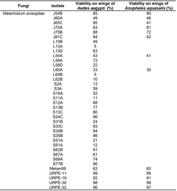
Table 2. Percentage conidial viability of the 83 fungal isolates used on wings of Aedesaegypti and 40 of these on wings of Anopheles aquasalis.
Fungi Isolate Viability on wings of
Aedes aegypti (%)
Viability on wings of
Anopheles aquasalis (%) Beauveria bassiana C76B
J57A J80B L2A L8A L46C L56A L72A S6C S33B S52C S55D S71B S71D S79A S79C Boveril® URPE-3 URPE-4 URPE-14 URPE-18 URPE-22 90 74 80 86 93 82 84 90 72 82 62 89 80 85 52 96 81 99 92 74 79 95 79 79 70 61 82 90 75
22
Table 2. Continue…
Fungi Isolate Viability on wings of
Aedes aegypti (%)
Viability on wings of
Anopheles aquasalis (%) Metarhizium anisopliae J54B
J60A J65C J75A J75B J81C L10B L13A L13D L54A L58A L58D L60A L60B L62B S2A S3A S10A S11A S12A S13B S13C S24C S31B S33C S34B S35B S51A S61A S62B S67A S68A S77B Metarril® URPE-11 URPE-19 URPE-30 URPE-32 85 49 85 83 88 94 49 5 63 43 72 22 33 5 10 13 59 52 11 88 77 80 96 24 93 94 86 21 12 91 61 74 86 62 99 92 98 96 85 46 41 81 72 42 41 30 60 99 91 99 97
4.2 Improving the screening technique
23
Figure 4. Selection of stains for observation of germinating conidia of Metarhizium anisopliae. (a) shows a slide made with lactophenol, (b) with lactophenol cotton blue, (c) lacto-fuchsin. The arrows indicate fungal conidia (Bar = 10 μm).
In pilot experiments inoculating fungi in Tenebrio wing cuticles, we determined 24 h post-inoculation was too soon to observe germination or
appressorium formation, compared with 30 and 48 h. However, at 48 h, the
hyphae had grown across the wing, making it difficult to observe conidia and
verify appressorium formation. Saprophytes also began to contaminate the
material at this point. Therefore, we decided to use 30 h post-inoculation to
24
Of the fungi tested on Tenebrio wings, isolate J27A presented the highest amount of apressorium formation so was subsequently used as a
positive control.
4.3 Germination and appressorium formation on Aedes wings
Of 83 isolates tested in Ae. aegypti between 3 did not germinate, 13 germinated but did not form appressoria while 67 germinated and formed
appressoria (Table 4), with a range of values for both variables.
4.4 Germination and appressorium formation on Anopheles wings
All of the 40 isolates tested in An. aquasalis germinated although 8 of these did not form appressoria (Table 3). There was no apparent correlation
between the values of these two variables. Note that here, isolate J43A on
Aedes wings was used as the positive control.
Table 3. Percentage of the isolates of entomopathogenic fungi Beauveria bassiana and Metarhizium anisopliae that germinate (G%) and formed appressorium (A%) on wing cuticles of Aedes aegypti and Anopheles aquasalis (G% is the percentage of number of germinated conidia/total of conidia; A% is the percentage of number of conidia that formed appressorium/number of germinated conidia).
Aedes aegypti Anopheles aquasalis
Fungi Isolate
G% A% G% A%
Metarhizium
anisopliae C76B
25
Table 3. Continue…
Aedes aegypti Anopheles aquasalis
Fungi Isolate
G% A% G% A%
Beauveria
bassiana S79A
S79C Boveril® URPE-3 URPE-4 URPE-14 URPE-18 URPE-22 62,07 87,50 29,17 86,49 77,78 28,95 61,70 61,29 11,11 14,29 28,57 40,63 64,29 9,09 44,83 15,79 0 0 78,05 0 77,78 91,89 0 0 0 0 43,75 0 14,29 8,82 0 0 Metarhizium
anisopliae C26A
26
Table 3. Continue…
Aedes aegypti Anopheles aquasalis
Fungi Isolate
G% A% G% A%
Metarhizium
anisopliae L54A
L58A L58D L60A L60B L62B S2A S3A S10A S11A S12A S13B S13C S24C S31B S33C S34B S35B S51A S61A S62B S67A S68A S77B Metarril® URPE-11 URPE-19 URPE-30 URPE-32 72,22 81,82 76,47 84,21 48,39 82,61 0,00 37,84 72,73 42,86 60,00 97,40 66,67 75,68 41,59 86,21 54,55 8,70 47,06 11,86 46,67 50,00 24,00 82,35 31,25 100,00 37,78 86,67 92,31 46,15 55,56 0,00 56,25 0,00 5,26 0,00 10,71 0,00 0,00 0,00 50,67 0,00 7,14 31,46 70,00 8,33 50,00 37,50 28,57 28,57 11,11 27,78 28,57 80,00 33,33 23,53 11,54 54,17 78,57 0,00 0,00 91,84 0,00 0,00 0,00 0,00 0,00 0,00 0,00 0,00 0,00 0,00 0,00 0,00 0,00 0,00 0,00 0,00 0,00 0,00 0,00 0,00 85,71 91,89 100,00 93,02 87,50 13,64 0,00 0,00 60,00 0,00 0,00 0,00 0,00 0,00 0,00 0,00 0,00 0,00 0,00 0,00 0,00 0,00 0,00 0,00 0,00 0,00 0,00 0,00 0,00 16,67 50,00 47,22 0,00 28,57
4.5 Test with live Aedes aegypti
The pilot bioassay showed promising initial results in that there was
generally a higher mortality in the fungus-treated insects than the control (Table
4). Between days 5 and 15, however, there were very few deaths, implying that
27
Table 4. Mortality of Aedes aegypti mosquitoes per day of total 10 adults females 1-8 days old, during 15 days, randomized in three blocks.
Number of dead mosquito per day Block Fungi Isolates Total number of
mosquitoes 1 2 3 4 5 6 7 8 9 10 11 12 13 14 15 Final number of mosquitoes
Water plus Tween® 10 10
J33C 10 10
J43A 10 1 9
J75A 10 1 9
Metarril® 10 10
URPE 11 10 10
URPE 32 10 1 9
URPE 4 10 10
URPE 14 10 1 9
Block 1
Boveril® 10 1 1 8
Water plus Tween® 10 1 9
J33C 10 10
J43A 10 1 1 8
J75A 10 1 1 1 7
Metarril® 10 1 9
URPE 11 10 1 2 7
URPE 32 10 2 8
URPE 4 10 1 2 7
URPE 14 10 1 1 8
Block 2
Boveril® 10 10
28
Table 4. Continue…
Number of dead mosquito per day Block Fungi Isolates Total number of
mosquitoes 1 2 3 4 5 6 7 8 9 10 11 12 13 14 15 Final number of mosquitoes
Water plus Tween® 10 1 9
J33C 10 10
J43A 10 1 1 8
J75A 10 10
Metarril® 10 10
URPE 11 10 1 9
URPE 32 10 1 1 8
URPE 4 10 1 1 8
URPE 14 10 10
Block 3
29 5. DISCUSSION
Due to problems with synthetic insecticides (Lacey & Undeen, 1986;
Hemingway & Ranson, 2000; Lacey et al., 2001; Shiff, 2002; Hargreaves et al., 2003), biological control of mosquitoes with entomopathogenic fungi is an
expanding area. Fungi infect all stages of mosquito development, having shown
satisfactory results against eggs (Luz et al., 2008), larvae (Mohanty et al., 2008) and adults (Mnyone et al, 2009; Leles et al., 2010). Meanwhile, biological control of larvae using bacterial pathogens is considered a cheap, easy and
environmentally friendly method (Kannan et al., 2008), but problems that can occur are the dissipation of the control agent in the aquatic environment and
killing of non-target organisms. Thus, more efforts have been done to develop
agents against adult mosquitoes (Kanzok & Jacobs-Lorena, 2006; Kannan et
al., 2008).
Several studies have demonstrated the potential of entomopathogenic
fungi, both in the control of vector populations and in acting on the vectorial
capacity of mosquitoes (Blanford et al. 2005; Scholte et al. 2004; 2005; 2006; 2007; Thomas & Read, 2007; de Paula et al., 2008; Kannan et al., 2008; Mohanty et al. 2008; Leles et al., 2010; Mnyone et al., 2011; Howard et al., 2010a). Reducing mosquito lifespan interferes with blood feeding, thus affecting
the pathogen acquisition and transmission (Scholte et al., 2007; Mohanty et al., 2008) and this is a key factor in the use of entomopathogenic fungi.
The traditional way to screen entomopathogenic fungi is to inoculate the
fungus in live organisms. In our study, we focused the technique that allows the
screening of some isolates based on the formation of infection structures called
appressoria, using the wing cuticle. Although we attempted to obtain isolates
from soil using larvae of Musca domestica (Diptera) as live bait (data not shown), this was not successful, probably due to rapid pupation of the larvae
prior to death from fungal infection.
For the assays of germination and appressorium formation on mosquito
wings, we found lactophenol alone to be inadequate for visualization of the
fungal structures. Although lactophenol cotton blue did stain the fungal
structures, it was not as clear as when we used lacto-fuchsin stain, especially
30
On the slides, we could see some long hyphae growing close to each
other, forming aggregations. We also noted some subterminal appressoria, as
described by Wang & St. Leger (2005). It could be that the two phenomena are
connected, perhaps resulting from unsuccessful attempts to invade the host or
as a means of obtaining nutrients to continue growth. Although cuticle
topography appears to influence the growth form of M. anisopliae (St. Leger et al., 1990a), our study was inconclusive in this respect. In line with Arruda et al. (2005), then, it is likely that other factors may have influenced fungal
development such as chemical clues linked to cuticle compounds.
According to Moino et al. (2002) the thickening of hyphae may be due to the translocation of the conidial cytoplasmatic content to facilitate the enzymatic
synthesis necessary for the penetration phase. However, B. bassiana in general had low percentages of appressorium formation. Some studies have shown that
the germ-tube can penetrate the host cuticle without appressorium formation
(Melo et al., 2007), as with plants where penetration occurred through a small hole (Quesada-Moraga et al., 2006); this may suggest that isolates that did not
form appressoria could still be of interest.
Our screening technique was faster than the conventional method.
Screening of isolates for mosquito control usually begins with assays of daily
survival of infected mosquitoes, in order to select isolates that kill quickly. One
disadvantage of this method is that the adults survive up to 20 days so the
experiment is prolonged as some fungi take several days to kill them. It also
leads to a selection of the most virulent isolates, discarding those that would
take longer to kill but might cause significant sublethal effects for the control of
mosquitoes and their vectorial capacity. Similarities in the two methods are to
be found in the need of adult mosquitoes and sporulating fungi. As mosquitoes
and fungi take the same time to develop in both set-ups, the time spent with this
is equal, as is the preparation of spore suspensions.
Thus, what really varies between the methods is the next step, between
inoculation and the final result. In the traditional method, we have to wait for
infected mosquitoes to die, while using our technique of making the observation
of infection in the cuticle structures of the wings, we can use the newly emerged
mosquitoes, and our results will be obtained in just 30 hours, when there is the
31
more effective screening (as many isolates that were discarded in the other
method are kept here) of fungal isolates able to infect these insects.
Furthermore, our results support the notion that the interaction between
host and pathogen can be influenced by differences in the cuticular components
on mosquito wings, since there were differences in the development of the fungi
on mosquito cuticle, in line with results of Wang & St. Leger (2005). Fungi need
an external resource of nutrients to germinate (Pedrini et al., 2007) and the medium interferes in their germination, viability and virulence (Rangel et al., 2008). Different expression of genes encoding cuticle-degrading enzymes, cell
wall proteins, toxins and toxin-producing enzymes have been observed on
different insect cuticles (Freimoser et al., 2005). Meanwhile, germination on non-host cuticle is limited (Wang & St. Leger, 2005; Jarrold et al., 2007), this perhaps being one of the factors that influenced the development of our isolates
on mosquito cuticles. Wang and St. Leger (2005) suggested that there could be
localized formation of appressoria on non-host cuticle. In line with this, some of
our isolates formed small numbers of appressoria in our study.
Analyzing the studies that deal with infection progression, we have a
pattern of development of these fungi in the host cuticle, demonstrating that the
time of infection process varies among fungi (Castrillo et al., 2005). Usually, the sequence of events is: conidia attachment; conidia germination and
development of a germ-tube that colonizes the host cuticle, 24-48 h
infection; germ-tube penetration or development of appressorium; 24-48 h
post-infection (Arruda et al., 2005). There is variation in the timing of these events: thus, in one study, for M. anisopliae on locust wings, 73% germination was achieved at 24 h, with some appressorium formation, while on cicada wings
there was 54% germination of M. anisopliae at 24h and appressoria were rarely observed. Meanwhile, in beetles, this value was just 4% with no appressoria.
(Wang & St. Leger, 2005). In fleas, adherence occurred 2 h after inoculation,
there was germ-tube germination 26 h post-inoculation and there was also
thickening and branching of hyphae, with no appressoria.
Observing these temporal differences, we can confirm that 30 h
post-infection was satisfactory to our study of screening fugal isolates based on
32
formed, suggesting that there was a dearth of nutrients available on the wing
cuticle.
The mosquito must come in contact with the conidia of the fungus and
the conidia have to adhere to its surface to be established infection (Charnley,
1994). Thus, the success of infection is determined by host contact with treated
surface and concentration of infective conidia (Vandenberg et al., 1998). Therefore, our survival bioassay suggests that there may have been an
insufficient concentration of fungal conidia or insufficient time for the assay,
unfavorable environmental conditions to fungi development or there may have
been a repulsion of mosquitoes in relation to the paper treated with the fungus.
Scholte et al. (2003) reported a moderate repellence of An. gambiae for dry conidia of M. anisopliae. However, when the suspension is formulated with oil, this effect disappears (Mnyone et al., 2010). The relative high mortality in the first four days suggests that mortality may have been caused by the change of
environment for testing.
The tests with Anopheles cuticles and with live mosquitoes are not yet
concluded. The Anopheles tests should continue when more adult insects are available to obtain more wings, since just 40 of the 67 isolates that were
pre-selected in Aedes were tested. The assay in live mosquitoes needs refining and repeating; of the 67 isolates able to form structures in infective Aedes, only 8 were tested (of 10 treatments, one control and one was an isolate that was not
formed appressoria). Our intention is to conduct the test with live mosquitoes of
both species and with all isolates that formed infective structure on the wing
cuticles, to confirm that our technique is really effective and determine if the
mortality test is indeed a less effective means of screening.
Among the isolates tested, 5 of B. bassiana and 27 of M. anisopliae
formed appressoria in both mosquitoes, indicating promise for their use in
control. The commercial products Boveril® and Metarril® show potential which
is of great interest as these are registered products with a market history,
besides presenting a formulation already developed and field tested appropriate
to conditions in Brazil.
Further studies are necessary to screen fungi as possible candidates.
33
the fungus in field tests, and studying the formulation and application for the
control of mosquitoes of different genera.
6. CONCLUSION
Improving the screening fungal technique based on the formation of
appressoria using mosquito wings, allowed us to streamline the selection of
potential fungi for control of insect vectors. Furthermore, this study allowed the
observation of fungi development in the mosquitoes cuticle and demonstrated
that these phases vary with fungal isolate and host cuticle, and are very
important in selecting isolates for biological control of insects. Some isolates of
34 APPENDICES
Number of conidia (C), number of germinated conidia (G) and number of appressoria formatted (A) of Beauveria bassiana isolates in each one of five wings of Aedes aegypti.
Aedes aegypti
Beauveria
bassiana 1º Wing 2º Wing 3º Wing 4º Wing 5º Wing
Isolate C G A C G A C G A C G A C G A
C76B 2 0 0 1 1 0 3 2 2 1 1 0 2 1 0
J57A 2 1 0 2 0 0 1 0 0 2 0 0 0 0 0
J80B 1 0 0 7 7 1 2 0 0 3 2 0 6 5 0
L2A 8 7 0 17 17 0 2 2 0 6 6 0 7 7 0
L8A 7 4 0 5 1 0 2 0 0 5 0 0 5 0 0
L46C 4 0 0 3 0 0 7 3 0 3 1 0 6 3 1
L56A 4 3 0 12 12 4 2 1 0 6 3 0 6 5 0
L72A 20 0 0 12 0 0 5 0 0 7 0 0 4 0 0
S6C 7 6 0 4 3 0 4 3 2 6 5 3 12 12 7
S33B 6 2 0 7 5 0 6 2 0 3 2 2 2 1 0
S52C 4 4 0 5 5 0 11 11 2 4 0 0 5 5 0
S55D 10 6 2 22 19 7 35 35 5 2 2 0 3 0 0
S71B 3 2 0 2 2 1 3 1 1 5 5 2 1 0 0
S71D 10 0 0 8 1 0 7 0 0 17 0 0 2 0 0
S79A 10 9 1 4 2 0 6 3 0 6 4 1 3 0 0
S79C 1 1 0 1 0 0 7 6 0 4 4 1 3 3 1
Boveril® 5 3 1 7 3 0 4 0 0 4 0 0 4 1 1
URPE-3 12 9 2 10 8 5 8 8 2 5 5 2 2 2 2
URPE-4 1 1 0 14 11 9 1 1 0 1 0 0 1 1 0
URPE-14 10 0 0 13 5 0 7 2 0 5 2 1 3 2 0
URPE-18 18 13 8 5 3 0 6 3 2 8 5 1 10 5 2
URPE-22 4 0 0 3 1 0 6 4 0 18 14 3 0 0 0
Number of conidia (C), number of germinated conidia (G) and number of appressoria formatted (A) of Metarhizium anisopliae isolates in each one of five wings of Aedes aegypti.
Aedes aegypti Metarhizium
anisopliae 1º Wing 2º Wing 3º Wing 4º Wing 5º Wing
Isolate C G A C G A C G A C G A C G A
C26A 9 6 0 7 7 0 19 14 6 3 3 0 0 0 0
C46A 4 4 2 7 6 0 5 4 2 8 8 2 4 3 1
C54B 22 3 0 33 4 0 38 3 0 2 0 0 19 0 0
C55A 6 2 0 0 0 0 3 2 1 3 3 0 6 2 0
C66A 16 11 2 17 13 7 8 8 3 2 2 0 2 2 0
C84A 7 7 2 3 3 0 7 7 1 0 0 0 3 3 3
C86C 5 5 0 13 8 0 5 5 1 10 5 0 7 4 0
J2B 6 6 4 8 8 2 2 0 0 0 0 0 5 5 0
J8C 4 1 0 4 2 0 9 7 0 1 0 0 1 0 0
J11B 11 8 2 26 26 5 2 2 0 15 15 5 30 30 5
J15D 6 6 2 3 3 1 2 0 0 4 0 0 0 0 0
J18A 1 0 0 4 0 0 2 2 2 17 17 3 1 1 0
J21C 33 33 16 8 8 2 1 1 0 10 10 2 0 0 0
J25A 4 3 0 4 4 0 13 12 4 0 0 0 1 1 1
J27A 7 7 0 3 3 0 2 2 0 2 2 0 4 2 0
J30B 11 9 0 5 4 0 14 14 6 12 12 0 4 4 2
J32A 12 7 0 14 7 3 30 28 18 16 16 5 22 21 13
J33C 17 0 0 18 0 0 4 0 0 10 0 0 4 0 0
J38A 1 1 0 13 7 0 10 6 1 6 2 1 14 4 0
J42A 3 3 0 3 3 0 1 1 0 1 0 0 4 4 1