©2012 Science Publication
doi:10.3844/ajisp.2012.146.153 Published Online 8 (4) 2012 (http://www.thescipub.com/aji.toc)
HELICOBACTER PYLORI INFECTION IN NEVER-SMOKING
MALE PATIENTS WITH CHRONIC OBSTRUCTIVE
PULMONARY DISEASE AND ITS RELATION TO LUNG
FUNCTION
1
Jordan Minov,
1Jovanka Karadzinska-Bislimovska,
2
Kristin Vasilevska,
1Snezana Risteska-Kuc,
1Saso Stoleski and
1Dragan Mijakoski
1
Department of Cardiorespiratory Functional Diagnostics, Institute for Occupational Health of R. Macedonia,
WHO Collaborating Center and GA2LEN Collaborating Center, Skopje, R. Macedonia
2
Institute for Epidemiology and Biostatistic, II Makedonska Brigada 431000 Skopje, R. Macedonia
Received 2012-08-29, Revised 2012-09-11; Accepted 2012-10-18
ABSTRACT
There is a recent epidemiologic and serologic evidence for relationship between Helicobacter pylori (H. pylori) infection and Chronic Obstructive Pulmonary Disease (COPD). In order to assess the relationship between H. pylori infection and COPD and its impact on lung function we performed a cross-sectional study including 84 never-smoking male patients with COPD and an equal number of never-smoking males without chronic respiratory disease matched to the COPD patients by age. Evaluation of the study subjects included evaluation of H. pylori serological status, baseline and post-bronchodilator spirometry. We found significantly higher H. pylori seropositivity in COPD patients than in controls (76.2 Vs 34.5%, p = 0.041). The prevalence of H. pylori seropositivity did not differ significantly between patients with mild, moderate and severe COPD. Borderline significance was registered for the difference of the forced expiratory volume in one second (FEV1) mean value between
seropositive and seronegative COPD patients (56.4 vs. 59.2, p = 0.063). The mean degree of FEV1
reversibility did not differ significantly between seropositive and seronegative COPD patients. Our findings indicate that in cross-sectional analysis there is higher prevalence of H. pylori seropositivity in COPD than in non-COPD patients, as well as that H. pylori infection has not significant impact on lung function in COPD patients.
Keywords: Baseline Spirometry, Chronic Obstructive Pulmonary Disease, Helicobacter Pylori, Never-Smokers, Post-Bronchodilator Spirometry
1. INTRODUCTION
Chronic Obstructive Pulmonary Disease (COPD) becomes one of the most important global public health problems in the last decades affecting 9-10% of the adults aged over 40 years (Halbert et al., 2006). COPD is the fourth leading cause of death in adults in the United States and is projected to be the third most common cause of death by 2020 (Petty, 2003). Between 1970 and 2002, in the United States death rates due to stroke and heart disease
decreased (63 and 52%, respectively), while death rates due to COPD increased 100% (Jemal et al., 2005).
represents the major cause of gastroduodenal pathologies (e.g., chronic active gastritis, peptic ulcer, B-cell lymphoma and gastric carcinoma) (Wotherspoon et al., 1999; Parsonnet et al., 1991; D’Elios et al., 1997). Some recent epidemiologic and serologic studies have reported a relationship between H. pylori seropositivity, especially of the high virulent cytotoxin-asssociated gene A (CagA) positive strains and extra-gastroduodenal diseases, such as vascular (coronary artery disease and stroke), metabolic (autoimmune atrophic thyroiditis), rheumatic (Henoch-Schönlein purpura), dermatologic (chronic urticaria and rosacea), as well as respiratory diseases (chronic bronchitis, COPD, bronchiectasis, asthma and lung cancer) (Whincup et al., 1996; Luis et al., 1998; Tsang et al., 1998; Roussos et al., 2006; Jun et al., 2006; Behroozian and Moradkhan, 2010). The activation of inflammatory mediators as a result of systemic immune response induced by H. pylori infection may be potential explanation for these associations (Kanbay et al., 2007).
In addition, results from some cross-sectional studies indicated gender-dependent difference in the decline in lung function associated with H. pylori infection. Fullerton et al. (2009) reported lower lung function, i.e., significantly lower Forced Expiratory Volume in one second (FEV1) and Forced Vital Capacity (FVC) values,
in men with positive serology for H. pylori as compared to seropositive women. On the other side, controversial results have been reported regarding H. pylori prevalence and smoking (considered as a major risk factor for COPD): Higher, normal and lower seropositivity were stated for smokers (Brenner et al., 1997; Parasher and Eastwood, 2000; Ogihara et al., 2000).
The present study is aimed at assessment of the relationship between H. pylori infection and COPD and its impact on lung function in never-smoking male patients.
2. MATERIALS AND METHODS
2.1. Study Design and Setting
A cross-sectional study was carried out in the Department of Cardiorespiratory Functional Diagnostics at the Institute for Occupational Health of R. Macedonia, Skopje-WHO Collaborating Center for Occupational Health and GA2LEN Collaborating Center in the period March 2011-June 2012.
The study protocol was approved by the ethics committee of the institution and each subject gave an informed consent before entering the study.
2.2. Study Subjects
The study protocol underwent 84 never-smoking males aged 39 to 74 years with COPD. Exclusion criteria for COPD patients were exacerbation of COPD in the last month, history of antibiotic use in the last month, history of H. pylori eradication and/or presence of other chronic respiratory disease.
In addition, an equal group of never-smoking males without chronic respiratory disease matched to the COPD patients by age was studied as a control. Exclusion criteria for controls were history of antibiotic use in the last month and history of H. pylori eradication. Never-smoker was defined as a non-smoker who has never smoked at all, or has never been daily smoker and has smoked less than 100 cigarettes in his lifetime (WHO, 1998; Leffondre et al., 2002).
2.3. Questionnaire
A questionnaire including demographic characteristics, family history of COPD and Chronic Bronchitis (CB) (taking into account the first-degree relatives), exposure to Environmental Tobacco Smoke (ETS), as well as presence of accompanying diseases, was completed by all study subjects.
Exposure to ETS or passive smoking was defined as an exposure to tobacco combustion products from smoking by others (at home, workplace), i.e., as a presence of at least one smoker in the household and/or in the workplace (DHHS, 1984; Janson et al., 2001). In addition, passive smokers were divided in two groups regarding the number of hours per day they were exposed to ETS (less or more than 4 h per day).
2.4. H. Pylori Serological Status
H. pylori serological status, i.e., quantitative
detection of serum Immunoglobuline G (IgG), was evaluated using the Siemens ImmuliteR 1000 assay (a solid-phase, chemiluminiscent IgG assay) (Siemens, Germany). Seropositivity was considered in the case of finding of specific IgG concentration equal or more than 1 U/mL, while the subjects with serum concentration of specific IgG equal or less than 0.9 U/mL were considered as seronegative ImmuliteR, 1000 Chemiluminiscent Technology, 2012.
2.5. COPD Diagnosis
dyspnea, chronic cough or sputum production and/or a history of exposure to risk factors for the disease (tobacco smoke, smoke from home cooking and heating fuels and/or occupational dusts and chemicals). The diagnosis was proved by the presence of a post-bronchodilator FEV1/FVC less than 0.70 suggesting
persistent airflow limitation.
The severity of the disease in the COPD patients was categorized according to the GOLD 2010 (GSD, 2012) spirometric classification as a mild COPD (FEV1/FVC <
0.70; FEV1 ≤ 80% predicted), a moderate COPD
(FEV1/FVC < 0.70; 50% ≤ FEV1 < 80% predicted) and
a severe COPD (FEV1/FVC < 0.70; 30% ≤ FEV1 < 50%
predicted).
2.6. Baseline Spirometry
The baseline spirometry, including measures of FVC, FEV1, FEV1/FVC and maximal expiratory flow at
25-75% of FVC (MEF25-75), was performed in all subjects
using spirometer Ganshorn SanoScope LF8 (Ganshorn Medizin Electronic GmbH, Germany) with recording the best result from three measurements the values of FEV1
of which were within 5% of each other. The results of spirometry were expressed as percentages of the predicted values according to the actual recommendations of European Repsiratory Society (ERS) and American Thoracic Society (ATS) (Miller
et al., 2005). The combined reference equations for
people aged 18 to 70 years, with a height range of 155-190 cm in males, published in the 1993 ERS statement (Quanjer et al., 1993) were used for deriving predicted values.
2.7. Bronchodilator Reversibility Testing
Bronchial reversibility testing was performed according to the actual GOLD spirometry guide. Spirometric measurements were performed before and 20 min after administration of 400 µg salbutamol by metered dose inhaler through spacer. Fixed airflow narrowing characteristic for COPD was considered if post-bronchodilator FEV1/FVC remained less than 0.70.
The degree of FEV1 reversibility was expressed as %
FEV1 reversibility ([post-bronchodilator FEV1
-pre-bronchodilator FEV1]/pre-bronchodilator FEV1×100).
Significant FEV1 improvement (a change more than 12%
and more than 200 mL) in the presence of fixed airflow limitation did not negate a diagnosis of COPD.
2.8. Statistical Analysis
Continuous variables were expressed as mean values with Standard Deviation (SD) and the nominal variables as numbers and percentages. Analyses of the data involved testing the differences in prevalence and comparison of the means. Chi-square test (or Fisher’s exact test where appropriate) was used for testing difference in the prevalence. Comparison of spirometric measurements was performed by independent-samples
T-test. A P-value less than 0.05 were considered as
statistically significant. Statistical analysis was performed using the Statistical Package for the Social Sciences (SPSS) version 11.0 for Windows.
3. RESULTS
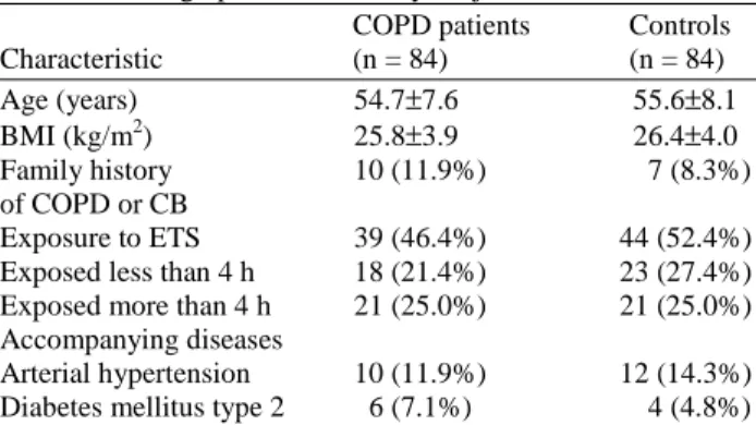
Demographic characteristics were similar in both examined groups (Table 1).
The mean baseline values of all spirometric parameters were significantly lower in COPD patients (Table 2).
Table 1. Demographics of the study subjects
COPD patients Controls
Characteristic (n = 84) (n = 84)
Age (years) 54.7±7.6 55.6±8.1
BMI (kg/m2) 25.8±3.9 26.4±4.0
Family history 10 (11.9%) 7 (8.3%)
of COPD or CB
Exposure to ETS 39 (46.4%) 44 (52.4%) Exposed less than 4 h 18 (21.4%) 23 (27.4%) Exposed more than 4 h 21 (25.0%) 21 (25.0%) Accompanying diseases
Arterial hypertension 10 (11.9%) 12 (14.3%) Diabetes mellitus type 2 6 (7.1%) 4 (4.8%) Numerical data are expressed as mean value with standard deviation; frequencies as number and percentage of study subjects with certain variable
Table 2. Mean baseline values of spirometric parameters in the study subjects
Spirometric COPD patients Controls
parameter (n = 84) (n = 84) P-value* FVC (%pred) 70.1±12.3 96.4±9.2 0.000 FEV1 (%pred) 57.3±9.2 88.1±11.2 0.000
FEV1/FVC 0.62±0.05 0.78±0.07 0.000
MEF25-75 (%pred) 44.7±14.8 73.6±16.7 0.000
Data are expressed as mean value with standard deviation. COPD: Chronic Obstructive Pulmonary Disease; FVC: Forced Vital Capacity; FEV1: Forced Expiratory Volume in one second;
MEF25-75: Maximal Expiratory Flow at 25-75% of FVC; % pred:
Table 3. Characteristics of the disease in the COPD patients COPD patients
Characteristic (n = 84)
Mean COPD duration (years) 9.2±3.4 COPD severity
Mild COPD 32 (38.1%)
Moderate COPD 28 (33.3%)
Severe COPD 24 (28.6%)
Numerical data are expressed as mean value with standard deviation; frequencies as number and percentage of study subjects with certain variable. COPD: chronic obstructive pulmonary disease.
Table 4. Mean baseline values of spirometric parameters in H. pylori seropositive and seronegative COPD patient
H. pylori H. pylori
Spirometric seropositive COPD seronegative COPD
parameter patients (n = 64) patients (n = 20) P-value*
FVC (%pred) 69.2±10.8 70.9±11.3 0.127
FEV1 (%pred) 56.4±8.4 59.2±10.6 0.063
FEV1/FVC 0.61±0.02 0.62±0.06 0.098
MEF25-75 (%pred) 45.3±12.9 43.9±15.8 0.104
Data are expressed as mean value with standard deviation. H. pylori: Helicobacter pylori; COPD: Chronic Obstructive Pulmonary Disease; FVC: Forced Vital Capacity; FEV1:
Forced Expiratory Volume in one second; MEF25-75: Maximal
Expiratory Flow at 25-75% of FVC; % pred: % of predicted value. *Compared by Independent-samples T-test
Table 5. Mean post-bronchodilator values of spirometric parameters in H. pylori seropositive and seronegative COPD patients
H. pylori H. pylori Seropositive seronegative Spirometric COPD patients COPD patients
Parameter (n = 64) (n = 20) P-value* FVC (%pred) 70.3±11.6 71.4±12.1 0.188 FEV1(%pred) 58.7±10.4 60.5±11.8 0.074
FEV1/FVC 0.62±0.04 0.62±0.07 0.109
MEF25-75(%pred) 46.9±14.2 45.2±13.9 0.131
Data are expressed as mean value with standard deviation.H. pylori: Helicobacter pylori; COPD: Chronic Obstructive Pulmonary Disease; FVC: Forced Vital Capacity; FEV1:
Forced Expiratory Volume in one second; MEF25-75: Maximal
Expiratory Flow at 25-5% of FVC; % pred: % of predicted value.*Compared by Independent-samples T-test
Characteristics of the disease in the COPD patients are presented on Table 3.
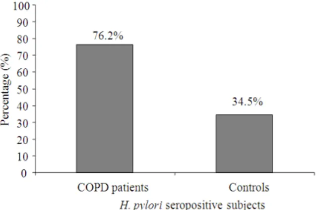
The prevalence of H. pylori seropositive subjects was significantly higher in the group of COPD patients (76.2% Vs. 34.5%, P = 0.041; Chi-square test) (Fig. 1).
Fig. 1. Prevalence of H. pylori seropositivity in the examined groups
Fig. 2. Prevalence of H. pylori seropositivity in the patients with mild, moderate and severe COPD
The prevalence of H. pylori seropositive subjects in the patients with mild, moderate and severe COPD is shown on Fig. 2. There was no significant difference in the prevalence of H. pylori seropositivity between patients with mild and moderate COPD (68.7% vs. 79.2%, p = 0.104; Chi-square test), mild and severe COPD (68.7% vs. 78.6%, p = 0.112; Chi-square test), as well as between patients with moderate and severe COPD (79.2% vs. 78.6%, p = 0.279; Chi-square test).
With exception of the mean FEV1 baseline value
where a borderline significance was registered, there was no significant difference in the mean baseline values of the other spirometric parameters between seropositive and seronegative COPD patients (Table 4). The mean baseline spirometric values did not differ significantly between seropositive and seronegative controls.
and seronegative COPD patients (Table 5). The mean post-bronchodilator spirometric values did not differ significantly between seropositive and seronegative controls.
The degree of FEV1 reversibility expressed as % FEV1
reversibility was significantly higher in COPD patients than in controls (10.2±2.3 Vs 4.7±2.1, p = 0.000; Independent-samples T-test). The mean value of % FEV1 reversibility in
H. pylori seropositive COPD patients was similar to its mean value in seronegative COPD patients (10.7±3.1 vs. 9.9±2.9, P = 0.119; Independent-samples T-test).
4. DISCUSSION
COPD remains frequent and costly disease representing one of the principal demands of the public health worldwide. Inhaled tobacco smoke and other noxious particles, such as occupational exposures and smoke from biomass fuels, are the most important exogenous factors that influence disease development and progression (GSD, 2012; Chatila et al., 2008). On the other side, H. pylori infection, a lifelong and often asymptomatic infection of the stomach, profoundly alters gastric immune response that may lead to systemic effects.
H. pylori persistence leads to chronic inflammation and
immune stimulation, which could contribute to several extra-gastroduodenal pathologies (Pellicano et al., 1999; Kowalski et al., 2006; Islami and Kamangar, 2008; Najafizadeh et al., 2007).
As it is mentioned above, the results of several studies emphasis the relationship between H. pylori infection and chronic bronchial inflammatory disorders, e.g., chronic bronchitis, COPD and bronchiectasis. It is believed that the release of proinflammatory cytokines, e.g., Interleukin-1 (IL-1), IL-8, IL-17, IL-23 and Tumor Necrosis Factor-ά (TNF-ά), stimulated by H. pylori infection plays a role in the chronic inflammation of bronchi (Roussos et al., 2006; Jafarzadeh et al., 2009; Cornwell et al., 2010). Moreover, serum levels of these cytokines normalize following eradication therapy of H.
pylori (Kountouras et al., 2000). There is a need of
further studies to assess whether eradication therapy of H. pylori may modify the course of COPD (Hashemi et al., 2011). The impact of chronic infections, e.g., H. pylori, Chlamidia pneumoniae (C.
pneumoniae) and Cytomegalovirus (CMV) infections,
on COPD development and severity actually is investigated in the United States in a large population-based sample in a MESA-Lung study, an extension of the Multi-Ethnic Study of Atherosclerosis (MESA) study (Hankinson et al., 2010).
The studies that investigated the relationship between
H. pylori infection and COPD is often difficult to compare
because of differences in the study design, as well as in the study population and study protocol. In the present study we assessed the relationship between H. pylori infection and COPD and its impact on lung function investigating a group of never-smoking COPD patients and a group of never-smoking males without chronic respiratory disease. The examined groups included subjects with similar demographic characteristics. In either group there was a large proportion of passive smokers that is similar to its prevalence in R. Macedonia documented in our previous studies (Minov et al., 2006; 2008).
We found significantly higher prevalence of seropositive subjects in the group of COPD patients than in the control group with no significant difference in the
H. pylori seropsotivity between the subjects with mild,
moderate and severe COPD. Significantly higher seropositivity to H. pylori was detected in several studies which investigated the relationship between COPD and
H. pylori infection. In the study including COPD patients
and healthy controls matched by sex and age, Gencer et al. (2007) reported significantly higher prevalence of H.
pylori seropositive subjects among COPD patients with
no significant difference in gender, age and smoking status between seropositive and seronegative COPD patients. Similarly, in the study including COPD patients and age- and sex- matched controls, Roussos et al. (2005) reported significantly higher prevalence of H.
pylori infection, as well as significantly higher
prevalence of CagA-positive H. pylori infection in COPD patients. In the study including patients with CB and controls matched by sex and social status, Caselli et al. (1999) reported significantly higher prevalence of H.
pylori seropositive subjects in the group of subjects with
CB. On the contrary, Hashemi et al. (2011) reported similar prevalence of H. pylori seropositivity in a case-control study including COPD patients and age and sex-matched controls with pulmonary diseases other than COPD (asthma, lung cancer and sarcoidosis) with no significant difference in H. pylori seropositivity between the patients with mild, moderate and severe COPD. The absence of significant difference in the prevalence of H.
pylori seropositivity between COPD and non-COPD
patients may be due to the increased prevalence of H.
With exception of borderline significant difference in the mean values of baseline and post-bronchodilator FEV1, we did not register significant difference in the
mean values of the other measured spirometric parameters between seropositive and seronegative COPD patients. There was non-significant FEV1 reversibility
between H. pylori seropositive and seronegative COPD patients. In our previous study in which we investigated the impact of H. pylori infection on lung function and severity of bronchial hyperresponsiveness in subjects with allergic asthma, we did not register significant difference in the mean values of measured spirometric parameters between seropositive and seronegative allergic asthma patients (Minov et al., 2011). In the study conducted by Gencer et al. (2007) mentioned above, they reported significantly lower FEV1 values in seropositive
as compared to seronegative COPD patients. The difference between these groups regarding FVC and FEV1/FVC was statistically non-significant (Gencer et al.,
2007). On the other side, Roussos et al. (2005) reported no statistically significant difference regarding the spirometric values between H. pylori seropositive and seronegative COPD patients. In addition, in a population-based study in adults which investigated the relationship between H. pylori infection and lung function, asthma, atopy and allergic disease mentioned above, Fullerton et al. (2009) reported significantly lower FEV1 and FVC values in H. pylori seropositive
men in a cross-sectional analysis, but after adjustment for either height or social class the size of these associations were reduced. Furthermore, in a longitudinal analysis they did not register significant association between H. pylori serological status and decline in the lung function over 9 years (Fullerton et al., 2009). On the contrary, in the study of Nottinghamshire miners, Siva et al. (2004) reported that a past history of peptic ulceration was present in more than 50% miners with severe COPD but only in 3% of miners with no respiratory symptoms and normal spirometric values.
The present study has some limitations. First, relatively small number of the subjects in the study groups could have certain implications on the data obtained and its interpretation. Second, the study design, i.e., cross-sectional analysis, could also have implications on the data obtained and its interpretation. Third, the serological data for exposure to H. pylori is unable to distinguish current from prior infection which limits interpretation of the associations observed. The strength of the study is the assessment of the impact of
H. pylori infection on COPD in never-smoking males
that, to our knowledge, so far has not been reported in published literature, as well as extensive lung function measurements performed in the study subjects.
5. CONCLUSION
In conclusion, in a cross-sectional study including never-smoking male COPD patients and non-COPD matched controls we found significantly higher prevalence of H. pylori seropositivity in COPD patients with no significant difference between patients with mild, moderate and severe COPD. Borderline significance was registered for the difference of the FEV1 mean value between seropositive and seronegative
COPD patients, while the mean values of other measured spirometric parameters, as well as the mean degree of FEV1 reversibility did not differ significantly. Our
findings support the need of further larger prospective studies in order to assess the complex relationship between H. pylori infection and COPD.
6. REFERENCES
Behroozian, R. and E. Moradkhan, 2010. The assessment of probable relationship between lung cancer and
Helicobacter pylori infection. Trop Gastroenterol.,
31: 34-36. PMID: 20860223
Brenner, H., D. Rothenbacher, G. Bode and G. Adler, 1997. Relation of smoking and alcohol and coffee consumption to active Helicobacter pylori infection: Cross sectional study. BMJ, 315: 1489-1492. PMID: 9420488
Caselli, M., E. Zaffoni, M. Ruina, S. Sartori and L. Trevisani et al., 1999. Helicobacter pylori and chronic bronchitis. Scand J. Gastronetrol., 34: 828-830. PMID: 10499486
Chatila, W.M., B.M. Thomashow, O.A. Minal, G.J. Crtiner and B.J. Make, 2008. Comorbidities in chronic obstructive pulmonary disease. Proc. Am. Thorac Soc., 5: 549-555. DOI: 10.1513/pats.200709-1 Cornwell, W.D., V. Kim, C. Song and T.J. Rogers, 2010.
Pathogenesis of inflammation and repair in advanced COPD. Semin Respir Crit. Care Med., 31: 257-266. PMID: 20496295
D’Elios, M.M., M. Manghetti, M. De Carli, F. Costa and C.T. Baldari et al., 1997. Th-1 effector cells specific for Helicobacter pylori in the gastric antrum of patients with peptic ulcer disease. J. Immunol., 158: 962-967. PMID: 8993017
Fullerton, D., J.R. Britton, S.A. Lewis, I.D. Pavord and T.M. McKeever et al., 2009. Helicobacter pylori and lung function, asthma, atopy and allergic disease-A population-based cross-sectional study in adults. Int. J. Epidemiol., 38: 419-426. PMID: 19109248
Gencer, M., E. Ceylan, F.Y. Zeyrek and N. Aksoy, 2007.
Helicobacter pylori seroprevalence in patients with
chronic obstructive pulmonary disease and its relation to pulmonary function tests. Respiration, 74: 170-175. PMID: 16369121
GSD, 2012. Management and prevention of chronic obstructive pulmonary disease.
Halbert, R.J., J.L. Natoli, A. Gano, E. Badamgarav and A.S. Buist et al., 2006. Global burden of COPD: systematic review and meta-analysis. Eur. Respir J., 28: 523-532. PMID: 16611654
Hankinson, J.L., S.M. Kawut, E. Shahar, L.J. Smith and K.H. Stikowsky, 2010. Performance of American Thoracic Society-recommended spirometry reference values in a multiethnic sample of adults. The Multi-Ethnic Study of Atherosclerosis (MESA) Lung Study Chest, 137: 138-145. PMID: 19741060 Hashemi, S.H., E. Nadi, M. Hajilooi, M.A. Seif-Rabiei
and U. Roustael, 2011. Relationship between
Helicobacter pylori infection and chronic obstructive pulmonary disease. Acta Med. Iranica, 49: 721-724. PMID: 22131241
Islami, F. and F. Kamangar, 2008. Helicobacter pylori and esophageal cancer risk: A meta-analysis. Cancer Prev. Res., 1: 329-338. PMID: 19138977
Jafarzadeh, A., V. Mizaee, H. Ahmad-Beygi, M. Nemati and M.T. Rezayati, 2009. Association of the CagA status of Helicobacter pylori and serum levels of interleukin (IL)-17 and IL-23 in duodenal ulcer patients. J. Dig. Dis., 10: 107-112. PMID: 19426392 Janson, C., S. Chinn, D. Jarvis, J.P. Zock and K. Toren et
al., 2001. Effect of passive smoking on respiratory
symptoms, bronchial responsiveness, lung function and total serum ige in the european community respiratory health survey: A cross-sectional study. Lancet, 358: 2103-2109. PMID: 11784622
Jemal, A., E. Ward, Y. Hao and M. Thun, 2005. Trends in the leading causes of death in the United States, 1970-2002. JAMA, 294: 1255-1259. DOI: 10.1001/jama.294.10.1255
Jun, Z.J., Y. Lei, Y. Shimizu, K. Dobashi and M. Mori, 2006. High seroprevalence of Helicobacter pylori in chronic bronchitis among Chinese population. Tohoku J. Exp. Med., 208: 327-331. PMID: 16565595
Kanbay, M., A. Kanbay and S. Boyaciogly, 2007.
Helicobacter pylori infection as a possible risk
factor for respiratory system disease. Respir Med., 101: 203-209. PMID: 16759841
Kountouras, J., P. Boura and N.J. Lygidakis, 2000. Omeprazole and regulation of cytokine profile in Helicobacter pylori-infected patients with duodenal ulcer disease. Hepatogastroenterology, 47: 1301-1304. PMID: 11100337
Kowalski, M., M. Pawlik, J.W. Konturek and S.J. Konturek, 2006. Helicobacter pylori infection in coronary artery disease. J. Physiol. Pharmacol., 57: 101-111. PMID: 11407666
Leffondre, K., M. Abrahamowicz, J. Siemiatycki and B. Rachet, 2002. Modeling Smoking history: A comparison of different approaches. Am. J. Epidemiol., 156: 813-823. DOI: 10.1093/aje/kwf122 Luis, D.A.D., C. Varela, de la H. Calle, R. Canton and C.M. Argila et al., 1998. Helicobacter pylori infection is markedly increased in patients with autoinmune atrophic tiroiditis. J. Clin. Gastroenterol., 26: 259-263. PMID: 9649006 Miller, M.R., J. Hankinson, V. Brusasco, F. Burgos and
R. Casaburi et al., 2005. Standardisation of spirometry. Eur. Respir J., 26: 319-338. PMID: 16055882
Minov, J., J. Karadzinska-Bislimovska, K. Vasilevska and S. Stoleski, 2006. Smoking status in exposed and unexposed workers. Mak Med. Pregled., 60: 128. Minov, J., J. Karadzinska-Bislimovska, K. Vasilevska
and S. Stoleski, 2008. Exposure to environmental tobacco smoke in the workplace in Macedonia: Where are we now. Arh. Hig. Rada Toksikol., 59: 103-109. PMID: 18573747
Minov, J., J. Karadzinska-Bislimovska, K. Vasilevska, S. Risteska-Kuc and S. Stoleski et al., 2011. The impact of Helicobacter pylori infection on lung function and severity of bronchial hyperresponsiveness in subjects with allergic asthma. Am. J. Immunol., 7: 62-67. DOI: 10.3844/ajisp.2011.62.67
Najafizadeh, K., S.F. Tafti, M. Shiehmorteza, M. Saloor and M. Jamali, 2007. H pylori seroprevalence in
patients with lung cancer. World J. Gastroenterol.,
13: 2349-2351.
Ogihara, A., S. Kikuchi, A. Hasegawa, M. Kurosawa and M. Kazumasa et al., 2000. Relationship between
Helicobacter pylori infection and smoking and
Parasher, G. and G.L. Eastwood, 2000. Smoking and peptic ulcer in the Helicobacter pylori era. Eur. J. Gastroeneterol. Hepatol., 12: 843-853. PMID: 10958211
Parsonnet, J., G.D. Friedman, D.P. Vandersteen, Y. Chang and J.H. Vogelman et al., 1991.
Helicobacter pylori infection and the risk of gastric
carcinoma. New Engl. J. Med., 325: 1127-1131. PMID: 1891020
Pellicano, R., N. Broutet , A. Ponzetto and F. Megraud, 1999. Helicobacter pylori: From the stomach to the heart. Eur. J. Gastroenterol. Hepatol., 11: 1335-1337. PMID: 10563551
Petty, T.L., 2003. Definition, epidemiology, course and prognosis of COPD. Clin. Cornerstone, 5: 1-10. PMID: 12739306
Quanjer, P.H., G.J. Tammeling, J.E. Cotes, O.F. Pedersen and R. Peslin et al., 1993. Lung volumes and forced ventilatory flows. report working party standardization of lung function tests, european community for steel and coal. official statement of the european respiratory society. Eur. Respir J., 6: 5-40. PMID: 8499054
Roussos, A., N. Philippou, G.J. Mantzaris and K.I. Gourgoulianis, 2006. Respiratory disease and
Helicobacter pylori infection: Is there a link.
Respiration, 73: 708-714. PMID: 16763382
Roussos, A., N. Philippou, V. Krietsepi, E. Anastasakou and D. Alepopoulou et al., 2005. Helicobacter
pylori seroprevalence in patients with chronic
obstructive pulmonary disease. Respir Med., 99: 279-284. DOI: 10.1016/j.rmed.2004.08.007
Siva, R., M. Berry, R. Green and I.D. Pavord, 2004. Peptic ulcer disease and COPD in Nottinghamshire miners. Am. J. Respir Crit. Care Med., 169: A617- A617. Tsang, K.W., S.K. Lam, W.K. Lam, J. Karlberg and B.C.
Wong et al., 1998. High seroprevalence of
Helicobacter pylori in active bronchiectasis. Am. J.
Respir Crit. Care Med., 158: 1047-1051. PMID: 9769259
Whincup, P.H., M.A. Mendall, I.J. Perry, D.P. Strachan and M. Walker, 1996. Prospective relations between Helicobacter pylori infection, coronary heart disease and stroke in middle aged men. Heart, 75: 568-572. DOI: 10.1136/hrt.75.6.568
WHO, 1998. Guidelines for controlling and monitoring the tobacco epidemic. WHO Press.