1157
MORPHOMETRIC CHARACTERISTICS OF THE NEURONS OF THE HUMAN SUBICULUM PROPER
MAJA VULOVIĆ1, IVANA ŽIVANOVIĆ-MAČUŽIĆ1, D. JEREMIĆ1, D. STOJADINOVIĆ1,
IRENA TANASKOVIĆ2, SMILJKA POPOVIĆ DEUŠIĆ3, A. PELJTO3 and J. TOŠEVSKI1
1 Department of Anatomy and Forensic Medicine, Faculty of Medical Sciences, University of Kragujevac,
34000 Kragujevac, Serbia
2 Department of Histology, Faculty of Medical Sciences, University of Kragujevac, 34000 Kragujevac, Serbia 3 Institute of Mental Health, 11000 Belgrade, Serbia
Abstract - he human subiculum is a signiicant part of the hippocampal formation positioned between the hippocampus proper and the entorhinal and other cortices. It plays an important role in spatial navigation, memory processing and control of the response to stress. he aim of our study was identiication of the morphometric characteristics of the neu-rons of the human subiculum proper: the maximum length and width of cell body and total dendritic length and volume of cell body. Comparing the measured parameters of diferent types of subicular neurons (bipolar, multipolar, pyramidal neurons with triangular-shaped soma and neurons with oval-shaped soma), we can conclude that bipolar neurons have the lowest values of the measured parameters: the maximum length of their cell body is 14.1 ± 0.2 µm, the maximum width is 13.9 ± 0.5 µm, and total dendritic length is 14597 ± 3.1 µm. he lowest volume value was observed in bipolar neurons; the polymorphic layer is 1152.99 ± 662.69 µm3. he pyramidal neurons of the pyramidal layer have the highest value for the maximal length of the cell body (44.43 ± 7.94 µm), maximum width (23.64 ± 1.89 µm), total dendritic length (1830 ± 466.3 µm) and volume (11768.65±4004.9 µm3) hese characteristics of the pyramidal neurons indicate their importance, because the axons of these neurons make up the greatest part of the fornix, along with the axons of neurons of the CA1 hippocampal ield.
Key words: Neurons, human subiculum, hippocampal formation, dendrites, morphometry
INTRODUCTION
he human hippocampal formation is a complex structure which consists of the hippocampus prop-er (ields CA1, CA2, and CA3), gyrus dentatus, the subicular complex and entorhinal cortex (Insauti and Amaral, 2004). he subiculum proper is a part of the subicular complex that includes the presubiculum and parasubiculum (Amaral and Insauti, 1990; Am-aral and Witter, 1995; O’Mara et al., 2001). It is one of the links of the trisynaptic circuit, is a major tar-get of CA1 projections and represents the principal
he subiculum proper plays a signiicant role in memory processes, the consolidation of memory and learning (O’Mara et al, 2009). Its role had been described in the processing of spatial information and in spatial memory (Morris et al, 1990), as in-dicated by lesion studies (Schenk and Morris, 1985; Taube et al, 1992; Sharp and Green, 1994). he dor-sal subiculum is relatively more related to space and memory, and the ventral subiculum is related to stress, anxiety and reward. he hypothalamo-pi-tuitary-adrenocortical (HPA) stress response is vi-tal for the survival and well-being of all mammals. Some of the products of HPA activation are gluco-corticoid hormones. In the brain, glucogluco-corticoids modulate the memory of aversive events and alter emotionality and mood. In stressful situations and the stress response, the ventral part of the subicu-lum proper (Herman and Mueller, 2006) inhibits the HPA axis trans-synaptically through GABAer-gic neurons that give projections directly to the par-aventricular nucleus or hypothalamic autonomous control system. However, the role of the subiculum in the integration of stress response is still unclear. he results of some studies indicate that it could exert an excitatory efect on the HPA axis under certain circumstances (Herman and Mueller, 2006; Jacobson and Sapolsky, 1991; Herman and Cull-inan, 1997).
Some changes are perceived in the subiculum proper in pathophysiological states such as epilepsy (Arellano et al., 2004, Dreier and Heinemann, 1991; Cohen et al. ,2002), schizophrenia (Weinberger, 1999; Steven, 2000; Shulman and Tibbo, 2005) and Alzheimer’s disease (Van Hoesen and Damasio, 1987; Falke et al., 2003).
For a better understanding of the function of the subiculum proper, it is necessary to determine the details of not only the morphological, but also the morphometric characteristics of its neurons.
MATERIALS AND METHODS
he current study included 10 human brains (20 hemispheres), of both gender, 20 to 70 years old. he
brains had no discernible pathological changes and no neuropsychiatric diseases. Ater ixation in 10% puferized solution of formaldehyde that lasted at least three months, the brains were subjected to the Golgi method of staining.
Blocks of tissue of dimensions of 2x2x1 cm were used for the Golgi method. Ater ixation in formal-dehyde, the blocks of tissue were treated with 2.5% KCrO5 at 37ºC in the dark, with frequent changes of
solution during 2 to 4 days. he block of tissue was rinsed in a 2.5% solution of AgNO3. he next step
was impregnation of the block in a 2.5% solution of AgNO3 during 4 days in the dark at room
tempera-ture. Ater transferring through a series of alcohols with rising concentrations (60%, 100%), the block of tissue was shaped in parain. he block was cut on a microtome into 80 to 100 μm-thick samples. Deparainization of a sample was performed on micro slides, and the tissue was covered with DPX and cover glasses. he Golgi method is highly se-lective and impregnates only a small proportion of the neurons. he neurons and ibers were extracted from the preparations and reproduced in camera lucida (Reichert-Jung, Polyvar) drawings. he to-tal number of Golgi impregnation neurons chosen from the human subiculum was 100. Photographs, made with an Olympus C-35 AP-4, of the select-ed neuronal types were also taken under diferent magniications. he drawings of the neuronal types were irst recorded by scanning, subsequently digi-talized and measured.
he classiication of neurons was performed ac-cording to the following criteria: a) shape of the cell bodies; b) dendritic organization-position, number, length and branching patterns, and c) density of the spines covering the dendrites.
RESULTS
he human subiculum proper is the laminar struc-ture represented by three archicortical layers. he sparse neurons, classiied into two types as bipolar and multipolar neurons respectively, were found in the molecular layer of the subiculum proper. Mor-phometric parameters of the neurons are shown in Table 1.
he presence of the pyramidal layer of the human subiculum proper is clearly visible deeper from the pial surface and the molecular layer. he neurons of this layer were classiied into two main types: type I – pyramidal cells and type II – nonpyramidal cells, interneurons which were divided in four subtypes of neurons: IIa – bipolar subtype; IIb – multipolar
sub-type; IIc – neurons with triangular-shaped soma; IId – neurons with oval-shaped soma (microphotographs 1, 2, 3 and 4). he pyramidal cells were the predomi-nant cells in this layer. hese neurons have the typical pyramidal shape of soma and thick apical and many thinner basal dendrites. he nonpyramidal cells, in-terneurons, represent a morphologically heterogene-ous type of neurons, situated among the pyramidal neurons in all portions of this layer (Table 2).
he polymorphic layer is the deepest layer of subiculum proper. We deined two types of neurons (Table 3) in this layer: multipolar and bipolar.
DISCUSSION
he subiculum is a very important output structure
Table 1. Morphometric analysis of neurons of molecular layer of subiculum proper.
Types of neurons Dmax ± SD (µm) Dmin ± SD (µm) TDL ± SD (µm) V ± SD (µm3)
Bipolar neurons 14.1 ± 0.2 13.9 ± 0.5 145.97 ± 3.1 1426 ± 1.3
Multipolar neurons 29.33 ± 1.13 15.71 ± 2.26 1202.01 ±215.4 3850.56 ± 0.2
Dmax (the maximum length of cell body), Dmin (the maximum width of cell body), TDL (total dendritic length) and V (volume of cell)
Table 2. Morphometric analysis of neurons of pyramidal layer of subiculum proper.
Types of neurons Dmax±SD (µm) Dmin±SD (µm) TDL±SD (µm) V±SD (µm3)
pyramidal neurons 44.43 ± 7.94 23.64 ± 1.89 1830 ± 466.3 11768.65 ± 4004.9
neurons with triangular-shaped soma
30.27 ± 6.82 20.32 ± 5.43 1275.41 ± 134.9 7514.19 ± 156.9
neurons with oval-shaped soma 29.35 ± 4.97 22.75 ± 2.67 1294.19 ± 377.36 8009.57 ± 234.7 multipolar neurons 33.99 ± 5.14 22.62 ± 3.64 1248.92 ± 344.91 7693.87 ± 148.9
bipolar neurons 24.36 ± 0.01 14.21 ± 1.06 803.36 ± 203.6 2583.2 ± 25.4
Dmax (the maximum length of cell body), Dmin (the maximum width of cell body), TDL (total dendritic length) and V (volume of cell)
Table 3. Morphometric analysis of neurons of polymorphic layer of subiculum proper.
Types of neurons Dmax±SD (µm) Dmin±SD (µm) TDL±SD (µm) V±SD (µm3)
Bipolar neurons 33.61 ± 10.4 20.76 ± 4.35 1152.99 ± 662.69 1152.99 ± 662.69
Multipolar neurons 26.54 ± 0.35 20.84 ± 0.46 815.65 ± 44.9 6031.5 ± 231
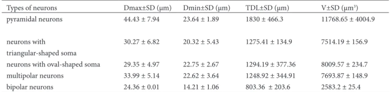
Fig. 1. (a) Microphotograph of Golgi impregnated pyramidal neuron in pyramidal layer, magniication x 400. (b) Drawing of Golgi impregnated pyramidal neuron in pyramidal layer
Fig. 2. (a) Microphotograph of neuron with oval-shaped soma in pyramidal layer, magniication x 400. (b) Drawing of Golgi
of the whole hippocampal formation. here are only a few studies of its anatomy, physiology and function as the interface between the hippocampal complex and cerebral cortex; there are no data about the cel-lular morphology and morphometrics of the human subiculum proper (O’Mara, 2005).
he human subiculum proper is a three-layer archicortex, in which the supericial layer is a mo-lecular layer that is continuous with the stratum lacunosum-moleculare and radiatum of the hip-pocampal CA1 ield. he bipolar neurons of the molecular layer have the lowest values of measured parameters: Dmax 14.1 ± 0.2 µm, Dmin 13.9 ± 0.5 µm and TDL 145.97 ± 3.1 µm. A wide pyramidal cell layer with large pyramidal neurons is deeper,
and the deepest is the polymorphic layer. he prin-cipal cell layer of the subiculum is populated with pyramidal neurons: these are consistent in their shape and size and extend their apical dendrites into the molecular layer and their basal dendrites into deeper portions of the pyramidal cell layer (In-sauti and Amaral, 2004; Amaral and In(In-sauti, 1990; Amaral and Witter, 1995; O’Mara et al., 2001). he pyramidal neurons were described as large neurons by Amaral and Insauti (1990), which we conirmed in our study. Comparing the measured parameters, we can conclude that the pyramidal neurons of the pyramidal layer have the highest value: Dmax (44.43 ± 7.94 µm), Dmin (23.64 ± 1.89 µm), TDL (1830 ± 466.3 µm) and V (11768.65 ± 4004.9 µm3).
Taking into account that the axons of the pyramidal
Fig. 3. (a) Microphotograph of Golgi impregnated triangular-shaped neuron in pyramidal layer, magniication x 400. (b) Drawing of
Golgi impregnated triangular-shaped neuron in pyramidal layer
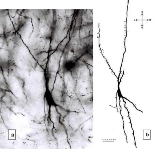
Fig. 4. (a) Microphotograph of Golgi impregnated multipolar neuron in pyramidal layer, magniication x 400. (b) Drawing of Golgi
neurons of the pyramidal layer make up the great-est part of the fornix postcommissuralis (Amaral and Insauti, 1990), these values can be expected. Among the pyramidal cells, there are many smaller neurons; these are considered the interneurons of the subiculum (Amaral and Insauti, 1990; Swanson et al, 1987). he bipolar neurons of the polymor-phic layer have the lowest value of the volume layer (1152.99 ± 662.69 µm3).
he dendritic and axonal characteristics of rat subicular neurons were described in electrophysio-logical studies. However, the morphoelectrophysio-logical charac-teristics of neurons of human and animal subiculum have not yet been suiciently investigated (Vulovic et al., 2010). It is particularly noticeable that there is al-most no information about the morphological char-acteristics of human and animal subicular neurons. For this reason, we could not compare our indings with the research of other scientists.
he results of this study, in which we have de-scribed the morphometric characteristics of neurons of the human subiculum proper, are just a part of the missing information. Future study of the human subiculum proper will provide further information which will improve our understanding of the com-plex functions of the subiculum proper.
REFERENCES
Amaral, D. G. and R. Insauti (1990). Hippocampal formation, In: he human nervous system, (Ed. G. Paxinos), 711–755. Academic Press, New York.
Amaral, D. G., and M. P. Witter (1995). Hippocampal formation. In: he rat nervous system, 2 (Ed. G. Paxinos), 344-393. Academic Press, New York
Anderson M. I. and S. M. O’Mara (2003). Analysis of recordings of single-unit iring and population activity in the dorsal subiculum of unrestrained, freely moving rats. J. Neuro-physiol.90(2), 655-65.
Arellano, J. I., Munoz, A., Ballesteros-Yanez, I., Sola, R. G., and
J. De Felipe (2004). Histopathology and reorganization of chandelier cells in the human epileptic sclerotic hip-pocampus. Brain, 127, 45–64.
Commins, S., Gigg, J., Anderson, M. and S. M. O’Mara (1998). he projection from hippocampal area CA1 to the
subic-ulum sustains long-term potentiation. NeuroReport, 9, 847–850.
Commins, S., Aggleton, J. P. and S. M. O’Mara (2002). Physiologi-cal evidence for a possible projection from dorsal subic-ulum to hippocampal area CA1. Exp.Brain Res. 146(2), 155-160.
Cohen, I., Vincent, N., Stephane, C., Michel, B. and M. Richard
(2002). On the Origin of interictal activity in human tem-poral lobe epilepsy in vitro. Science, 298, 1418-1421.
Dreier, J. P. and U.Heinemann (1991). Regional and time depen-dent variations of low Mg2 induced epileptiform activity in rat temporal cortex slices. Exp. Brain Res.87, 581–596.
Falke, E., Jonathan, N., Mitchell, T. W., Bennett, D. A., Trojanows-ki, J. Q., and E. A. Steven (2003). Subicular dendritic ar-borization in correlates with neuroibrillary tangle density Alzheimer’s disease. American Journal of Pathology, 163
(4), 1615-1621.
Herman, J. P. and W. E. Cullinan (1997). Neurocircuitry of stress: central control of the hypothalamo-pituitary-adrenocorti-cal axis. Trends in Neuroscience, 20, 78-84.
Herman, J. P. and N. K. Mueller (2006). Role of the ventral subic-ulum in stress integration. Behavioural Brain Research,
174, 215–224.
Insausti, R. and D. G. Amaral (2004). Hippocampal Formation. In: he human nervous system, 2 (Ed. G. Paxinos), 871-913. Academic Press, New York.
Jacobson, L. and R. M. Sapolsky (1991). he role of the hippocam-pus in feedback regulation of the hypothalamo-pituitary-adrenocortical axis. Endocr. Rev.12, 118-134.
Naber, P. A. and M. P. Witter (1998). Subicular eferents are or-ganized mostly as parallel projections: a double-labeling, retrograde-tracing study in the rat. J. Comp. Neurol. 393, 284–297.
Morris, R. G. M., Schenk, F., Tweedie, F. and L. E. Jarrard (1990). Ibotenate lesions of the hippocampus and/or subiculum: dissociating components of allocentric spatial learning.
Eur. J. Neurosci.2, 1016–1028.
O’Mara, S .M., Comins, S., Anderson, M., and J. Gigg (2001). he subiculum: a review of form, physiology and function.
Progr. Nerobiol.64, 129–55.
O’Mara S. (2005). he subiculum: what it does, what it might do, and what neuroanatomy has yet to tell us. J. Anat. 207, 271–282.
Sharp, P. E. and C. Green (1994). Spatial correlates of eating pat-terns of single cells in the subiculum of the freely moving rat. J. Neurosci.14, 2339-2356.
Schenk, F. and R. G. M. Morris (1985). Dissociation between components of spatial memory in rats ater recovery from the efects of retrohippocampal lesions. Exp. Brain Res. 58, 11-28.
Shulman, Y. and P. Tibbo (2005). GABA-ergic deicits in schizo-phrenia: evidence and implications. Reviews UAHSJ, 2, 23-27.
Steven, E. A. (2000). Cellular and molecular neuropathology of the parahippocampal region in schizophrenia. Annals of the New York Academy of Sciences. 911, 275-292.
Swanson, L. W., Wyss, J. M. and W. M. Cowan (1978). An auto-radiographic study of the organization of intrahippocam-pal association pathways in the rat. J. Comp. Neurol. 181, 681–716.
Swanson, L. W., Kohler, C. and A. Bjorklund (1987). he limbic region. I. he septohippocampal system. In: Handbook of Chemical Neuroanatomy, Vol. 5, Integrated Systems of the
CNS, Part I (Eds. T. Hökfelt, A. Björklund and L.W. Swan-son), 125-227. Elsevier, Amsterdam
Tamamaki, N. and Y. Nojyo (1990). Disposition of slab-like mod-ules formed by axon branches originating from single CA1 pyramidal cell neurons in the rat hippocampus. J. Comp. Neurol. 291, 509-519.
Taube, J. S., Kesslak, J. P. and C. W. Cotman (1992). Lesions of the rat post-subiculum impair performance on spatial tasks.
Behav. Neural Biol. 57, 131-143.
Van Hoesen, G. W. and A. R. Damasio (1987). Neural correlates of cognitive impairment in Alzheimer s disease. In: Hand-book of Physiology (Ed: F. Plum), 871-898. Williams & Wilkins, Baltimore.
Vulović, M., Živanovic-Mačužić, I., Sazdanović, P., Jeremić, D. and J. Toševski (2010). Morphology of neurons of human subiculum proper. Med. Pregl. LXIII (5-6), 356-360.
Weinberger, R. D. (1999). Cell biology of the hippocampal for-mation in schizophrenia. Society of Biological Psychiatry,