Learning to Predict miRNA-mRNA
Interactions from AGO CLIP Sequencing and
CLASH Data
Yuheng Lu, Christina S. Leslie*
Computational Biology Program, Memorial Sloan Kettering Cancer Center, New York, New York, United States of America
Abstract
Recent technologies like AGO CLIP sequencing and CLASH enable direct transcriptome-wide identification of AGO binding and miRNA target sites, but the most transcriptome-widely used miRNA target prediction algorithms do not exploit these data. Here we use discriminative learning on AGO CLIP and CLASH interactions to train a novel miRNA target prediction model. Our method combines two SVM classifiers, one to predict miRNA-mRNA duplexes and a sec-ond to learn a binding model of AGO’s local UTR sequence preferences and positional bias in 3’UTR isoforms. The duplex SVM model enables the prediction of non-canonical target sites and more accurately resolves miRNA interactions from AGO CLIP data than previous methods. The binding model is trained using a multi-task strategy to learn context-specific and common AGO sequence preferences. The duplex and common AGO binding models together outperform existing miRNA target prediction algorithms on held-out binding data. Open source code is available athttps://bitbucket.org/leslielab/chimiric.
Author Summary
MicroRNAs (or miRNAs) are a family of small RNA molecules that guide Argonaute (AGO) to specific target sites within mRNAs and regulate numerous biological processes in normal cells and in disease. Despite years of research, the principles of miRNA targeting are incompletely understood, and computational miRNA target prediction methods still achieve only modest performance. Most previous target prediction work has been based on indirect measurements of miRNA regulation, such as mRNA expression changes upon miRNA perturbation, without mapping actual binding sites, which limits accuracy and precludes discovery of more subtle miRNA targeting rules. The recent introduction of CLIP (UV crosslinking followed by immunoprecipitation) sequencing technologies enables direct identification of interactions between miRNAs and mRNAs. However, the data generated from these assays has not been fully exploited in target prediction. Here, we present a model to predict miRNA-mRNA interactions solely based on their sequences, using new technologies to map AGO and miRNA binding interactions with machine
a11111
OPEN ACCESS
Citation:Lu Y, Leslie CS (2016) Learning to Predict miRNA-mRNA Interactions from AGO CLIP Sequencing and CLASH Data. PLoS Comput Biol 12 (7): e1005026. doi:10.1371/journal.pcbi.1005026
Editor:Kevin Chen, Rutgers University, UNITED STATES
Received:January 3, 2016
Accepted:June 21, 2016
Published:July 20, 2016
Copyright:© 2016 Lu, Leslie. This is an open access article distributed under the terms of the Creative Commons Attribution License, which permits unrestricted use, distribution, and reproduction in any medium, provided the original author and source are credited.
Data Availability Statement:All relevant data are within the paper and its Supporting Information files.
Funding:This work was supported in part by National Institute of Health (http://www.nih.gov/) grant U01-CA164190 received by CL. The funders had no role in study design, data collection and analysis, decision to publish, or preparation of the manuscript.
learning techniques. Our algorithm produces more accurate predictions than state-of-the-art methods based on indirect measurements. Moreover, interpretation of the learned model reveals novel features of miRNA-mRNA interactions, including potential coopera-tivity with specific RNA-binding proteins.
Introduction
Recent high-throughput technologies like AGO CLIP sequencing [1,2] and CLASH (crosslink-ing, ligation, and sequencing of miRNA-RNA hybrids [3]) enable direct biochemical identifica-tion of AGO binding and miRNA target sites transcriptome-wide. The miRNA field has a strong tradition of computationally leveraging transcriptome-wide data to improve target site prediction, but the leading miRNA target prediction methods today do not exploit these new biochemical data. Here we present a systematic approach to learn both the rules of miRNA-tar-get site pairing and a binding model of AGO’s local sequence preferences and positional bias in alternative 3’UTR isoforms in order to accurately predict miRNA-target interactions.
Before it became possible to map AGO-mRNA and miRNA-mRNA interactions directly, the major advance in miRNA target prediction came from restricting to predefined classes of miRNA seed matches in 3’UTRs and training a model to predict mRNA expression changes in miRNA overexpression experiments. TargetScan was the first algorithm to introduce the strat-egy of correlating context features of miRNA seed sites—including flanking AU content, posi-tion in the 3’UTR, and complementarity to the 3’end of the miRNA—with extent of target down-regulation in miRNA transfection experiments [4]. Similar observations were encapsu-lated in the TargetRank method [5], and these studies established that rules of miRNA target-ing could be statistically decoded from transcriptome-wide data.
However, new data from AGO CLIP sequencing and CLASH challenge some of the assump-tions of existing prediction strategies. These data confirm the prevalence of non-canonical tar-get sites lacking complementarity to the miRNA 2–7 (6-mer) seed region and conversely show that even exact miRNA 2–8 (7-mer) seed matches are often not AGO bound [6,7]. Meanwhile, most target prediction methods require strong seeds to avoid false positives. For example, downloadable predictions from the most recent version of TargetScan still require either per-fect 2–8 seed complementarity (7-mer-m8 site) or a 2–7 seed with A across from miRNA posi-tion 1 (7-mer-1A site), although AGO CLIP data suggests that 7-mer and 8-mer seeds are found in only about half of AGO binding sites [6]. The mirSVR method [8], which also trains on miRNA overexpression experiments, allows up to one mismatch or G:U wobble in the 6-mer seed region, but in practice few non-canonical sites are assigned even moderate scores. Therefore, current target prediction methods may focus on detecting the most effective miRNA sites at the cost of missing a large proportion of miRNA-mRNA interactions. Further-more, training on non-physiological miRNA overexpression experiments may obscure more subtle targeting rules.
A few studies have developed algorithms to resolve which highly expressed miRNAs are associated with individual AGO CLIP peaks. For example, microMUMMIE is an algorithm for analysis of AGO PAR-CLIP that uses the location of T-to-C mutations—indicative of the site of cross-linking of the RNA-binding protein to the RNA in the PAR-CLIP assay—to assign the most likely canonical seed [9]. Other methods use energy-based duplex prediction to associate miRNAs with CLIP-mapped target sequences [10–13]. In particular, MIRZA uses an unsuper-vised probabilistic approach to learn parameters of a duplex alignment model from AGO CLIP peaks, and the duplex model can be used to makede novomiRNA target site predictions from
3’UTR sequence [12]. Note that the MIRZA study used the term“non-canonical”to refer to sites lacking 7 or 8 nucleotides of perfect complementarity to the 5’end of the miRNA; there-fore, their reported non-canonical sites included both perfect 6-mer and many 7-mer-1A sites. (We will use“non-canonical”exclusively for sites lacking full complementarity in the 2–7 6-mer seed region.) More recently, MIRZA-G combined MIRZA duplex quality scores with known context features like flanking AU content and predicted secondary structure accessibil-ity as well as conservation, once again to predict extent of down-regulation in miRNA overex-pression experiments [14].
Here we present a novel model for miRNA target prediction through discriminative learn-ing on transcriptome-wide AGO CLIP and CLASH profiles. Our goal was to learn to accurately predict biochemical miRNA-target site interactions, rather than the extent of regulation, in order to increase the sensitivity of miRNA target prediction and learn physiological targeting rules. As the CLASH protocol captures direct interactions between miRNAs and mRNAs by ligation, it provides a partially labeled training set of miRNA-mRNA interactions including many non-canonical pairings, which we combined with canonical AGO binding sites identified by CLIP. We trained one support vector machine (SVM) classifier to model the miRNA-mRNA duplexes and a second SVM to learn AGO’s local sequence preferences in the UTR and positional bias in 3’UTR isoforms. The duplex SVM model enables the prediction of both canonical and non-canonical pairings between miRNA and target sequences and outperforms existing methods for assignment of miRNAs to AGO binding sites. The AGO binding model is trained using a multi-task strategy to distinguish between cell type and protocol specific sequence signals and common AGO sequence preferences. The duplex SVM and common AGO binding SVM together outperform existing target prediction approaches when evaluated on held out interaction data. Our prediction method, called chimiRic, is available as open source code athttps://bitbucket.org/leslielab/chimiric.
Results
ChimiRic learns both miRNA-mRNA duplex structures and AGO binding
preferences from CLIP and CLASH data
ChimiRic’s duplex model is trained on chimeric reads from CLASH data, which associates a miRNA with a target sequence via chimeric reads and can identify non-canonical binding sites, and AGO CLIP binding sites containing a 6-mer seed match (or longer seed) for a single highly expressed miRNA (Fig 1A). In the latter case, differential AGO CLIP-seq analysis suggests that an AGO bound site that can be associated with a unique miRNA by a canonical 6-mer seed is likely a binding site for that miRNA [6].
We trained a structural SVM [16] on positive and negative (miRNA, site) training examples to learn a model for predicting miRNA-site duplex alignments. Here, the model vectorwof the
SVM represents the scoring parameters for local pairwise alignment. SVM training proceeds iteratively, alternating between obtaining optimal alignments of all training examples given the current SVM parameterswand updating the model vectorwgiven the current duplex
align-ments (Materials and Methods,S1 Fig). The model update step involves solving the SVM large-margin optimization problem so that the discriminant scores assigned to positive and negative (miRNA, site) examples have the correct sign and obey margin constraints, with a hinge loss function to control margin violations (seeMaterials and Methods). To define the local align-ment scoring system and convert the alignalign-ment score into an SVM discriminant function, we used a parameterization similar to the energy-based scoring system in MIRZA, namely a match/ mismatch score that depends on the position in the miRNA sequence together with the nucleo-tides being aligned and penalties for loop opening and for symmetric and asymmetric loop
Fig 1. Overview of the chimiRic prediction model.(A) The first component of the chimiRic model is the duplex SVM, which learns
to predict and score miRNA-mRNA duplex alignments from CLASH and CLIP-seq data. Positive (miRNA, site) training examples comprise canonical and non-canonical pairings identified by chimeric reads in CLASH data (top left) as well as sites with canonical miRNA seeds supported by AGO CLIP data (bottom left). Negative (miRNA, site) training examples include sites that are paired with a different miRNA based on CLASH chimeric read data (top right) or miRNA seed matches with no AGO CLIP evidence (bottom right). The duplex SVM learns the parameters for local duplex sequence alignment and predicts optimal alignments for (miRNA, site) pairs through an iterative training procedure (seeMaterials and Methods). (B) The second component of chimiRic is the AGO binding SVM, which uses features encoding the positional bias of AGO binding sites relative to (possibly multiple) 3’ends of transcripts as well as the local positionalk-mer sequence features. Mouse and human ApA atlases based on 3’end sequencing data (bottom) provide the coordinates of 3’ends used in the analysis.
doi:10.1371/journal.pcbi.1005026.g001
extensions (seeMaterials and Methods). One important difference with MIRZA is that the chi-miRic alignment can only start at position 1 of the miRNA if is it matched against nucleotide A, which more accurately reflects known determinants of miRNA targeting [17].
The second component of chimiRic’s scoring system is an SVM classifier that learns to dis-criminate the local sequence features and positional bias in 3’UTR isoforms of true AGO bind-ing sites versus sites that contain 6-mer seed matches of highly expressed miRNAs but are not AGO-bound, as determined by CLIP data (Fig 1B). Here we considered two AGO CLIP sequencing data sets, the human HEK293 PAR-CLIP data set [2] as well as a HITS-CLIP data set in activated mouse CD4+ T cells [6]. The local sequence context of the upstream and down-stream 30 nt regions flanking the 6-mer seed match are represented using weighted degree ker-nels [18], which encode position specifick-mers fork= 1. . .6 (seeMaterials and Methods).
The positions of 3’ends of alternative 3’UTR isoforms were identified from a human 3’-seq tis-sue atlas [19] and a mouse PolyA-seq atlas [20]. For each site in human or mouse, positional information was encoded by a vector of distance values (measured in nucleotides) to the anno-tated stop codon and to the nearest mapped 3’ends and transformed using a radial basis kernel (seeMaterials and Methods), and the sum of the weighted degree kernels and positional radial basis kernel was used to train the SVM. In order to model differences in AGO binding prefer-ences between the two data sets—both due to protocol differprefer-ences and potentially due to type specific factors influencing AGO occupancy—we used multi-task learning to train cell-type specific AGO preference models together with a common AGO binding model (Fig 1B, Materials and Methods). The cell-type specific models are intended to absorb sequence signals that predict AGO binding in a context-dependent manner, while the common model can be used for target prediction in any new context.
ChimiRic
’
s duplex model outperforms existing methods for predicting
miRNA-mRNA interactions supported by chimeric reads
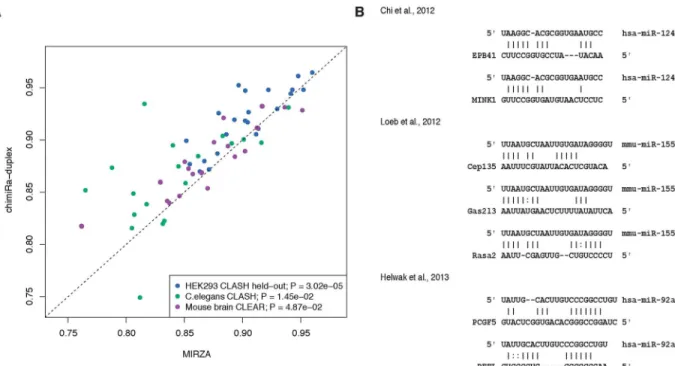
To evaluate chimiRic’s duplex model, we held out from training all HEK293 CLASH interac-tions for a single miRNA seed family (positive test examples) together with a collection of tar-gets sites that interact with other miRNAs based on chimeric read evidence (negative test examples), and we assessed whether the model could rank the held-out miRNA family’s true target sites above these other sites. For each held-out miRNA family in turn, we used chimiRic to generate and score the duplexes between miRNAs in the seed family and mRNA site sequences in the test set. We found that the duplex model could more accurately discriminate true from false interactions compared to MIRZA, an existing method for learning miRNA-mRNA interactions from CLIP data, based on area under the ROC curve (auROC) analysis (Fig 2A, blue points,p<3.02e-5, signed rank test). Note that the original MIRZA model was trained on the same HEK293 PAR-CLIP data set as we used to train the duplex model. To fur-ther evaluate the performance on independent data sets, we then used the duplex model trained on HEK293 CLIP and CLASH data to predict miRNA-mRNA interactions supported by chi-meric reads from iPAR-CLIP inC.elegans[21] and CLEAR-CLIP in mouse brain [22]. Again, chimiRic’s duplex model outperformed MIRZA for the task of ranking observed interactions for each miRNA seed family above interactions with targets sites of other miRNAs in bothC. elegans(Fig 2A, green points,p<1.45e-2, signed rank test) and mouse brain (Fig 2A, purple
points,p<4.87e-2, signed rank test) data sets. These results suggest that chimiRic’s
mRNA duplex model can generalize across organisms and protocols for mapping miRNA-mRNA interactions.
within seed sequences, and interactions relying on 3’base pairing instead of seed pairing [3,6, 7]. In order to test whether our duplex model captures some of these known patterns of non-canonical binding, we predicted duplexes for a variety of non-non-canonical miRNA target sites that have been validated by luciferase assays in previous studies (Fig 2B). Our model have not only correctly identified the correct interacting miRNA above the other highly expressed miR-NAs, despite the lack of exact 6-mer seed matches, but also produced duplex structures repre-sentative of the previously described interaction modes, including GU wobbles, mismatches and bulges in the seed region, and complementary base pairings in the 3’region (Fig 2B).
The full chimiRic model outperforms traditional target prediction for
discriminating CLIP-supported miRNA binding sites
Next we combined the duplex model with the AGO binding model, which is trained to dis-criminate between true AGO bound sites containing 6-mer seeds for highly expressed miRNAs and sites with 6-mer seeds that are not supported by AGO CLIP read evidence, based both on local sequence context and positional bias within 3’UTR isoforms. We used a multi-task strat-egy to train on AGO-bound versus unbound canonical seed sites for highly expressed miRNAs in two AGO CLIP data sets, HEK293 PAR-CLIP [15] and HITS-CLIP in mouse CD4+ T cells [6]. This procedure learned both task-specific SVM models of AGO binding and a common SVM model. The task-specific SVMs may capture protocol-specific CLIP biases and/or cell-type specific AGO binding preferences. For target prediction in a new context where no CLIP
Fig 2. Performance of chimiRic’s duplex model for predicting miRNA-mRNA interactions supported by chimeric reads.(A)
Duplex model’s performance for predicting the correct interacting miRNA seed family among miRNA-mRNA interactions supported by CLASH chimeric reads. For each miRNA seed family tested, all CLASH-supported interactions for miRNAs in the family are held out from training and form the positive test set; negative test examples consist of interactions for a collection of miRNAs that are held out from training in all experiments. Each point represents the held-out auROC for one of the top 23 miRNA seed families in HEK293 (blue), top 19 miRNA seed families inC.elegans(green) and top 20 miRNA seed families in mouse brain (purple). (B) Examples of duplexes predicted by the model for previously validated non-canonical mRNA interactions. Various non-canonical miRNA-mRNA interaction modes were represented, including GU wobbles, bulges and mismatches within seed sequences and interactions relying on 3’base pairing instead of seed pairing.
doi:10.1371/journal.pcbi.1005026.g002
data is available, the common SVM provides a“cell-type agnostic”model of AGO sequence and position preferences.
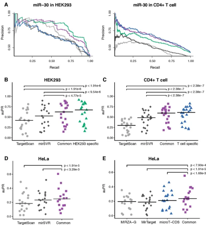
To evaluate the combined chimiRic model, for each miRNA seed family, we held out all HEK293 positive target site sequences—both canonical and non-canonical sites supported by chimeric reads from CLASH as well as canonical sites with AGO CLIP read evidence that can be unambiguously assigned to the seed family—and negative site sequences, for training both the duplex and AGO binding models. We then asked how well the combined model performs at dis-criminating AGO-bound from unbound canonical sites relative to TargetScan [4,23] and mirSVR [8], two widely used miRNA target prediction algorithms.Fig 3Ashows precision-recall curves for the combined chimiRic duplex and HEK293-specific AGO binding model as well as for TargetScan and mirSVR for prediction of canonical sites for several miRNA families. Since TargetScan requires greater seed complementarity than the canonical 6-mer seed (either 7-mer 1A or complementary at miRNA positions 2–8), its overall recall of biochemically-defined sites is limited (note that while the TargetScan 7.0 release discusses 6-mer seeds and non-canonical seeds [23], only a very small fraction of sites were non-canonical in the prediction download files). Evaluating performance by area under the precision-recall curve (auPR) across held-out miRNA seed families showed that this performance advantage was significant over TargetScan (Fig 3B,p<1.91e-6, signed rank test) and mirSVR (Fig 3B,p<9.54e-6, signed rank test). More-over, even measuring performance up to 50% recall (auPR50), where there are still AGO-bound 7-mer sites to detect, chimiRic still outperformed TargetScan on held-out miRNAs in the HEK293 and T cell data sets (S2 Fig). We then tested the combination of chimiRic’s duplex model and the common AGO binding model. Again we found that chimiRic significantly out-performed TargetScan (Fig 3B,p<1.91e-6, signed rank test) and mirSVR (Fig 3B,p<4.77e-5,
signed rank test) on held-out miRNA seed families in HEK293, with minor difference in chimi-Ric’s performance compared to the HEK293-specific model. Similarly, when predicting the bio-chemically defined target sites of held-out miRNA families in CD4+ T cells, chimiRic’s duplex model combined with either the T cell specific or the common AGO binding model outper-formed TargetScan (Fig 3C,p<2.38e-7 andp<2.38e-7, signed rank tests) and mirSVR (Fig 3C,p<2.38e-7 andp<2.38e-7, signed rank tests). As an independent validation, we also
evalu-ated chimiRic’s performance in a third cellular context using two HITS-CLIP data sets in HeLa cells [1,7]. Again, we found that the common AGO binding model combined with duplex model had a significant advantage over TargetScan (Fig 3D,p<1.91e-5, signed rank test) and mirSVR (Fig 3D,p<3.29e-3, signed rank test). Evaluation using auPR50, which favors
TargetS-can by allowing reduced recall, still showed a signifiTargetS-cant performance advantage of the common chimiRic model over TargetScan and mirSVR in HEK293 and T cells, with a statistical tie on the HeLa cells (S2 Fig). We also evaluated the performance of three additional methods, MIRZA-G [24], MirTarget [13] and DIANA-microT-CDS [10], all of which are trained on AGO CLIP data and provide one a single prediction score for each miRNA-gene interaction. When we compared the performance on the same HeLa data set, the common chimiRic model outperformed all three methods measured by auPR (Fig 3E,p<7.90e-4,p<1.91e-5 andp<1.68e-3, signed rank test), partly due to chimiRic’s better recall. When measured by auPR50, chimiRic still achieved a statistical tie against these methods (S2 Fig), showing that chimiRic’s top-ranked predictions are at least as accurate as other methods trained on AGO CLIP data sets.
Fig 3. Performance comparison between chimiRic and other methods for discriminating AGO bound sites from unbound sites.(A) Examples of precision-recall curves for discriminating AGO-bound canonical target sites from seeds with no AGO support for a single miRNA family (miR-30) in HEK293 and CD4+ T cell. Curves correspond to task-specific (T cell: blue; HEK293: green) and common (purple) AGO binding models, TargetScan (grey) and mirSVR (black). (B,C) Performance of TargetScan, mirSVR and task-specific/common AGO binding models on held-out miRNA families in HEK293 and CD4+ T cells measured by auPR. Crossbars represent the median auPR of each model. (D) Performance of TargetScan, mirSVR and the common AGO binding model on the top miRNA families in an independent HeLa CLIP-seq data set measured by auPR. Crossbars represent the median auPR of each model. (E) Performance of MIRZA-G (grey), MirTarget (black), DIANA-microT-CDS (blue) and the common AGO binding model (purple) on the top miRNA families in an independent HeLa CLIP-seq data set measured by auPR. Crossbars represent the median auPR for each model.
doi:10.1371/journal.pcbi.1005026.g003
AGO-binding model learns 3
’
UTR positional preferences and
RNA-binding motifs associated with miRNA targeting
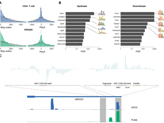
Previous studies have suggested that 3’UTR miRNA target sites tend to reside near the stop codons or near the 3’end of the transcript rather than the middle of 3’UTRs [4]. We confirmed a positional enrichment of AGO-bound sites near the stop codons (Fig 4A, top) and near the end of the 3’UTR compared to miRNA seeds with no AGO binding in CD4+ T cells across mouse transcripts. Additionally, for multi-UTR transcripts, we observed an enrichment of AGO-bound sites in the region upstream of internal 3’cleavage sites (as mapped by
PolyA-Fig 4. Interpretation of the AGO-binding model learned from CLIP-seq data.(A) Positional distribution of AGO binding sites (blue/
green) and unbound sites (grey) within 3’UTRs in CD4+ T cell (top) and HEK293 (bottom), showing enrichment of bound sites near the start of the 3’UTR (left) and in the region upstream of internal 3’cleavage sites of multi-UTR transcripts (right). There is also enrichment of AGO-bound sites ~200nt downstream of internal 3’cleavage sites, suggesting that the resolution of the PolyA-seq peaks can be limited and/or that clusters of nearby 3’cleavage sites confound the analysis. All distances were between the position aligned against nucleotide 2 of the miRNA and the start/end of the corresponding 3’UTR. (B) RBPs with motifs that match the most discriminativek-mers in the common sequence model. Positions with the highest differential POIM for 6-mers upstream and downstream of the miRNA seeds were chosen, and then a signed rank test was used to assess the enrichment of POIMk-mers in RNAcompete array probes. False discovery rates (FDRs) were estimated using the empiricalp-value distribution from 1,000 SVMs trained on random permutations of the +/- labels. Motif logos summarized from the original RNAcompete assays are shown for the top 5 RBPs as ranked by FDR. (The same RBP symbol may appear multiple times since in some cases several constructs of the same protein were assayed by RNAcompete.) (C) An example of co-binding of Pumilio and Argonaute at miRNA target sites. Two miR-17/ 20/106 seed matches within the 3’UTR of UBNX2A are shown, one with AGO2 binding and one without, along with the coverage profiles of AGO2 and PUM2 CLIP in HEK293. For each site, the prediction scores from the SVM sequence model are decomposed into positional scores and displayed. Sequence features near the target site including the Argonaute motif, Pumilio motif, m1A and m9U are also highlighted.
seq) that was absent for the negative site examples (Fig 4A, top,p<2.2e-16, KS test). We also
observed an enrichment of positive site examples ~200ntdownstreamof internal cleavage sites, suggesting that the resolution of the mapped 3’ends in the mouse atlas is limited and/or that clusters of nearby 3’cleavage sites confound the analysis. Likewise, we found HEK293 AGO binding sites enriched upstream of internal 3’cleavage sites based on the human 3’end atlas (mapped by 3’-seq), with more modest downstream enrichment (Fig 4A, bottom). These posi-tional biases are encoded in the feature representation for the AGO binding model (see Materi-als and Methods) and lead to a significant performance improvement for the full chimiRic model (mean auROC on held-out miRNA families of 0.775 without positional bias information vs. 0.849 in the full model,p<2.38e-7, signed rank test;S4 Fig).
To further interpret the sequence features in the AGO binding model, we used the posi-tional oligomer importance matrix (POIM) [26] approach to identify the significant positional k-mers. From the 1-mer POIMs, we observed not only high AU content flanking the miRNA seed matches in general but also specific positional signals like m1A and m8/9U (S5 Fig), which are consistent with findings from previous studies [5,17]. Moreover, the representation allowed us to go beyond single nucleotide composition, which is the extent of sequence contex-tual information used in most previous miRNA target prediction methods, to explore more complex sequence features.
Previous studies have suggested that various RNA binding proteins (RBPs) can bind to regions proximal to miRNA target sites in order to enhance or repress miRNA-mediated regu-lation [27–29]. Therefore, one potential explanation for the long positionalk-mers that dis-criminate between AGO binding sequences and unbound sequences is that they correspond to the motifs of co-binding RBPs that mediate AGO occupancy. To explore this hypothesis, we matched the 6-mers from positions with top differential POIM scores to RNAcompetein vitroaffinity data for a compendium of RBPs [30,31]. By measuring the enrichment of these k-mers in RNAcompete data across all RBPs and assessing significance relative to an empirical null model based on training SVMs on random permutations of the class labels (seeMaterials and Methods), we found that the position-specifick-mers in upstream and downstream sequences were indeed consistent with several known RBP motifs (Fig 4B). In the common AGO-binding model, we identified an AC-rich motif upstream of the seed match that matched an AGO RNAcompete experiment and has been proposed to be the miRNA-inde-pendent binding signal for Argonaute [31]. Meanwhile, in the downstream component of the common model, Pumilio was identified as the most significant RBP motif. It has been previ-ously suggested that Pumilio has a role in regulating miRNA site accessibility of specific target genes [28,32,33]. Our analysis suggests that Pumilio may play a transcriptome-wide role in mediating AGO binding. We compared the HEK293 AGO CLIP to PUM2 PAR-CLIP in the same cell type [2] and found that 16.4% of AGO sites in HEK293 overlapped with PUM2 binding sites.Fig 4Cshows one example of a miR-17/20/106 target site in the 3’UTR of UBNX2A together with sequence signals identified by the model. After decomposing the SVM sequence scores into positional prediction scores (seeMaterials and Methods), we found that the positions with positive contribution overlapped exactly with the Pumilio binding motif and Pumilio CLIP coverage. In contrast, another miR-17/20/106 seed match site in the same 3’UTR was not bound by AGO and lacked significant positionalk-mers from the sequence model.
Discussion
We have presented an integrative model for predicting miRNA binding sites by training on sequencing assays that map biochemical interactions via AGO cross-linking and
mRNA ligation. We demonstrated that chimiRic can detect non-canonical miRNA-mRNA binding modes and significantly outperforms MIRZA for predicting the interacting miRNA for both canonical and non-canonical mRNA target sites. Moreover, chimiRic out-performs TargetScan, a leading target prediction method, for discriminating canonical seed sites that are bound by AGO from unbound sites. The feature representation of our AGO binding model exploits recent 3’-end sequencing data that identifies alternative 3’UTR iso-forms and enables analysis of mRNA sequence signals in the vicinity of the miRNA binding sites, suggesting that other RBPs may collaborate with AGO to mediate miRNA-mRNA interactions.
ChimiRic directly predicts miRNA targeting by learning from miRNA binding data, whereas most existing algorithms infer miRNA targets and model their efficiency using mRNA expression changes in miRNA overexpression experiments in cell culture [4,8]. One major issue with methods trained solely on gene expression changes is that the direct effects of miRNA regulation are confounded with secondary effects, leading to label noise in the learning problem. Since the true binding sites that mediate direct regulation are unknown in this setting, inference of miRNA targets involves“bootstrapping”from an initial set of assumptions of what constitutes a viable target. Furthermore, miRNA transfections in cell culture represent a non-physiological context for miRNA activity and may not accurately reflect endogenous targeting rules. Finally, miRNA binding can inhibit translational efficiency of target mRNAs in addition to or instead of reducing mRNA abundance [34]. While previous global studies suggest that miRNA-mediated changes at the mRNA and protein levels are correlated, these data also depend on miRNA overexpression in cell lines [35,36]. For all these reasons, it is possible that what we have already exhausted what can be learned indirectly from mRNA expression changes due to miRNA perturbations—and from miRNA overexpression experiments in par-ticular—and that new AGO CLIP and CLASH technologies for mapping direct interactions are required to advance our understanding of miRNA targeting in cells.
Materials and Methods
Processing of the sequencing data
Argonaute PAR-CLIP data in HEK293 cells [15], HITS-CLIP in mouse CD4+ T cells [6] and HITS-CLIP in HeLa cells [1,7] were used to define the miRNA target sites in mRNA
sequences. Reads from the wild-type libraries were aligned to hg19 and mm9 genome using the bwa aligner [41]. Argonaute binding sites were then identified from the coverage profile of uniquely aligned reads using a previously described peak calling algorithm [19]. Chimeric reads from CLASH in HEK293 cells [3], iPAR-CLIP inC.elegans[21], and CLEAR-CLIP in mouse brain [22] were also used for training (HEK293) and testing (C.elegansand mouse brain) the duplex model. We used the list of miRNA-mRNA interactions provided in the origi-nal publications with additioorigi-nal filtering. Interactions were chosen according to following cri-teria: (1) binding sites were located in the 3’UTR; (2) binding sites contained complementary matches to the interacting miRNA 6-mer seed with edit distance of 0 or 1; (3) interactions were also supported by non-chimeric reads.
3’-seq data in human tissues [19] and PolyA-seq data in mouse tissues [20] were used to construct 3’UTR isoform atlases in human and mouse. The processing procedure was the same as previously described [19].
Processing of the mRNA expression data
The array data set included gene expression changes in eight individual miRNA transfection experiments in HCT116 cells (miR-15a, miR-16, miR-215, miR-17, miR-20a, let-7c, miR-106b and miR-103a, corresponding to GEO data sets GSM156545, GSM156546, GSM156548, GSM156553, GSM156554, GSM156557, GSM156576 and GSM156580) [25]. To reduce the noise from genes with baseline expressions, we restricted our analysis to probes with signal intensities above median in the control experiments. We also only included the genes with a single potential target site of the transfected miRNA in order to simplify the analysis. The extent of downregulation was represented by the log2 fold change between 24 h post-transfec-tion and control, while genes with multiple probes were represented by the median of all probes.
Composition of training and testing sets
For the duplex model, each example was a pair of miRNA and mRNA site sequences. If the same interaction was identified by chimeric reads in the CLASH data, then we considered the interaction to be positive. Otherwise, if the site was not interacting with the miRNA or another miRNA from the same seed family, then we considered it as a negative example. Due to the limited number of interactions identified by CLASH, we also added interactions inferred from CLIP data by assuming that Argonaute binding sites containing 6-mer seed matches interacted with the corresponding miRNAs, while sites without Argonaute binding were unlikely to inter-act with miRNAs. These assumptions provided another set of positive and negative examples of miRNA-mRNA interactions.
For the AGO binding model, each example was a 3’UTR site matched to the 6-mer seed of one of the highly expressed miRNAs in the corresponding cell type (HEK293: 59 miRNAs from 21 miRNA families; CD4+ T cells: 58 miRNAs from 24 miRNA families). If a seed match overlapped with an Argonaute binding site in the CLIP data, we identified it as a positive exam-ple for the corresponding miRNA. Otherwise, if a seed overlapped with no Argonaute CLIP reads, we considered it to be a negative example.
Feature representation for duplex and context models
We adapted the feature representation from MIRZA [12] to describe the duplex structures formed between interacting (miRNA, site) pairs. Three types of features were included in the representation: (1) the type of base pair (GU, UG, AU, UA, GC, CG) at each position in the alignment; (2) the bases where a loop is opened, symmetrically extended or asymmetrically extended in the duplex structure; (3) binary variables for each position in the miRNA sequence representing whether it is paired to an mRNA base or not. One major change we made to the original representation was that the only permissible base pairing of the first base in the miRNA was with an A in mRNA sequence, so that only an A across from position 1 would contribute positively to the score. This restriction is derived from the observations in previous studies [4].
We described the mRNA sites with two types of UTR features: local sequence context and global positional context. The sequence context was represented by positionalk-mer features (k= 1,. . ., 6) from 30 nt sequences upstream and downstream of the miRNA seed match and
implemented using two weighted degree string kernels [18]. Three positional context features for each site were computed as (i) the distance to the nearest stop codon, (ii) the distance to the next end of a 3’UTR isoform, and (iii) the distance to the previous end of a 3’UTR isoform and were renormalized with a radial basis kernel. These local sequence kernel and positional kernel were then combined by summing kernel matrices.
Training and testing of duplex and context models
We trained the duplex model both on (miRNA, site) examples directly derived from CLASH interactions and on examples with interactions inferred from CLIP based on 6-mer seed com-plementarity. One major advantage of the miRNA-mRNA duplex representation described above is that the model weightswcan also be used as the parameters for local pairwise
align-ment [12]: given the feature descriptionφ(miRNA, site) for a duplex alignment, the alignment
score can be described by the additive scoring functionwφ(miRNA, site). Therefore, by
itera-tively optimizing the model weights given the currents alignments and then computing the optimal alignments given current model weights, we can simultaneously optimize the duplexes and the scoring model. The initial duplex structure for each (miRNA, site) pair was predicted byduplexfoldin the ViennaRNA package [42], and the corresponding duplex feature vectors were then used to train a linear support vector machine (SVM) classifier. The model weightsw
were then used as local alignment parameters to update the duplex structure between the miRNA and mRNA site sequences. The same process was repeated for 12 iterations, by which point the model vector had converged, and the final duplex structures and model weights were used as the duplex model’s output. To compensate for the class imbalance, in each iteration we only used a fraction of negative examples randomly sampled from the whole set while using all positive examples. Specifically, we sampled 15 times as many CLASH negatives as CLASH pos-itives, and the same number of CLIP negatives as CLIP positives.
We applied a regular SVM classifier to the UTR kernel matrix when we trained the AGO binding model using CLIP training data from a single cell type. When we combined data sets from multiple cell types, we applied the multi-task learning approach [43] and treated the dif-ferent cell types as difdif-ferent but related learning tasks to address the possibility of cell type spe-cific miRNA targeting and AGO binding rules as well as protocol spespe-cific biases. We
implemented the multi-task SVM as a modification to the kernel matrix:
If two examples x and z belong to the same task (in other words, two sites were from the same cell type), then an extra weight is added to their product in the kernel matrix to reflect the relationship. The free parameterμcontrols the closeness of task-specific models to the average
model, and its optimal value was determined by five-fold cross-validation.
All the machine learning procedures described above were implemented with Numpy (http://www.numpy.org) and the Shogun machine learning tool box ( http://www.shogun-toolbox.org).
Method comparison
The latest TargetScan 7.0 predictions for human and mouse (context++ scores) were down-loaded fromhttp://www.targetscan.organd mirSVR predictions for human and mouse were downloaded fromhttp://www.microrna.org. For both methods, if any target site had multiple possible interacting miRNAs, we used the interaction with the highest prediction score. Predic-tions for human genes from MIRZA-G (seed-MIRZA-G-C variant), DIANA-microT-CDS and MirTarget were downloaded fromhttp://www.clipz.unibas.ch/index.php?r=tools/sub/mirza_g, http://diana.imis.athena-innovation.gr/DianaTools/index.php?r=microT_CDS/indexand http://mirdb.org. Since these methods provide one single prediction score for each miRNA-gene interaction, for this comparison we also simplified our predictions by using the highest score for genes with multiple sites for the same miRNA.
Search for potential RNA binding protein motifs near miRNA target sites
In order to interpret the local sequence context features captured by the learned AGO binding SVM, we computed the positional oligomer importance matrices (POIMs) [26] for the upstream and downstream weighted degree string kernels, which represent the positional k-mer features enriched in Argonaute binding sequences. For both POIMs, we chose the posi-tional 5-mer or 6-mer with the highest differential POIM weight and used 15k-mers with highest POIM weights from that position to represent the most significant motifs within the positive sequences. We then matched them to potential RNA binding protein motifs identified by RNAcompete assays [30,31]. Normalized array probe intensities for 208 RBPs were down-loaded from the supplemental websites (http://cisbp-rna.ccbr.utoronto.ca;http://www.cs. toronto.edu/~taehyung/gr_ago.html). For each RNAcompete experiment, we selected the top 1000 probe sequences with highest intensity and performed a one-sided Wilcoxon rank-sum test comparing the probes with and without the topk-mers to test the significance of enrichment.
Due to the biased nucleotide content near miRNA targets, it is necessary to estimate false discovery rates (FDRs) for the statistical tests. For each RNAcompete experiment, we generated an empirical null distribution ofp-values by training the AGO-binding SVM models 1,000 times with randomly permutated labels, extracting the top POIMk-mers with the samekand position as in the real model, and testing the enrichment of the topk-mers from the random models within the top probes. The FDRs were then computed by converting the enrichment p-values from the real model to empiricalp-values from the 1,000 rounds of permutations. To better relate enrichedk-mer signals to the biological context, we also filtered out RBPs with no homologs in mouse and human according to cisBP-RNA (http://cisbp-rna.ccbr.utoronto.ca) or with low mRNA abundance according to RNA-seq data in the same cell types [3,6]. Of the remaining RBPs, we considered the top 5 as ranked by FDR as the ones with potential motif enrichment near the miRNA target sites.
To examine the contribution of positionalk-mer features at specific binding sites, we decomposed the SVM score by summing up the SVM weights for allk-mers from the same
position in the upstream/downstream sequence model. One example was visualized inFig 4C to show the overlap between RBP binding and the corresponding sequence signals.
Supporting Information
S1 Fig. Illustration of the iterative learning process of the duplex model.An advantage of the miRNA-mRNA duplex representation is that the model weightswcan be used as the
param-eters for local pairwise alignment: given the feature descriptionφ(miRNA, site) for a duplex
alignment, the alignment score can be described by the additive scoring functionwφ(miRNA,
site). The initial duplex structure for each (miRNA, site) pair was predicted byduplexfoldin the ViennaRNA package, and the corresponding duplex feature vectors were used to train a linear support vector machine (SVM) classifier. The model weightswwere then used as local
align-ment parameters to update the duplex structure between the miRNA and mRNA site sequences. The same iterative process was repeated until convergence of the duplex model.
(PDF)
S2 Fig. Performance comparison between chimiRic and other methods for discriminating AGO bound sites from unbound sites.Area under the precision-recall curve up to 50% recall (auPR50) was used as an alternative metric to compensate for the fact that TargetScan in gen-eral has lower recall due to omission of 6-mer seed match sites. (A,B) Performance of TargetS-can, mirSVR and task-specific/common chimiRic AGO binding models on held-out miRNA families in HEK293 and CD4+ T cells measured by auPR50. Crossbars represent the median auPR50 of each model. (C) Performance of TargetScan, mirSVR and the common chimiRic AGO binding model on the top miRNA families in an independent HeLa CLIP-seq data set measured by auPR50. Crossbars represent the median auPR50 of each model. (D) Performance of MIRZA-G, MirTarget, DIANA-microT-CDS and the common chimiRic AGO binding model on the top miRNA families in an independent HeLa CLIP-seq data set measured by auPR50. Crossbars represent the median auPR50 of each model.
(PDF)
S3 Fig. Performance comparison of chimiRic, TargetScan and mirSVR for predicting tar-get downregulation in miRNA transfection assays.The extent of mRNA downregulation for the top N predictions of each method (chimiRic: purple; TargetScan: grey; mirSVR: black), including(A)or excluding(B)predicted 6-mer seed sites. The extent of downregulation was represented by the median log2 fold changes between transfection and control, while the varia-tion between eight data sets was represented by standard error bars.
(PDF)
S4 Fig. Contribution of positional features to the performance of AGO binding model. Each data point represents the performance on one held-out miRNA family in HEK293 CLIP data set, where the x-axis represents the auROC of the chimiRic AGO binding model without positional features and the y-axis represents the auROC of the full chimiRic AGO binding model.
(PDF)
S1 Table. Top miRNA families used in CLIP-seq training and testing data sets. (XLSX)
Acknowledgments
The authors would like to thank Steve Lianoglou for assistance with his 3’-seq and CLIP-seq processing pipeline, which was adapted for use in this project, along with Gunnar Rätsch and Michael Moore for helpful discussions.
Author Contributions
Conceived and designed the experiments: CSL. Performed the experiments: YL. Analyzed the data: YL. Wrote the paper: YL CSL.
References
1. Chi SW, Zang JB, Mele A, Darnell RB. Argonaute HITS-CLIP decodes microRNA-mRNA interaction maps. Nature. 2009; 460(7254):479–86. doi:10.1038/nature08170PMID:19536157
2. Hafner M, Landthaler M, Burger L, Khorshid M, Hausser J, Berninger P, et al. Transcriptome-wide iden-tification of RNA-binding protein and microRNA target sites by PAR-CLIP. Cell. 2010; 141:129–41. doi:
10.1016/j.cell.2010.03.009PMID:20371350
3. Helwak A, Kudla G, Dudnakova T, Tollervey D. Mapping the Human miRNA Interactome by CLASH Reveals Frequent Noncanonical Binding. Cell. 2013; 153:654–65. doi:10.1016/j.cell.2013.03.043
PMID:23622248
4. Grimson A, Farh KK-H, Johnston WK, Garrett-Engele P, Lim LP, Bartel DP. MicroRNA targeting speci-ficity in mammals: determinants beyond seed pairing. Molecular Cell. 2007; 27:91–105. PMID:
17612493
5. Nielsen CB, Shomron N, Sandberg R, Hornstein E, Kitzman J, Burge CB. Determinants of targeting by endogenous and exogenous microRNAs and siRNAs. RNA. 2007; 13:1894–910. PMID:17872505
6. Loeb GB, Khan Aa, Canner D, Hiatt JB, Shendure J, Darnell RB, et al. Transcriptome-wide miR-155 binding map reveals widespread noncanonical microRNA targeting. Molecular Cell. 2012; 48:760–70. doi:10.1016/j.molcel.2012.10.002PMID:23142080
7. Chi S, Hannon G, Darnell R. An alternative mode of microRNA target recognition. Nature Structural & Molecular Biology. 2012; 19:321–7.
8. Betel D, Koppal A, Agius P, Sander C, Leslie C. Comprehensive modeling of microRNA targets predicts functional non-conserved and non-canonical sites. Genome biology. 2010; 11(8):R90. doi: 10.1186/gb-2010-11-8-r90PMID:20799968
9. Majoros WH, Lekprasert P, Mukherjee N, Skalsky RL, Corcoran DL, Cullen BR, et al. MicroRNA target site identification by integrating sequence and binding information. Nature Methods. 2013; 10:630–3. doi:10.1038/nmeth.2489PMID:23708386
10. Reczko M, Maragkakis M, Alexiou P, Grosse I, Hatzigeorgiou AG. Functional microRNA targets in pro-tein coding sequences. Bioinformatics. 2012; 28(6):771–6. doi:10.1093/bioinformatics/bts043PMID:
22285563
11. Liu C, Mallick B, Long D, Rennie WA, Wolenc A, Carmack CS, et al. CLIP-based prediction of mamma-lian microRNA binding sites. Nucleic acids research. 2013; 41(14):e138. doi:10.1093/nar/gkt435
PMID:23703212
12. Khorshid M, Hausser J, Zavolan M, van Nimwegen E. A biophysical miRNA-mRNA interaction model infers canonical and noncanonical targets. Nature Methods. 2013; 10:253–5. doi:10.1038/nmeth.2341
PMID:23334102
13. Wang X. Improving microRNA target prediction by modeling with unambiguously identified microRNA-target pairs from CLIP-ligation studies. Bioinformatics. 2016:btw002.
14. Gumienny R, Zavolan M. Accurate transcriptome-wide prediction of microRNA targets and small inter-fering RNA off-targets with MIRZA-G. Nucleic acids research. 2015; 43(3):1380–91. doi:10.1093/nar/ gkv050PMID:25628353
15. Kishore S, Jaskiewicz L, Burger L, Hausser J, Khorshid M, Zavolan M. A quantitative analysis of CLIP methods for identifying binding sites of RNA-binding proteins. Nature Methods. 2011; 8:559–64. doi:
10.1038/nmeth.1608PMID:21572407
16. Tsochantaridis I, Joachims T, Hofmann T, Altun Y. Large margin methods for structured and interde-pendent output variables. Journal of Machine Learning Research. 2005; 6:1453–84.
17. Lewis BP, Burge CB, Bartel DP. Conserved seed pairing, often flanked by adenosines, indicates that thousands of human genes are microRNA targets. Cell. 2005; 120(1):15–20. PMID:15652477
18. Sonnenburg S, Rätsch G, Rieck K. Large scale learning with string kernels. Large Scale Kernel Machines2007. p. 73–103.
19. Lianoglou S, Garg V, Yang JL, Leslie CS, Mayr C. Ubiquitously transcribed genes use alternative polya-denylation to achieve tissue-specific expression. Genes & Development. 2013; 27:2380–96.
20. Derti A, Garrett-Engele P, Macisaac KD, Stevens RC, Sriram S, Chen R, et al. A quantitative atlas of polyadenylation in five mammals. Genome Research. 2012; 22:1173–83. doi:10.1101/gr.132563.111
PMID:22454233
21. Grosswendt S, Filipchyk A, Manzano M, Klironomos F, Schilling M, Herzog M, et al. Unambiguous Identification of miRNA: Target site interactions by different types of ligation reactions. Molecular Cell. 2014; 54:1042–54. doi:10.1016/j.molcel.2014.03.049PMID:24857550
22. Moore MJ, Scheel TK, Luna JM, Park CY, Fak JJ, Nishiuchi E, et al. miRNA-target chimeras reveal miRNA 3'-end pairing as a major determinant of Argonaute target specificity. Nature communications. 2015; 6:8864. doi:10.1038/ncomms9864PMID:26602609
23. Agarwal V, Bell GW, Nam JW, Bartel DP. Predicting effective microRNA target sites in mammalian mRNAs. eLife. 2015; 4.
24. Breda J, Rzepiela AJ, Gumienny R, van Nimwegen E, Zavolan M. Quantifying the strength of miRNA-target interactions. Methods. 2015.
25. Linsley PS, Schelter J, Burchard J, Kibukawa M, Martin MM, Bartz SR, et al. Transcripts targeted by the microRNA-16 family cooperatively regulate cell cycle progression. Molecular and cellular biology. 2007; 27(6):2240–52. PMID:17242205
26. Sonnenburg S, Zien A, Philips P, Rätsch G. POIMs: Positional oligomer importance matrices— Under-standing support vector machine-based signal detectors. Bioinformatics. 2008; 24:i6–14. doi:10.1093/ bioinformatics/btn170PMID:18586746
27. Kedde M, Strasser MJ, Boldajipour B, Oude Vrielink JaF, Slanchev K, le Sage C, et al. RNA-binding protein Dnd1 inhibits microRNA access to target mRNA. Cell. 2007; 131:1273–86. PMID:18155131
28. Kedde M, van Kouwenhove M, Zwart W, Oude Vrielink JaF, Elkon R, Agami R. A Pumilio-induced RNA structure switch in p27-3' UTR controls miR-221 and miR-222 accessibility. Nature Cell Biology. 2010; 12:1014–20. doi:10.1038/ncb2105PMID:20818387
29. Mukherjee N, Corcoran DL, Nusbaum JD, Reid DW, Georgiev S, Hafner M, et al. Integrative regulatory mapping indicates that the RNA-binding protein HuR couples pre-mRNA processing and mRNA stabil-ity. Molecular Cell. 2011; 43:327–39. doi:10.1016/j.molcel.2011.06.007PMID:21723170
30. Ray D, Kazan H, Cook KB, Weirauch MT, Najafabadi HS, Li X, et al. A compendium of RNA-binding motifs for decoding gene regulation. Nature. 2013; 499:172–7. doi:10.1038/nature12311PMID:
23846655
31. Li J, Kim TH, Nutiu R, Ray D, Hughes TR, Zhang Z. Identifying mRNA sequence elements for target recognition by human Argonaute proteins. Genome Research. 2014; 24:775–85. doi:10.1101/gr. 162230.113PMID:24663241
32. Nolde MJ, Saka N, Reinert KL, Slack FJ. The Caenorhabditis elegans pumilio homolog, puf-9, is required for the 3' UTR-mediated repression of the let-7 microRNA target gene, hbl-1. Developmental Biology. 2007; 305:551–63. PMID:17412319
33. Miles WO, Tschöp K, Herr A, Ji JY, Dyson NJ. Pumilio facilitates miRNA regulation of the E2F3 onco-gene. Genes & Development. 2012; 26:356–68.
34. Fabian MR, Sonenberg N, Filipowicz W. Regulation of mRNA translation and stability by microRNAs. Annual review of biochemistry. 2010; 79:351–79. doi:10.1146/annurev-biochem-060308-103103
PMID:20533884
35. Baek D, Villen J, Shin C, Camargo FD, Gygi SP, Bartel DP. The impact of microRNAs on protein output. Nature. 2008; 455(7209):64–71. doi:10.1038/nature07242PMID:18668037
36. Selbach M, Schwanhausser B, Thierfelder N, Fang Z, Khanin R, Rajewsky N. Widespread changes in protein synthesis induced by microRNAs. Nature. 2008; 455(7209):58–63. doi:10.1038/nature07228
PMID:18668040
37. Friedersdorf MB, Keene JD. Advancing the functional utility of PAR-CLIP by quantifying background binding to mRNAs and lncRNAs. Genome biology. 2014; 15(1):R2. doi:10.1186/gb-2014-15-1-r2
38. Erhard F, Haas J, Lieber D, Malterer G, Jaskiewicz L, Zavolan M, et al. Widespread context depen-dency of microRNA-mediated regulation. Genome Res. 2014; 24(6):906–19. doi:10.1101/gr.166702. 113PMID:24668909
39. Nam JW, Rissland OS, Koppstein D, Abreu-Goodger C, Jan C, Agarwal V, et al. Global analyses of the effect of different cellular contexts on microRNA targeting. Molecular Cell. 2014; 53:1031–43. doi:10. 1016/j.molcel.2014.02.013PMID:24631284
40. Lu LF, Gasteiger G, Yu IS, Chaudhry A, Hsin JP, Lu Y, et al. A Single miRNA-mRNA Interaction Affects the Immune Response in a Context- and Cell-Type-Specific Manner. Immunity. 2015.
41. Li H, Durbin R. Fast and accurate long-read alignment with Burrows-Wheeler transform. Bioinformatics. 2010; 26:589–95. doi:10.1093/bioinformatics/btp698PMID:20080505
42. Package ViennaRNA 2.0, (2011).
43. Evgeniou T, Pontil M. Regularized multi-task learning. Proceedings of the tenth ACM SIGKDD interna-tional conference on Knowledge discovery and data mining. 2004.