Integrin-Mediated Monocyte Adhesion, Transendothelial
Migration and Phagocytosis
Dan-Qing Liu1, Li-Min Li1,2, Ya-Lan Guo1, Rui Bai1, Chen Wang1, Zhen Bian1, Chen-Yu Zhang1, Ke Zen1,2*
1Jiangsu Diabetes Research Center, State Key Laboratory of Pharmaceutical Biotechnology, School of Life Sciences, Nanjing University, Nanjing, Jiangsu, China,2Jiangsu CDC-Nanjing University Joint Institute for Virology, Nanjing, Jiangsu, China
Abstract
Background:Signal regulate proteina(SIRPa) is involved in many functional aspects of monocytes. Here we investigate the role of SIRPain regulatingb2integrin-mediated monocyte adhesion, transendothelial migration (TEM) and phagocytosis.
Methodology/Principal Findings:THP-1 monocytes/macropahges treated with advanced glycation end products (AGEs) resulted in a decrease of SIRPaexpression but an increase ofb2integrin cell surface expression andb2integrin-mediated adhesion to tumor necrosis factor-a (TNFa)–stimulated human microvascular endothelial cell (HMEC-1) monolayers. In contrast, SIRPaoverexpression in THP-1 cells showed a significant less monocyte chemotactic protein-1 (MCP-1)–triggered cell surface expression ofb2integrins, in particular CD11b/CD18. SIRPaoverexpression reducedb2integrin-mediated firm
adhesion of THP-1 cells to either TNFa–stimulated HMEC-1 monolayers or to immobilized intercellular adhesion molecule-1 (ICAM-1). SIRPa overexpression also reduced MCP-1–initiated migration of THP-1 cells across TNFa–stimulated HMEC-1 monolayers. Furthermore,b2integrin-mediated THP-1 cell spreading and actin polymerization in response to MCP-1, and
phagocytosis of bacteria were both inhibited by SIRPaoverexpression.
Conclusions/Significance:SIRPanegatively regulatesb2integrin-mediated monocyte adhesion, transendothelial migration and phagocytosis, thus may serve as a critical molecule in preventing excessive activation and accumulation of monocytes in the arterial wall during early stage of atherosclerosis.
Citation:Liu D-Q, Li L-M, Guo Y-L, Bai R, Wang C, et al. (2008) Signal Regulatory ProteinaNegatively Regulatesb2Integrin-Mediated Monocyte Adhesion,
Transendothelial Migration and Phagocytosis. PLoS ONE 3(9): e3291. doi:10.1371/journal.pone.0003291
Editor:Dominik Hartl, Yale University School of Medicine, United States of America
ReceivedJune 3, 2008;AcceptedSeptember 7, 2008;PublishedSeptember 29, 2008
Copyright:ß2008 Liu et al. This is an open-access article distributed under the terms of the Creative Commons Attribution License, which permits unrestricted use, distribution, and reproduction in any medium, provided the original author and source are credited.
Funding:This work was supported in part by China Basic Science 973 program (CYZ, KZ).
Competing Interests:The authors have declared that no competing interests exist.
* E-mail: [email protected]
Introduction
Recruitment of monocytes from circulation to inflamed tissues plays a pivotal role in the initiation and progression of atherosclerosis [1,2,3]. After migrated to lesion region, monocytes are rapidly differentiated into macrophage which engulf lipids and form the fatty streak [4]. Although the mechanisms that govern the delivery of monocytes from circulation to inflammatory site are not fully understood, the process of monocyte diapedesis has been regarded as a multi-step event that is sequentially regulated by a panel of adhesion molecules and signaling pathways. E- and P-selectins are involved in the initial reversible adherence of monocytes to the endothelial cell monolayers [5]. The following firm adhesion is mediated by monocyte b2 integrins,including CD11a/CD18 and CD11b/CD18,that recognize vascular cell adhesion molecule-1(VCAM-1) and intercellular adhesion mole-cule-1 (ICAM-1) on endothelial cells [6]. Firm adhesion of monocytes requires activation of integrins, which can be triggered by agonist-induced activation of G protein–coupled chemokine receptors [7]. Monocytes express CC chemokine receptor 2 (CCR2), which binds monocyte chemoattractant protein-1 (MCP-1), leading tob2integrin-mediated firm adhesion and subsequent
transmigration of adhered monocytes through the vascular endothelium [8].
Recently signal regulatory proteina(SIRPa) (also designate as SHPS-1[9], p84[10], BIT[11], MFR[12], MyD-1[13], etc.) has been reported to serve as an important modulator for controlling leukocyte inflammatory responses [14,15]. As an immunoglobulin superfamily member (IgSF), SIRPa is expressed mainly by myeloid. SIRPa has a long intracellular domain that contains four tyrosine residues to form two immunoreceptor tyrosine-based inhibition motifs (ITIMs) and this type of signaling structure is highly conserved between mice, rats and humans. Studies have suggested that binding of SIRPawith its extracellular ligand CD47 results in phosphorylations of SIRPaITIMs, which in turn, leads to their association with SH2-domain-containing protein tyrosine phosphotases SHP-1 and SHP-2 [16,17] to delivers signals that regulate a variety of cellular functions [14]. Ligation of SIRPaby antibody or CD47 recombinant inhibits many leukocyte functions, including phagocytosis[18,19], tumour-necrosis factor produc-tion[20] andin vitro transmigration[21,22]. Activation of SIRPa
and actin stress fibres in response to interaction with extracellular matrix, suggesting that SIRPa also plays a role in integrin-mediated cytoskeletal organization [24]. Negative regulatory role of SIRPahas also been found in tumor metastasis, survival, and cell transformation [25].
In the present study, to further explore the negative regulatory role of SIRPain various functional aspects of monocytes, we examined the correlation between expression level of SIRPain THP-1 cells and THP-1 cell transmigratory capacity. By overexpressing SIRPa
in THP-1 cells, we also determined the alteration of b2 integrin expression andb2integrins-mediated cellular functions of monocytes in response to chemoattractant stimulation.
Materials and Methods
Reagents and Antibodies
Recombinant human MCP-1 and TNFa, were purchased from PeproTech (Rocky Hill, NJ). AGEs-BSA (AGEs) was prepared according to a method previously described [26]. Briefly, 50 mg/ ml bovine serum albimin (BSA) (Fraction V, sterile filtered, Sigma-Aldrich) was incubated with 0.6 M D-ribose or 0.5 M D-glucose and 0.3 M lysine in PBS containing 100 units/ml penicillin and streptomycin for 4 weeks. The unincorporated sugars were removed by dialysis against PBS. Polyclonal anti-human CD11b were generated against C-terminal peptide of CD11b [27]. Polyclonal anti-SIRPa antibody (SIRPa-ct) was obtained from Chemicon (Temecula, CA). Monoclonal anti-CD11b (OKM-1), and anti-CD11a (TIB-217), prepared in our laboratory from hybridoma, were obtained from American Type Culture Collec-tion (ATCC) (Manassas, VA). Monoclonal anti-CD11c (S-HCL-3, IgG2b) and anti-CCR2 (clone 48607) were obtained from BD Biosciences (San Diego, CA).
Cells
THP-1 cells (Chinese Cell Culture Center, Shanghai, China) were cultured and maintained as described [8]. In separated experiments, THP-1 cells were treated with AGEs-BSA (AGEs) at various concentrations overnight. Cells treated with BSA of the same concentration served as controls. Immortalized HMEC-1 was kindly provided by Dr. E.W. Ades (Centers for Disease Control and Prevention, Atlanta, GA)[28] and were grown in MCDB-131 (Invitrogen) supplemented with 10 ng/ml epidermal growth factor (Becton-Dickinson), 1mg/ml hydrocortisone (Sig-ma-Aldrich) and 10–15% fetal bovine serum (Hyclone). HMEC-1 cells were seeded on collagen (Sigma-Aldrich)-coated tissue culture plates or permeable Transwell filters (5.0mm pore size, 0.33 cm2 surface areas, Costar, NY).
SIRPaOverexpression
Complete sequence of human SIRPawas amplified, inserted into the expression vector pcDNA3.1 (Invitrogen, Carlsbad, CA), and confirmed by DNA sequencing. The transfection of THP-1 cells was conducted via Lipofectmin 2000 (Invitrogen). Briefly, Lipofectmin-SIRPapcDNA3.1 complex (250mL) was added dropwise to 56106 THP-1 cells in 20 mL RPMI medium 1640 containing 2% heat-inactivated fetal bovine serum. After 24 h incubation, THP-1 cells were harvested and used for further analysis.
Immunofluorescence, Confocal Microscopy and Flow Cytometry
Surface expressions of b2-integrins were detected using flow cytometry as described [8,29]. The relative surface expression was estimated by subtracting the mean fluorescence intensity (MFI) of cells labeled with the nonspecific antibody from that of cells
labeled with the antibodies detecting b2-integrins. All studies consisted of at least three independent experiments. Flow cytometry was performed and data were analyzed using CELLQUEST software (BD Biosciences). The polymerization of actin filaments in THP-1 cells, induced by pretreatment with 10 nM MCP-1 or 25 ng/mL TNFa for 30–60 minutes, was determined using rhodamine-conjugated phalloidin staining (Molecular Probes) according to the manufacturer’s protocol. Briefly, cells treated with cytokines were fixed with 3.7% paraformaldehyde in PBS for 5 minutes, gently permeabilized with 0.1% Triton X-100, and blocked with 1% BSA in PBS for 30 min followed by rhodamine-conjugated phalloidin staining. In some experiments, cells were treated with cytochalasin D (Sigma-Aldrich) to inhibit actin polymerization [30]. Coverslips were mounted with antifade mounting medium (Molecular Probes). Images were captured and analyzed by a laser scanning confocal microscope equipped with an image processing system (Olympus Microsystems).
Cell Adhesion Assays
Confluent HMEC-1 cell monolayers cultured on gelatin (Difco)–coated tissue culture plates or permeable Transwell filters. Monolayers were treated with 25 ng/mL TNFafor 6 h to induce VCAM-1 and ICAM-1 expression prior to the adhesion assay [31,32]. In separate experiments, 24-well plates were coated for 2 h with 10mg/mL human recombinant VCAM-1 or ICAM-1. THP-1 cells were briefly labeled with 29,79 -bis-(2-carboxyethyl)-6-carboxyfluorescein acetoxymethyl ester (BCECF-AM, Molecular Probes)[27] and then suspended in RPMI 1640 medium containing 0.1% BSA and stimulated with 10 nM MCP-1 for 30 min to activate integrins [30]. In a subset of experiments, monocytes were pre-incubated with 50mM dibutyl-cAMP (Bt2cAMP) for 30 min to inhibit chemokine-mediated integrin activation. Fluorescently labeled monocytes (,26105cells/well) were then added to HMEC-1 monolayers or 24-well plates coated with VCAM-1 or ICAM-1, and incubated for 30 min at 37uC. Nonadherent monocytes were removed by gentle washing with PBS and the bound cells were measured by a fluorescence plate reader at excitation/emission wavelengths of 485/535 nm (Milli-pore, Milford, MA)[27].
Transendothelial Migration (TEM)
Migration of THP-1 cells across TNFa–pre-activated HMEC-1 monolayers was performed as previously described [30] with minor modification. Prior to migration assay, HMEC-1 monolay-ers cultured on gelatin (Difco)–coated transwell filtmonolay-ers were treated with 25 ng/mL TNFa for 6 h. THP-1 cells (5.06105/per well) were added to the upper chamber of Transwell inserts containing 200ml HBSS. 700ml HBSS containing 10 nM MCP-1 was placed in the bottom chamber. After 90 min and 180 min incubation at 37uC under 5% CO2, cells that had transmigrated to the lower chamber were harvested in 1 ml PBS containing 0.1% BSA and labeled with phycoerythrin (PE)-conjugated anti-human CD14 antibody. 106FITC-conjugated standard beads (PharMingen, La Jolla, CA) were added to the cell suspension and the number of THP-1 cells was counted until 10,000 beads were counted by flow cytometry. All experiments were repeated as triplicated fashion in at least three independent studies.
solution devoid of Ca2+
and Mg2+
(HBSS2), cell associated fluorescence was observed under microscopy or measured using a SpectraMax microtiter plate reader (Molecular Devices).
Western Blot
THP-1 cells were solubilized in lysis buffer containing 1% Triton X-100 and a panel of protease inhibitors at 4uC. Pellet was removed after centrifuged at 13,0006gfor 5 minutes. Supernatant was normalized for total protein, and loaded on 10% SDS-PAGE. After electrophoresis and transfer onto Hybond membranes, membranes were blocked with 5% non-fat milk. Antigens were detected using suitable primary antibodies followed by incubation with HRP-conjugated antibodies and ECL (Amer-sham) detection.
Statistical Analysis
Data were analyzed by the Studentt test; P values of,0.05 were regarded as significant differences (*, p,0.05; **, p,0.01).
Results
Downregulation of SIRPain THP-1 cells is correlated with enhancedb2integrin-mediated cell adhesion
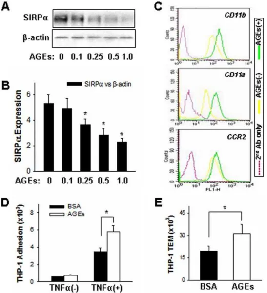
It has been reported that advanced glycation end products (AGEs) are involved in tissue damage associated with diabetic complications and aging [34,35]. Although the mechanism is still not clear, monocytes tend to be activated by AGEs and show an enhanced chemotaxis under such inflammatory conditions. As shown by Western blot analysis in Figure 1A, the expression of SIRPa in THP-1 cells was decreased after AGEs treatment. Served as controls, b-actin level was not altered. The down-regulation of SIRPain AGEs–treated THP-1 cells is contrast to that of receptor for advanced glycation end products (RAGE) and junctional adhesion molecule-like protein (JAML), which expres-sion levels are both increased after AGEs treatment (Zen et al, unpublished). Fig. 1B showed the quantitative analysis of SIRPa
downregulation by AGEs in a dose-dependent fashion.
Interest-Figure 1. Down-regulation of SIRPain THP-1 cells treated with BSA-AGEs is correlated to enhanced THP-1 cell surface expression of leukocyteb2integrins andb2integrins-mediated THP-1 cell inflammatory responses.In these experiments, THP-1 cells were treated with
BSA-AGEs (AGEs) or BSA overnight at 37uC.A:SIRPaprotein level in THP-1 cells;B:reduction of SIRPaprotein level by AGEs in dose-dependent fashion;C:Cell surface expression level ofb2integrins in THP-1 cells; D and E: THP-1 cell adhesion to and migration across TNFa–pre-activated
HMEC-1 monolayers, respectively. All data are mean6SD (n= 3) of three independent experiments.
ingly, AGEs–treated THP-1 cells showed a significant enhanced cell surface expression ofb2integrins, in particular CD11b/CD18, in response to the stimulation of MCP-1 (Fig. 1C). Also, compared to BSA–treated THP-1 cells, AGEs– treated THP-1 cells had a higher percentage of cell adhesion to TNFa–activated HMEC-1 monolayer (Fig. 1D). In response to MCP-1, AGEs–treated THP-1 cells also showed an increased transmigration across TNFa– activated HMEC-1 monolayers compared to THP-1 cells treated with BSA (Fig. 1E). Together, these results suggest that AGEs treatment can activate THP-1 cells and enhance cell chemotaxis.
Overexpression of SIRPain THP-1 cells inhibits MCP-1– induced cell surface expression of b2 integrins
To define the role of SIRPa in regulating monocyte inflammatory response, we characterized the alteration of b2 integrin expression and b2 integrin-mediated cell adhesion, migration and phagocytosis in THP-1 cells after significantly increase SIRPa expression level. As shown in Figure 2A, immunoblot analysis showed that the delivery of the pcDNA3.1 vector encoding human SIRPa into THP-1 cells profoundly enhanced SIRPa expression. Compared to mock–transfected THP-1 cells, SIRPa–transfected THP-1 cells also showed a significantly enhanced expression of SIRPa on cell surface, as indicated by flow cytometry (Fig. 2B).
Similar to circulating monocytes, THP-1 cells normally expressb2 integrins [8,36,37] and their cell surface expression levels are rapidly up-regulated by chemoattractants during inflammatory response. We next determined the alteration of cell surface expression of CD11b/CD18 and CD11a/CD18 in SIRPa– or mock–transfected THP-1 cells. In these experiments, SIRPa– or mock–transfected THP-1 cells were treated with or without 10 nM MCP-1 for 30 min and then directly labeled with PE-conjugated mouse IgG specific for human CD11a, CD11b, and CC chemokine receptor 2 (CCR2), and surface expression was analyzed using flow cytometry. The results showed that SIRPa overexpression in THP-1 cells did not affect the basal level ofb2 integrin expression on cell surface but significantly reduced the up-regulation of cell surfaceb2 integrin expression by MCP-1 (Fig. 2C).
SIRPaoverexpression affects CD11b/CD18-mediated THP-1 cell functions in response to MCP-1 stimulation
Chemokines such as MCP-1 has been reported to trigger integrin–mediated firm adhesion and subsequent transmigration of monocytes [8]. As shown in Figure 3A, MCP-1–stimulated THP-1 cells showed a significant integrin-mediated firm adhesion to TNFa–activated HMEC-1 monolayers. However, this firm adhesion was largely reduced in THP-1 cells with SIRPa
overexpression. Since TNFa–stimulated HMEC-1 monolayers express both VCAM-1 and ICAM-1, the specific ligands forb1 and b2 integrins, respectively, additional adhesion assays were performed using plates coated with human recombinant ICAM-1 or VCAM-1, respectively. To estimate background adhesion, control adhesion assays were performed using THP-1 cells pretreated with Bt2cAMP, a permeable analogue of cAMP that blocks integrin-dependent firm adhesion triggered by MCP-1 [30]. SIRPa–transfected THP-1 cells did not show MCP-1–induced firm adhesion to plates coated with ICAM-1, while adhesion to plates coated with VCAM-1 was intact (Fig. 3B).
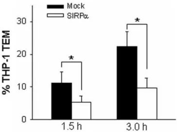
Previous studies have reported that chemokine-mediated activation ofb2 integrins was essential for THP-1 cell adhesion and subsequent transmigration through endothelial monolayers [7,8,38]. Therefore, the effect of SIRPa overexpression on transendothelial migration (TEM) of THP-1 cells was examined by transmigration assay. In mock-transfected THP-1 cells, MCP-1 triggered strong THP-1 cell migration across HMEC-1 monolay-ers. As shown in Figure 4, more than 20% of total applied THP-1 cells were migrated across TNFa–activated HMEC-1 monolayers after 3 h incubation. In contrast, MCP-1–triggered transmigration of THP-1 cells that were overexpressed with SIRPawas strongly reduced (Fig. 4). In both mock-transfected and SIRPa overex-pressed THP-1 cells, spontaneous migration of THP-1 cells in the absence of MCP-1 was minimal (data not shown).
Leukocyteb2integrin–mediated cell firm adhesion initiates cell shape changes and spreading of monocytes, events that must occur for subsequent cellular locomotion and transmigration. Next we investigated the effect of SIRPa overexpression on TNFa– and MCP-1–stimulated actin polymerization and cell spreading in THP-1 cells. SIRPa– or mock– transfected THP-1 cells were stimulated with TNFaor MCP-1 for 30 minutes, then fixed, and labeled with rhodamine-conjugated phalloidin to visualize actin filaments. As a control, mock–transfected THP-1 cells were pretreated with cytochalasin D to inhibit actin polymerization [39]. Labeled monocytes were mounted on coverslips and images were obtained using confocal microscopy. As shown in Figure 5, confocal microscope images showed that mock– transfected THP-1 cells exposed to TNFa or MCP-1 underwent morphological changes resulting in multiple pseudopods (arrowheads) with abundant actin filaments, and that this process was inhibited by
Figure 2. SIRPaoverexpression in THP-1 cells and its effect on cell surface expression ofb2integrins and CCR2.THP-1 cells were
transfected with the empty pcDNA3.1 vector (Mock) or the SIRPa -encoding pcDNA3.1 vector (SIRPa).A:SIRPaprotein level in SIRPa– or mock– transfected THP-1 cells; B: Cell surface SIRPa expression in Mock– and SIRPa–transfected THP-1 cells; C: THP-1 cell surface expression of CD11a, CD11b, CD11c and CCR2. Note that SIRPa
overexpression significantly suppressed MCP-1–induced up-regulation of THP-1 surface expression of CD11b and CD11a but not CCR2, and that SIRPaoverexpression did not affect the basal level ofb2integrins.
cytochalasin D. In contrast, significantly less TNFa– or MCP-1– stimulated actin polymerization and cell spreading had occurred in THP-1 cells with SIRPaoverexpression (Fig. 5).
The phagocytic function of THP-1 cells is also dependent onb2 integrins [40,41,42]. Next we examined the effect of SIRPa
overexpression on the capacity of THP-1 cells to engulf fluorescein–labeledE. coliK12 bioparticles. As shown by confocal images in Figure 6A, the mock-transfected THP-1 cells showed a
Figure 3. SIRPaoverexpression in THP-1 cells decreases MCP-1–stimulated adhesion. A:Micrographs and histograms show THP-1 cells adhered to HMEC-1 monolayers. Bar = 20mm.B: MCP-1–depen-dent adhesion of SIRPa– or mock–transfected THP1 monocytes to plates coated with ICAM-1 or VCAM-1. In separate experiments, monocytes were pretreated with 50mM Bt2cAMP to determine background integrin-independent nonspecific adhesion. All data are mean6SD (n= 3) of three independent experiments.
doi:10.1371/journal.pone.0003291.g003
Figure 4. SIRPa overexpression reduces the MCP-1–induced migration of THP-1 cells across HMEC-1 monolayers pre-activated with 25 ng/mL TNFa. All data are mean6SD (n= 3) of
three independent experiments. doi:10.1371/journal.pone.0003291.g004
Figure 5. TNFaand MCP-1–induced cell spreading and actin polymerization in THP-1 cells.SIRPa– or mock–transfected THP-1 cells were stimulated with TNFaor MCP-1 for 30 minutes, fixed, and labeled with rhodamine-conjugated phalloidin to visualize actin filaments. As a negative control, mock–transfected THP-1 cells were pretreated with cytochalasin D (Mock+Cyt. D). Bar = 20mm.
significant phagocytosis of fluorescein–conjugated bacteria parti-cles after 3 h incubation (Fig. 6A, arrows), while in SIRPa– transfected THP-1 cells, uptake of fluorescein–conjugated bacteria particles was strongly reduced. The quantitative analysis of uptaking fluorescein–conjugated bacteria particles by THP-1 cells was shown in Figure 6B. Taken together, these results clearly show that SIRPa overexpression in THP-1 cells reduces various inflammatory responses mediated by leukocyteb2integrins.
Discussion
Recent studies have demonstrated that SIRPa is involved in regulating various inflammatory responses of leukocytes, in particular leukocyte chemotaxis and phagocytosis. By studying the leukocyteb2 integrin-mediated functional changes in THP-1 cells after downregulation or overexpression of SIRPa level, we show that SIRPanegatively regulatesb2integrin-mediated THP-1 cell inflammatory responses, such as adhesion, transendothelial migration and phagocytosis.
Correlation between SIRPaprotein level and CD11b/ CD18-mediated cellular functions in THP-1 cells
Ligation of SIRPawith its extracellular ligand CD47 results in phosphorylations of SIRPa ITIMs, which in turn, leads to their association with SH2-domain-containing protein tyrosine
phos-photases SHP-1 and SHP-2 [16,17] to delivers signals that regulate a variety of cellular functions [14]. Binding of SIRPaby antibody or CD47 recombinant inhibits many leukocyte functions, including phagocytosis [18,19], tumour-necrosis factor production [20] andin vitro transmigration [21,22]. Activation of SIRPa by arterial elastic laminae also inhibits monocyte adhesion [23]. Fibroblasts expressing a SIRPamutant lacking ITIMs-containing cytoplasmic tail showed increased formation of focal adhesions and actin stress fibres in response to interaction with extracellular matrix, suggesting that SIRPa is also involved in mediating outside-in signal transduction during cell-matrix interaction. Using THP-1 cell as model cell line, here we show that SIRPaprotein level is downregulated by AGEs treatment, which is also correlated to an enhanced cell surface expression of b2 integrins and b2 integrins-mediated cell adhesion (Fig. 1). The finding of SIRPa
reduction in AGEs-treated THP-1 cells is supported by a recent report that mouse macrophages have lower SIRPa expression level following LPS stimulation [43]. The correlation between SIRPa expression level and chemoattractant-induced cell surface upregulation ofb2integrins andb2integrins-mediated THP-1 cell inflammatory responses is further characterized in THP-1 cells overexpressed with SIRPa(Fig. 2–6). The results not only confirm the inhibitory function of SIRPaon THP-1 inflammatory responses, but also indicated that the role of SIRPain THP-1 cells is through affecting the functions ofb2integrins, particularly CD11b/CD18. It is worthy to note that overexpression of SIRPadoes not alter the basal level of b2 integrin expression but the upregulation of b2 integrins by MCP-1 stimulation, suggesting that SIRPa is one of essential molecules along the signal pathways that may regulate the synthesis, transportation and translocation process ofb2 integrins. Moreover, if AGEs and other inflammatory factors can affectb2 integrin expression and function through down-regulating SIRPa, it might be reasonable to conclude that SIRPacan mediate an inside-out signal in regulatingb2integrin function.
SIRPaas a negative regulator in monocyte recruitment during inflammation
The expression of b2 integrins and adhesion molecules in monocytes is regulated by chemokines such as MCP-1, SDF-1 alpha and RANTES [32,44,45,46]. The positive correlation between CD11b expression in circulating monocytes and the degree of monocyte infiltration into the proatherogenic vascular wall has been well-documented [8,47,48]. The increased expres-sion of monocyte CD11b under pro-inflammatory conditions enhanced MCP-1–mediated chemotaxisin vitro[8], induced excess monocyte adhesion to vascular endothelium, and increased formation of neointima and atherosclerotic plaques [48]. Although SIRPaoverexpression did not affect surface expression of CCR2, the receptor for MCP-1, it resulted in a profound reduction of MCP-1–mediated upregulation of THP-1 cell cell surface b2 integrins and THP-1 cell TEM. In addition to reduction of CD11b and other b2 integrins, our study has also demonstrated that overexpressing SIRPa in THP-1 cells display less cell spreading and actin polymerization in response to chemokine stimulation. The mechanism by which SIRPa modulates chemokine-induced cell spreading and actin polymerization is unknown although several possibilities exist: a) directly activates protein phosphatase and initiates signal pathways that attenuate filament actin polymerization and cell spreading, and b) binding to integrin-associated protein CD47 and modulating the integrin functions. Since SIRPais a cellular ligand of CD47, which can augment the functions of integrins of theb1, b2 and b3 families via initiating heterotrimeric Gi protein signaling [49], thus modulating a range of cell activities including cell motility and adhesion, and leukocyte
Figure 6. SIRPa overexpression in THP-1 cells impairs cell phagocytic function.Fluorescently labeled bacteria were incubated with SIRPa– or mock–transfected THP-1 cells for 3 h. A: Images of phagocytosis of bacteria by THP-1 cells; B: Quantitative analysis of bacteria phagocytosis by THP-1 cells. Bar = 20mm. All data are mean6SD (n= 4) of four independent experiments.
adhesion, migration and phagocytosis. Indeed, phagocytosis of bacteria by THP-1 cells, an event that is largely dependent onb2 integrin and actin polymerization, was significantly reduced by overexpression of SIRPa. This result was in agreement with the previous finding that SIRPa contributes to down-regulating the macrophage phagocytic response [18]. In summary, the present study demonstrates for the first time that SIRPa overexpression potently inhibits the various inflammatory responses of THP-1 monocytes/macrophages mediated byb2integrins. The induction of SIRPa expression in THP-1 cells led to a reduction of chemokine-induced cell surface expression ofb2integrins, which eventually resulted in less cell adhesion, cellular spreading, cell transmigration and phagocytosis. This observation suggests that SIRPa may function to decrease transendothelial migration of monocytes or other circulating leukocytes, reduce the burden of inflammatory cells in atheroma, and ultimately decrease plaque mass under atherogenic conditions. Since migration of monocytes across blood vessel lining endothelial monolayers is a key
component during early stage of atherosclerosis, such an outcome would indicate that SIRPa overexpression in monocytes or macrophages has an anti-atherogenic effect and that SIRPais a potential target in therapeutical implications.
Acknowledgments
The authors thank Dr. E.W. Ades (Centers for Disease Control and Prevention, Atlanta, GA) and Dr. Yuan Liu (Department of Biology, Georgia State University, Atlanta, GA) for kindly gift of the immortalized HMEC-1 and SIRPaexpression vectors, respectively. The authors also thank Mr. Bihn Ha and Ms Celia Chen for excellent technical assistance.
Author Contributions
Conceived and designed the experiments: CYZ KZ. Performed the experiments: DQL LML YLG RB CW ZB KZ. Analyzed the data: DQL LML YLG RB KZ. Contributed reagents/materials/analysis tools: CYZ. Wrote the paper: KZ.
References
1. Gerrity RG (1981) The role of the monocyte in atherogenesis: I. Transition of blood-borne monocytes into foam cells in fatty lesions. Am J Pathol 103: 181–190.
2. Ross R (1993) The pathogenesis of atherosclerosis: a perspective for the 1990s. Nature 362: 801–809.
3. Rosenfeld ME (2002) Leukocyte recruitment into developing atherosclerotic lesions: the complex interaction between multiple molecules keeps getting more complex. Arterioscler Thromb Vasc Biol 22: 361–363.
4. Boisvert WA, Santiago R, Curtiss LK, Terkeltaub RA (1998) A leukocyte homologue of the IL-8 receptor CXCR-2 mediates the accumulation of macrophages in atherosclerotic lesions of LDL receptor-deficient mice. J Clin Invest 101: 353–363.
5. Dong ZM, Chapman SM, Brown AA, Frenette PS, Hynes RO, et al. (1998) The combined role of P- and E-selectins in atherosclerosis. J Clin Invest 102: 145–152.
6. Springer TA (1994) Traffic signals for lymphocyte recirculation and leukocyte emigration: the multistep paradigm. Cell 76: 301–314.
7. Campbell JJ, Hedrick J, Zlotnik A, Siani MA, Thompson DA, et al. (1998) Chemokines and the arrest of lymphocytes rolling under flow conditions. Science 279: 381–384.
8. Han KH, Chen Y, Chang MK, Han YC, Park JH, et al. (2003) LDL activates signaling pathways leading to an increase in cytosolic free calcium and stimulation of CD11b expression in monocytes. J Lipid Res 44: 1332–1340. 9. Oshima K, Ruhul Amin AR, Suzuki A, Hamaguchi M, Matsuda S (2002)
SHPS-1, a multifunctional transmembrane glycoprotein. FEBS Lett 519: 1–7. 10. Jiang P, Lagenaur CF, Narayanan V (1999) Integrin-associated protein is a
ligand for the P84 neural adhesion molecule. Journal of Biological Chemistry 274: 559–562.
11. Timms JF, Carlberg K, Gu H, Chen H, Kamatkar S, et al. (1998) Identification of major binding proteins and substrates for the SH2-containing protein tyrosine phosphatase SHP-1 in macrophages. Mol Cell Biol 18: 3838–3850. 12. Saginario C, Sterling H, Beckers C, Kobayashi R, Solimena M, et al. (1998)
MFR, a putative receptor mediating the fusion of macrophages. Mol Cell Biol 18: 6213–6223.
13. Patel V, Smith RE, Serra A, Brooke G, Howard CJ, et al. (2002) MyD-1 (SIRPalpha) regulates T cell function in the absence of exogenous danger signals, via a TNFalpha-dependent pathway. Eur J Immunol 32: 1865–1872. 14. Barclay AN, Brown MH (2006) The SIRP family of receptors and immune
regulation. Nat Rev Immunol 6: 457–464.
15. Liu Y, Soto I, Tong Q, Chin A, Buhring HJ, et al. (2005) SIRPbeta1 is expressed as a disulfide-linked homodimer in leukocytes and positively regulates neutrophil transepithelial migration. J Biol Chem 280: 36132–36140.
16. Fujioka Y, Matozaki T, Noguchi T, Iwamatsu A, Yamao T, et al. (1996) A novel membrane glycoprotein, SHPS-1, that binds the SH2-domain-containing protein tyrosine phosphatase SHP-2 in response to mitogens and cell adhesion. Mol Cell Biol 16: 6887–6899.
17. Kharitonenkov A, Chen Z, Sures I, Wang H, Schilling J, et al. (1997) A family of proteins that inhibit signalling through tyrosine kinase receptors. Nature 386: 181–186.
18. Yamao T, Noguchi T, Takeuchi O, Nishiyama U, Morita H, et al. (2002) Negative regulation of platelet clearance and of the macrophage phagocytic response by the transmembrane glycoprotein SHPS-1. J Biol Chem 277: 39833–39839.
19. Okazawa H, Motegi S, Ohyama N, Ohnishi H, Tomizawa T, et al. (2005) Negative regulation of phagocytosis in macrophages by the CD47-SHPS-1 system. J Immunol 174: 2004–2011.
20. Smith RE, Patel V, Seatter SD, Deehan MR, Brown MH, et al. (2003) A novel MyD-1 (SIRP-1alpha) signaling pathway that inhibits LPS-induced TNFalpha production by monocytes. Blood 102: 2532–2540.
21. Liu Y, Buhring HJ, Zen K, Burst SL, Schnell FJ, et al. (2002) Signal regulatory protein (SIRPalpha), a cellular ligand for CD47, regulates neutrophil transmigration. J Biol Chem 277: 10028–10036.
22. de Vries HE, Hendriks JJ, Honing H, De Lavalette CR, van der Pol SM, et al. (2002) Signal-regulatory protein alpha-CD47 interactions are required for the transmigration of monocytes across cerebral endothelium. J Immunol 168: 5832–5839.
23. Liu SQ, Alkema PK, Tieche C, Tefft BJ, Liu DZ, et al. (2005) Negative regulation of monocyte adhesion to arterial elastic laminae by signal regulatory protein alpha and Src homology 2 domain-containing protein-tyrosine phosphatase-1. J Biol Chem 280: 39294–39301.
24. Inagaki K, Yamao T, Noguchi T, Matozaki T, Fukunaga K, et al. (2000) SHPS-1 regulates integrin-mediated cytoskeletal reorganization and cell motility. Embo J 19: 6721–6731.
25. Wu CJ, Chen Z, Ullrich A, Greene MI, O’Rourke DM (2000) Inhibition of EGFR-mediated phosphoinositide-3-OH kinase (PI3-K) signaling and glioblas-toma phenotype by signal-regulatory proteins (SIRPs). Oncogene 19: 3999–4010.
26. Valcourt U, Merle B, Gineyts E, Viguet-Carrin S, Delmas PD, et al. (2007) Non-enzymatic glycation of bone collagen modifies osteoclastic activity and differentiation. J Biol Chem 282: 5691–5703.
27. Zen K, Liu Y, Cairo D, Parkos CA (2002) CD11b/CD18-dependent interactions of neutrophils with intestinal epithelium are mediated by fucosylated proteoglycans. J Immunol 169: 5270–5278.
28. Ades EW, Candal FJ, Swerlick RA, George VG, Summers S, et al. (1992) HMEC-1: establishment of an immortalized human microvascular endothelial cell line. J Invest Dermatol 99: 683–690.
29. Zen K, Cui LB, Zhang CY, Liu Y (2007) Critical Role of Mac-1 Sialyl Lewis X Moieties in Regulating Neutrophil Degranulation and Transmigration. J Mol Biol.
30. Ryu JW, Hong KH, Maeng JH, Kim JB, Ko J, et al. (2004) Overexpression of uncoupling protein 2 in THP1 monocytes inhibits beta2 integrin-mediated firm adhesion and transendothelial migration. Arterioscler Thromb Vasc Biol 24: 864–870.
31. Luscinskas FW, Ding H, Tan P, Cumming D, Tedder TF, et al. (1996) L- and P-selectins, but not CD49d (VLA-4) integrins, mediate monocyte initial attachment to TNF-alpha-activated vascular endothelium under flow in vitro. J Immunol 157: 326–335.
32. Zhang H, Issekutz AC (2002) Down-modulation of monocyte transendothelial migration and endothelial adhesion molecule expression by fibroblast growth factor: reversal by the anti-angiogenic agent SU6668. Am J Pathol 160: 2219–2230.
33. Sharlow ER, Paine CS, Babiarz L, Eisinger M, Shapiro S, et al. (2000) The protease-activated receptor-2 upregulates keratinocyte phagocytosis. J Cell Sci 113 (Pt 17): 3093–3101.
34. Miyata T, Inagi R, Iida Y, Sato M, Yamada N, et al. (1994) Involvement of beta 2-microglobulin modified with advanced glycation end products in the pathogenesis of hemodialysis-associated amyloidosis. Induction of human monocyte chemotaxis and macrophage secretion of tumor necrosis factor-alpha and interleukin-1. J Clin Invest 93: 521–528.
36. Schober JM, Chen N, Grzeszkiewicz TM, Jovanovic I, Emeson EE, et al. (2002) Identification of integrin alpha(M)beta(2) as an adhesion receptor on peripheral blood monocytes for Cyr61 (CCN1) and connective tissue growth factor (CCN2): immediate-early gene products expressed in atherosclerotic lesions. Blood 99: 4457–4465.
37. Simon DI, Chen Z, Xu H, Li CQ, Dong J, et al. (2000) Platelet glycoprotein ibalpha is a counterreceptor for the leukocyte integrin Mac-1 (CD11b/CD18). J Exp Med 192: 193–204.
38. Sotiriou SN, Orlova VV, Al-Fakhri N, Ihanus E, Economopoulou M, et al. (2006) Lipoprotein(a) in atherosclerotic plaques recruits inflammatory cells through interaction with Mac-1 integrin. Faseb J 20: 559–561.
39. Mateo V, Brown EJ, Biron G, Rubio M, Fischer A, et al. (2002) Mechanisms of CD47-induced caspase-independent cell death in normal and leukemic cells: link between phosphatidylserine exposure and cytoskeleton organization. Blood 100: 2882–2890.
40. Gafa V, Manches O, Pastor A, Drouet E, Ambroise-Thomas P, et al. (2005) Human cytomegalovirus downregulates complement receptors (CR3, CR4) and decreases phagocytosis by macrophages. J Med Virol 76: 361–366.
41. Shi C, Zhang X, Chen Z, Sulaiman K, Feinberg MW, et al. (2004) Integrin engagement regulates monocyte differentiation through the forkhead transcrip-tion factor Foxp1. J Clin Invest 114: 408–418.
42. Capo C, Moynault A, Collette Y, Olive D, Brown EJ, et al. (2003) Coxiella burnetii avoids macrophage phagocytosis by interfering with spatial distribution of complement receptor 3. J Immunol 170: 4217–4225.
43. Kong XN, Yan HX, Chen L, Dong LW, Yang W, et al. (2007) LPS-induced down-regulation of signal regulatory protein {alpha} contributes to innate immune activation in macrophages. J Exp Med 204: 2719–2731.
44. Shahabuddin S, Ponath P, Schleimer RP (2000) Migration of eosinophils across endothelial cell monolayers: interactions among IL-5, endothelial-activating cytokines, and C-C chemokines. J Immunol 164: 3847–3854.
45. Papayannopoulou T, Priestley GV, Bonig H, Nakamoto B (2003) The role of G-protein signaling in hematopoietic stem/progenitor cell mobilization. Blood 101: 4739–4747.
46. Weber KS, Klickstein LB, Weber C (1999) Specific activation of leukocyte beta2 integrins lymphocyte function-associated antigen-1 and Mac-1 by chemokines mediated by distinct pathways via the alpha subunit cytoplasmic domains. Mol Biol Cell 10: 861–873.
47. Aiello RJ, Bourassa PA, Lindsey S, Weng W, Freeman A, et al. (2002) Leukotriene B4 receptor antagonism reduces monocytic foam cells in mice. Arterioscler Thromb Vasc Biol 22: 443–449.
48. van Royen N, Hoefer I, Bottinger M, Hua J, Grundmann S, et al. (2003) Local monocyte chemoattractant protein-1 therapy increases collateral artery formation in apolipoprotein E-deficient mice but induces systemic monocytic CD11b expression, neointimal formation, and plaque progression. Circ Res 92: 218–225.