Clinical and polysomnographic assessment of the
BRD Appliance in the treatment of Obstructive
Sleep Apnea Syndrome
Cibele Dal-Fabbro*, Cauby Maia Chaves Junior**, Lia Rita Azeredo Bittencourt***, Sergio Tufik****
Objectives: This research was designed to perform a clinical and polysomnographic evalu-ation of the effect of an oral appliance (OA) for the treatment of Obstructive Sleep Apnea Syndrome (OSAS), developed and tested by two Brazilian federal universities. Methods: The sample consisted of 50 patients (aged between 18 and 65 years, 33 men and 17 women) with initial polysomnographic diagnoses of mild to moderate OSAS. All patients underwent a new polysomnographic assessment during an entire night (using the OA) approximately six months after the first evaluation. Based on the reduction of apnea and hypopnea index (AHI) obtained with the use of the OA, the patients were divided into good responders (reduction of 50% or more of the AHI, remaining below 10 events/hour) and poor responders (AHI
remained greater than or equal to 10 events/hour). Results and Conclusions: In 54% of the
sample, the AHI decreased to less than five events/hour with the use of the OA; in 38% the reduction in AHI was greater than 50% but remained above the five events/hour; and in 6% of the sample the AHI decreased less than 50%. The good responders accounted for 86% of the studied sample, while the other 14% were poor responders. There were significant improve-ments in the sleepiness scale, the AHI, the micro-arousals and the minimum oxyhemoglobin saturation with the utilized therapy. A high Body Mass Index seemed to negatively interfere in the performance of the device under study.
Abstract
Keywords: Obstructive Sleep Apnea. Polysomnography. Respiratory protection devices. Snoring.
* Masters in Oral Rehabilitation-FOB-USP. Doctorate in Medicine and Sleep Biology-UNIFESP.
** Masters and Doctor in Orthodontics-UMESP-UNICAMP. Post-Doctorate in Medicine and Sleep Biology-UNIFESP. Adjunct Professor IV- Orthodontics- Uni-versidade Federal do Ceará-UFC.
INTRODUCTION AND LITERATURE REVIEW
Obstructive Sleep Apnea Syndrome (OSAS) is a respiratory disorder related to sleep character-ized by repeated partial or complete obstructions of the upper airway during sleep.3,4 It is one of the
most common clinical entities among sleep disor-ders, affecting 3 to 5% of the adult population.30
Among the classical clinical findings are heavy snoring, intermittent respiratory pauses during sleep, breathless and recurrent awakenings, non-restorative (fragmented) sleep and excessive day-time sleepiness.3,5 Snoring broken by respiratory
pauses is a typical account of the companions of these patients.5,22 However, snoring can occur in
the absence of OSAS, and as such, it is character-ized as primary snoring, which affects about 40% of the adult male population.5
Precise diagnosis of sleep-disordered breath-ing is given by means of polysomnography exam (PSG), which permits the quantification of the events reported above and the temporal relation-ship of the parameters measured during a full night of sleep. The quantification of respiratory events per hour of sleep, given by the apnea-hy-popnea index (AHI), confirms the diagnosis and defines the severity of OSAS: mild = between 5 and 15; moderate = between 15 and 30; and se-vere = above 30 events.3 There are still other
pa-rameters that can be altered in these patients and viewed in the polysomnography, such as oxyhe-moglobin desaturation, alteration in the percent-ages of sleep stpercent-ages, reduced sleep efficiency and sleep fragmentation.3,4,7
The pathophysiology of OSAS appears to be multifactorial, and there are anatomical, func-tional and neuromuscular factors involved. The soft tissues, adipose tissue, muscles and cranio-facial skeleton directly affect the configuration and dimension of the pharynx. In this manner, we frequently observe patients with OSAS with lingual hypotonia, macroglossia, mandibular and/or maxillary retrognathia, micrognathia, ogival palate, transverse maxillary deficiency
and crossbite.10,18,22,25,27
OSAS is commonly associated with cogni-tive and cardiovascular complications as a result of intermittent hypoxia and sleep fragmentation, which significantly increases the morbidity and mortality of this syndrome.21 Recent studies have
established that OSAS is an independent risk fac-tor for systemic arterial hypertension (SAH) and contributes to the onset and progression of other cardiovascular diseases.26 The cognitive
conse-quences are principally related to attention, mem-ory and executive function.1,13
Because this syndrome is a chronic and long lasting condition, an effective approach to
treat-ment is mandatory.15 OSAS treatments consist of
clinical and surgical procedures. The choice of the clinical modality of treatment, using positive pres-sure devices (CPAP, auto-adjusting CPAP, BiPAP) or oral appliances (OA), is directly related to the severity of the disease.
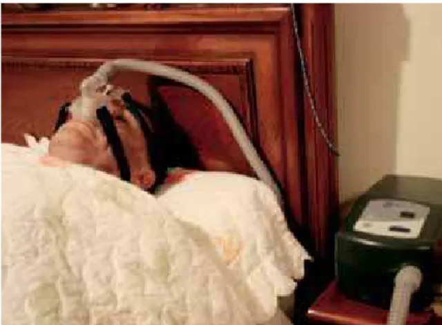
The nasal CPAP (“continuous positive airway pressure”) device is considered the gold standard in the treatment of OSAS14 (Fig 1). It consists of
a non-invasive method of continuous positive air pressure application in the airway, generating a continuous airflow that, through a flexible tube, reaches a nasal mask that is adjusted on the face with straps. Thus, a pneumatic cushion is created
in its interior that tends to dislocate the soft pal-ate in the direction of the lingual base and dilpal-ate the sectional area of the whole pharynx.13 While
it is an extremely effective treatment, there are problems with the adhesion of positive pressure devices and their acceptance in the long term.
Oral Appliances (OA) are devices used in the oral cavity during sleep, with the objective to pre-vent the collapse between the tissues of the oro-pharynx and the lingual base, i.e., the obstruction of the upper airway.2
OA constitute an effective and well-accepted form of treatment, and they have been an increas-ing line of treatment of OSAS and snorincreas-ing for more than 20 years. Today there are more than 80 described types of OA, which specifically fit into the categories of lingual retainers and mandibular repositioners. Only some have been approved by the FDA (Food and Drugs Administration), and there are few controlled studies available. There are different types of OA, differentiated by man-ufacturing method (prefabricated or made in a laboratory), retention, titration of the mandibu-lar position, anterior vertical opening, freedom of mandibular movement and construction material, among others. The effectiveness of the OA seems to be related to some of these aspects. When the precautions related to these factors are not
observed during the planning of the device, there is a greater probability of side effects, including those related to temporomandibular joint (TMJ) and occlusal alterations, as well as a reduction in adherence to the treatment.2
Treatment with OA is prescribed in patients with primary snoring and mild OSAS, as well as in cases of moderate or severe OSAS in which there was intolerance or refusal of CPAP use, surgical contraindication or the need for a short-term sub-stitute therapy.7,17
The objective of the present study was to evaluate the performance of this new design of OA for OSAS, while attempting also to compare responding and non-responding patients with the use of this mode of treatment.
THE INTRA-ORAL DEVICE
The Brazilian Dental Appliance (BRD) was developed from the experience of orthodon-tics researchers in the area of sleep medicine at UNIFESP-EPM and UFC who had previously used numerous other devices, mainly North American and Canadian, many of which had important limi-tations, high costs or a lack of consistent studies endorsing their use in patients with OSAS.
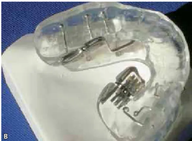
The BRD is an adjustable mandibular repo-sitioning OA (Fig 2, A and B). It is comprised
A B
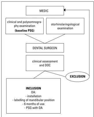
FIGURE 4 - Study design.
MEDIC
DENTAL SURGEON clinical and
polysomnogra-phy examination
(baseline PSG)
clinical assessment and DOC
EXCLUSION
INCLUSION
OA: - installation - labelling of mandibular position
- 6 months of use - PSG with OA
otorhinolaringological examination
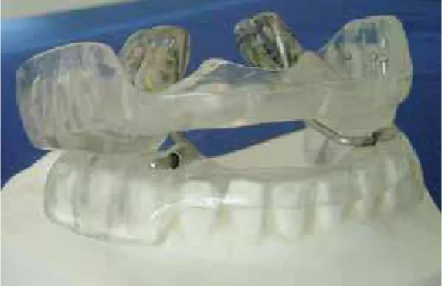
of retaining clamps on the posterior teeth and two acrylic support bases (one superior and an-other inferior) covering all teeth externally and internally (anterior and posterior). It also pos-sesses two independent (one right and another left in the posterior palate region of the superi-or acrylic base) expansion mechanisms (screws located with their long axle in the anterior-pos-terior direction). Two independent palate rods (one right and another left) protrude from these expansion mechanisms and are inserted inferi-orly into two small tubes located in the ante-rior portion (distal of the infeante-rior canines) of the inferior acrylic support base. This proposed design allows successive advances in the man-dibular position without, however, impeding lateral mandibular movements. That is, with the device in position, even when the jaw is in an anterior position, the patient can make lateral movements and a small buccal opening (Fig 3).
METHODOLOGY
This prospective study included 50 patients with OSAS prescribed for treatment with an OA, following the study design (Fig 4). All the patients had been previously diagnosed by a standardized polysomnography examination in the Department of Psychobiology at UNIFESP (Universidade Federal de São Paulo) and the Universidade Federal do Ceará.
The inclusion criteria consisted of patients with mild and moderate OSAS, aged between 25 and 70 years, with body mass index (BMI)
values less than 30 Kg/m2 and with at least 10
teeth in each dental arch. Excluded patients were comprised of those who had undergone previous surgeries for OSAS, CPAP users, those currently using any drug that could interfere with the sleep architecture, shift workers and patients with important anatomical alterations in the superior airway, such as deviated septum, turbinate and/or tonsil hypertrophy.
Volunteers who were diagnosed with extensive
periodontal disease, severe temporomandibular joint disorder or dental units with insufficient anat-omy for the necessary OA retention were excluded from the study.
FIGURE 5 - BRD device used in the study. Odontological documentation (DOC)
All of the patients underwent a basic orth-odontic documentation, consisting of panoramic x-ray, cephalometric analysis, intra and extra-buccal photographs and study models.
Polysomnography (PSG)
All of the patients were subjected to a baseline polysomnography (PSG) for OSAS diagnosis and another one with the device (BRD) in the man-dibular position of maximum comfortable protru-sion. The average time between the first and the second polysomnographies was six months.
The full night of PSG was performed in a sleep laboratory using a system of 13 channels, which included the electroencephalogram, electrocar-diogram, electrooculogram, electromyogram of the submentonian muscles and tibialis anterior, nasal and oral airflow (measured by thermistor and/or nasal cannula), thoracic and abdominal movements, body position (supine) and oxyhe-moglobin saturation measured by pulse oximetry.
Clinical and laboratory odontologic procedures
Impressions of the dental arches were made with alginate type II and immediately cast with special plaster type IV. The record of the ini-tial mandibular position was obtained with the “George Gauge”2 device and condensation
sili-cone (dense). The plaster models and records were then used for the manufacture of an indi-vidualized BRD device. All devices in the study were made of thermopolymerizable acrylic, with independent upper and lower parts and retention obtained by interproximal clips placed between all the posterior teeth. The connection between the two arches was made by two expanders posi-tioned at the palate region, just below the cervi-cal region of the superior molars. The purpose of these expanders is to allow the advancement of the mandible to move gradually over 11 mm, in 44 increments of 0.25 mm (Fig 5).
To obtain the mandibular position of maxi-mum comfortable protrusion, progressive adjust-ments were made in the expanders of the devices during a period of three to four 4 months, ac-cording to the presence of snoring and additional symptoms (drowsiness, fatigue, fragmented or non-restorative sleep).
A limit of 70% of maximum mandibular pro-trusion was observed.29 At each consultation, the
patients were asked about episodes of nasal ob-struction and weight change.
Statistical Analysis
The Kolmogorov-Smirnov test for normality was applied and was followed by the paired t-test for comparison between the baseline and OA, in cases when the baseline data displayed a normal distribution, or by the Wilcoxon test when the distribution was not normal.
BOX 1 - Percentage of success, partial success and failure for the total sample treated with the OA (BRD).
n %
success 28 54
partial success 19 38
failure 03 06
RESULTS
The data are presented as means and standard deviations. The sample was comprised of 33 men and 17 women with a mean age of 48.6 ± 12.3
and a mean BMI of 26.0 ± 2.8 Kg/m2. The mean
of the final protruding mandibular position was 8.8 ± 1.3 mm.
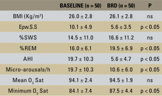
The results of the comparison between the baseline (initial) and the with-OA measurements for all patients are shown in Table 1 and, specifi-cally in relation to the number of apneas and hy-popneas per hour of sleep (AHI), are illustrated in Graph 1. Statistically significant improvements were noted in several of the parameters studied, like the reduction in sleepiness measured by the Epworth scale, increased percentage of REM sleep, decrease in AHI, increased minimum oxyhemoglo-bin saturation as well as the decrease in the number of micro-arousals per hour of sleep (Table 1).
In 28 patients (56% of the sample), the AHI normalized with the use of the OA, i.e., it was less than 5 events/hour (success); in 19 patients (38% of the sample), the AHI was more than 5 events/ hour with the use of the OA but decreased over 50% compared to the baseline condition (partial success); and in 3 patients (6% of the sample), the AHI was reduced less than 50% from the baseline or not reduced (failure) (Box 1).
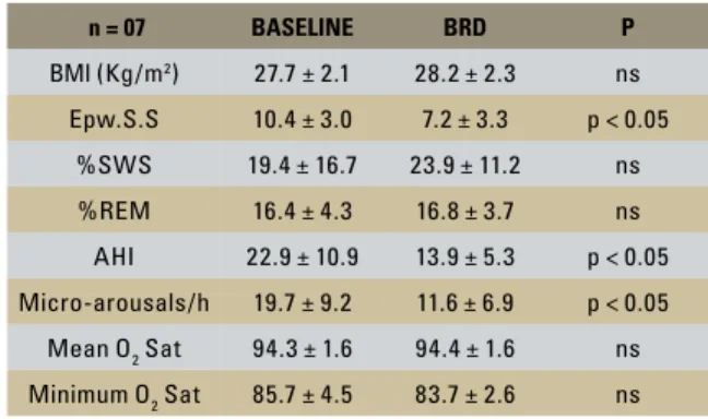
According to the AHI results, the patients were divided into two groups: good and poor re-sponders to the treatment. A patient was consid-ered a good responder when the AHI with OA reduced more than 50% from the baseline and was lower than 10 events/hour (n = 43); other-wise, they were considered as poor responders (n = 7) (Tables 2 and 3). It is shown in Table 2 that there were statistically significant improvements in various polysomnographic parameters evalu-ated in isolation when studying the group of good responders, but it is also important to note that even the group of poor responders showed signifi-cant improvement in sleepiness, the AHI and the number of micro-arousals during sleep (Table 3).
A comparison of the results between good and poor responders showed that those who re-sponded poorly to treatment had a greater body mass index (p < 0.05), including weight gained during treatment (Table 4, Graph 2, A and B). On the other hand, two other parameters showed a
GRAPH 1 - Baseline AHI and AHI with the use of the OA for all patients. 70
60
50
40
30
20
10
0
-10
baseline AHI AHI OA
baseline AHI and AHI with OA
TABLE 1 - Comparison between the baseline parameters and those with use of the OA (BRD) (mean ± standard deviation).
BASELINE (n = 50) BRD (n = 50) P
BMI (Kg/m2) 26.0 ± 2.8 26.1 ± 2.8 ns
Epw.S.S 10.1 ± 4.9 5.6 ± 3.5 p < 0.05
%SWS 14.5 ± 11.0 16.6 ± 11.2 ns
%REM 16.0 ± 6.1 19.5 ± 6.9 p < 0.05
AHI 19.7 ± 10.3 5.6 ± 4.7 p < 0.05
Micro-arousals/h 19.7 ± 10.3 10.6 ± 6.0 p < 0.05
Mean O2 Sat 94.1 ± 2.4 94.5 ± 1.9 ns
Minimum O2 Sat 84.1 ± 7.4 87.5 ± 4.4 p < 0.05
BMI = body mass index (kg/m2); Epw.S.S = Epworth Sleepiness Scale;
%SWS = % slow-wave sleep; % REM = % REM sleep; AHI = number of apnea and hypopnea events per hour of sleep; Mean O2 Sat = mean
oxy-hemoglobin saturation; Minimum O2 Sat = minimum oxyhemoglobin
tendency to differentiate, although not in a statis-tically significant manner; these were age (greater in poor responders) and the severity of OSAS (more severe in poor responders), the latter being insignificant for AHI and oxyhemoglobin satura-tion. It is noteworthy that the final position (pro-trusion) with the OA was not different between responders and non-responders (Table 4).
TABLE 2 - Comparison between the baseline parameters and those with the use of the OA (BRD) (mean ± standard deviation) in the group of good responders.
n = 43 BASELINE BRD P
BMI (Kg/m2) 25.7 ± 2.8 25.8 ± 2.8 ns
Epw.S.S 10.1 ± 5.2 5.3 ± 3.5 p < 0.05
%SWS 13.7 ± 9.9 15.4 ± 10.9 ns
%REM 15.9 ± 6.4 19.9 ± 7.2 p < 0.05
AHI 19.2 ± 10.2 4.3 ± 2.9 p < 0.05
Micro-arousals/h 19.7 ± 10.5 10.5 ± 6.0 p < 0.05
Mean O2 Sat 94.1 ± 2.5 94.6 ± 2.0 ns
Minimum O2 Sat 83.8 ± 7.8 88.1 ± 4.3 p < 0.05
BMI = body mass index (kg/m2); Epw.S.S = Epworth Sleepiness Scale;
%SWS = % slow-wave sleep; % REM = % REM sleep; AHI = number of apnea and hypopnea events per hour of sleep; Mean O2 Sat = mean
oxy-hemoglobin saturation; Minimum O2 Sat = minimum oxyhemoglobin satu-ration, ns = not statistically significant.
TABLE 3 - Comparison between the baseline parameters and those with the use of the OA (BRD) (mean ± standard deviation) in the group of poor responders.
n = 07 BASELINE BRD P
BMI (Kg/m2) 27.7 ± 2.1 28.2 ± 2.3 ns
Epw.S.S 10.4 ± 3.0 7.2 ± 3.3 p < 0.05
%SWS 19.4 ± 16.7 23.9 ± 11.2 ns
%REM 16.4 ± 4.3 16.8 ± 3.7 ns
AHI 22.9 ± 10.9 13.9 ± 5.3 p < 0.05
Micro-arousals/h 19.7 ± 9.2 11.6 ± 6.9 p < 0.05
Mean O2 Sat 94.3 ± 1.6 94.4 ± 1.6 ns
Minimum O2 Sat 85.7 ± 4.5 83.7 ± 2.6 ns
BMI = body mass index (kg/m2); Epw.S.S = Epworth Sleepiness Scale;
%SWS = % slow-wave sleep; % REM = % REM sleep; AHI = number of apnea and hypopnea events per hour of sleep; Mean O2 Sat = mean
oxyhemoglobin saturation; Minimum O2 Sat = minimum oxyhemoglobin saturation, ns = not statistically significant.
TABLE 4 - Comparison between good and poor responders.
GOOD RESPONDERS
POOR
RESPONDERS P
AGE 47.5 ± 11.6 55.3+15.4 ns
baseline BMI 25.7 ± 2.8 27.7 ± 2.1 p < 0.05
BMI w/ OA 25.8 ± 2.8 28.2 ± 2.3 p < 0.05
baseline
Epw.S.S 10.1 ± 5.2 10.4 ± 3.0 ns
baseline AHI 19.2 ± 10.2 22.9 ± 10.9 ns
baseline
mi-cro-arousals 19.7 ± 10.5 19.7 ± 9.2 ns
baseline
mean O2 Sat 94.1 ± 2.5 94.3 ± 1.6 ns
baseline
mini-mum O2 Sat 83.8 ± 7.8 85.7 ± 4.5 ns
OA
advance-ments (mm) 8.8 ± 1.3 8.6+1.9 ns
ns = not statistically significant.
good poor
Box & Whisker Plot baseline BMI good x baseline BMI poor
mean mean ± SD mean ±1,96*SD
A
GRAPH 2 - A) Baseline BMI for the good and poor response groups.
B) BMI with the OA for the two groups. good poor
Box & Whisker Plot BMI OA good x BMI OA poor
B
DISCUSSION
The mandibular repositioners for OSAS and snoring treatment may be of two subtypes: ad-justable and non-adad-justable. This latter category is on the decline due to difficulty of use and worse patient adherence to use. Of the OA, those that seem to be more effective are those of progressive or adjustable mandibular advancement, showing optimal retention as much on the maxilla as on the mandible, in addition to increasing comfort due to a reduced size.8
The differences in the BRD compared to other devices are that it contains a buccal opening and that there are two joining elements between the arches (two expansion mechanisms) positioned on the internal part of the superior arch. Devices that only possess retention in the maxilla cannot maintain a stable mandibular position,2
allow-ing buccal openallow-ing durallow-ing sleep and a clockwise turning of the mandible that may adversely in-fluence the pharyngeal dimensions. The fact that there are two independent expanders causes the device to position the lower arch in a more stable manner, allowing an anterior position without permitting much buccal opening. Making these devices with a single expander permits greater opening, which does not allow stable mandibu-lar positioning, letting the tongue obstruct the superior airflow passage. Furthermore, a single expander does not allow asymmetric mandibu-lar advancements. In devices made with two ex-panders positioned externally, the arches have important limitations to mandibular freedom in the lateral direction and the possibility of injury to the jugal mucosa.
This new design, by incorporating two internal expanders, permits asymmetric activation when necessary without impeding small lateral mandib-ular movements during sleep. Besides this, it does not cause injuries to the dorsal tongue.
The design of the BRD was developed to ad-dress these issues, as the device provides a stable mandibular position both vertically (opening) and
in the anterior-posterior direction (mandibular protrusion). It also promotes greater comfort, as it is individualized, measurable and extremely ver-satile with respect to the possibility of progressive mandibular advancements, making its use more physiological.
The results of 30 different studies in 456 pa-tients with OSAS and 224 with snoring during a period of 10 years (1982-1992) shows that intra-oral devices have the potential for signifi-cant airway increases, with a 54.3% reduction in the means of OSAS indices and an improve-ment of snoring in 87.5% of cases.23 In 1995,
Nowara et al20 selected 21 publications with
which they reviewed the results of 320 patients treated for OSAS and snoring with OAs. They observed that snoring was completely eliminat-ed in almost all patients and that others showeliminat-ed at least great improvements. For OSAS, there was an improvement of 60% of the apnea-hy-popnea index, AHI, on average (AHI before = 47 and after = 19), and approximately half of patients achieved an AHI <10, which is consid-ered successful by many authors.20 Liu et al19
observed a mean reduction in AHI from 40.3 to 17.1 and an insignificant mean improvement
in SaO2 from 76% to 80% with the KlearwayTM
device. However, it is noted that these results are not uniform, with some patients responding very quickly to therapy and others in whom the results are not as encouraging. This can be at-tributed to the lack of standardization of these studies, mainly in relation to selected samples and the process for evaluating results.
In the present study, there was a careful selec-tion of those individuals who actually had a need for OA therapy, and the monitoring of the indi-viduals followed the updated guidelines of the Brazilian consensus of snoring and apnea.7 For this
of slow-wave sleep and REM sleep). Valid and extremely important clinical criteria such as the Epworth Sleepiness Scale were also used. Statisti-cally significant improvements were observed in several evaluated parameters, like the reduction of sleepiness via the Epworth scale, increased per-centage of REM sleep, decreased apnea-hypopnea index, increased minimum oxyhemoglobin satu-ration as well as the decrease in the number of micro-arousals per hour of sleep (Table 1). These findings corroborate the research of Bernhold and Bondemark,6 whom reported an improvement in
SaO2 and a decrease in daytime sleepiness in the majority of their patients with OA. There was an observed improvement in the sleep quality of the patients, with increased percentages of REM stage, but little influence on the duration of NREM sleep,
as also found by Henke; Frantz and Kuna.16
How-ever, Rose et al24 reported that the sleep
architec-ture did not significantly change during their study with the Karwetzky-type activator OA.
With regard to obstructive events per hour of sleep (AHI), the results showed that in 56% of the sample the AHI with OA normalized, i.e., it was less than 5 events/hour (success); in 38% of the sample, the AHI was above 5 events/hour with the OA, but it had reduced more than 50% compared to baseline (partial success); and in 6% of the sample the AHI was reduced less than 50% from baseline or not reduced (failure). Generally, the effectiveness is reported based on an improve-ment in rates reaching 50% to 80% of those ob-tained initially, but there is still a big controversy on how to standardize the rates of success or the effectiveness of therapy.19
When the patients were separated into good responders (AHI with OA reduced more than 50% and less than 10 events/hour) and poor re-sponders to treatment (AHI with OA greater than 10 events/hour), it was found that forty-three pa-tients fell into the first group and seven fell into the second group. Significant improvements in the majority of the evaluated parameters were
detected in the group of good responders (Table 2), but it is worth mentioning here that even the group of poor responders showed significant im-provements in drowsiness, the AHI and number of micro-arousals during sleep (Table 3).
Liu et al19 studied patients with OSAS divided
into three groups according to the degree of im-provement in AHI: a group with good responses (reductions of AHI > 75%), a group with moder-ate responses (reductions in AHI from 25 to 75%) and a group with poor responses (reductions in AHI < 25%) in relation to treatment with intra-oral devices. This study showed that there was a significant difference in age between the group with poor response and the other groups. In older patients and those with higher BMI values, the OA was less effective. The study presented here also found a greater body mass index, including weight gain during treatment (Table 4, Graph 2, A and B), in patients who responded poorly to treat-ment with OA. There also seems to be a trend of greater age among the poor responders.
An important aspect is that the final position (protrusion) with the OA was not different be-tween the responders and non-responders. This presupposes that extreme mandibular advance-ments, without other criteria, will not improve the effectiveness of intra-oral devices. Exaggerated increases in the amount of mandibular advance-ment do not seem to have a great influence on the
improvement of OSAS.29
Intra-oral devices offer the most practical and logical way to start a treatment for the ma-jority of OSAS cases. A well-prescribed device that is properly manufactured and periodically monitored is effective, does not represent major expenses for patients and is easily accepted by most of them, while also being associated with other treatment modalities.9,12 The device tested
CONCLUSION
With the utilized therapy, there were signifi-cant improvements in the sleepiness scale, AHI, micro-arousals and minimum oxyhemoglobin saturation. Elevated Body Mass Index appears to unfavorably affect the performance of the device under study, suggesting that most patients who
are obese or who gain weight during treatment may become an unsuccessful group for this thera-peutic approach to OSAS.
Submitted: November 2008 Revised and accepted: February 2009
1. Adams N, Strauss M, Schluchter M, Redline S. Relation of measures of sleep-disordered breathing to neuropsy-chological functioning. Am J Respir Crit Care Med. 2001 Jun;163(7):1626-31.
2. Almeida FR, Dal-Fabbro C, Chaves Jr CM. Síndrome da apnéia e hipopnéia obstrutiva do sono (SAHOS): tratamento com aparelhos intra-orais. In: Tufik, S. Medicina e biologia do sono. 1ª ed. São Paulo: Manole; 2008. p. 263-80.
3. American Academy of Sleep Medicine. Sleep-related breath-ing disorders in adults: recommendations for syndrome definition and measurement techniques in clinical research. The Report of an American Academy of Sleep Medicine Task Force. Sleep. 1999 Aug;22(5):667-89.
4. American Academy of Sleep Medicine. International clas-sification of sleep disorders. 2nd ed. Diagnostic and coding manual. Westchester: Illinois; 2005.
5. Bassiri AG, Guilleminault C. Clinical features and evaluation of obstructive sleep apnea-hypopnea syndrome. In: Kryger MH, Roth T, Dement WC. Principles and Practice of Sleep Medi-cine. 3rd ed. Philladelphia: W.B. Saunders; 2000. p. 869-78. 6. Bernhold M, Bondemark L. A magnetic appliance for
treatment of snoring patients with and without obstruc-tive sleep apnea. Am J Orthod Dentofacial Orthop. 1998 Feb;113(2):144-55.
7. Bittencourt LRA. Diagnóstico e tratamento da Síndrome da Apnéia Obstrutiva do Sono (SAOS)-Guia Prático. 1ª ed. São Paulo: Médica Paulista; 2008.
8. Chaves CM Jr, Dal-Fabbro C, Almeida FR. Aparelhos intrabu-cais de avanço mandibular para tratamento da apnéia obstru-tiva do sono. In: Dominguez GC. Nova visão em Ortodontia e Ortopedia Funcional dos Maxilares. 1ª ed. São Paulo: Ed. Santos; 2007. p. 355-60.
REFERENCES
9. Clark GT, Nakand M. Dental appliances for the treatment of obstructive sleep apnea. J Am Dent Ass. 1989; 118:611-9. 10. Davies SF, Iber C. Obstructive sleep apnea associated with
adult-acquired micrognathia from rheumatoid arthritis. Am Rev Respir Dis. 1983 Feb;127(2):245-7.
11. George PT. A new instrument for functional appliance bite registration. J Clin Orthod. 1992 Nov;26(11):721-3. 12. George PT. Treatment of snoring and obstructive
sleep apnea with a dental device. Gen Dent. 1993 Jul/ Aug;41(4):294-8.
13. Gozal D, Kheirandish L. Sleepiness and neurodegen-eration in sleep-disordered breathing: convergence of signaling cascades. Am J Respir Crit Care Med. 2005 Jun 15;171(12):1325-7.
14. Grunstein R, Sullivan C. Continuous positive airway pressure for sleep breathing disorders. In: Kryger MH, Roth T, De-ment WC. Principles and practice of sleep medicine. 3rd ed. Philladelphia: W.B. Saunders; 2000. p. 894-912.
15. Guilleminault C, Simmons FB, Motta J, Cummiskey J, Rosekind M, Schroeder JS, Dement WC. Obstructive sleep apnea syndrome and tracheostomy. Long-term follow-up experience. Arch Intern Med. 1981 Jul;141(8):985-8. 16. Henke KG, Frantz DE, Kuna ST. An oral elastic mandibular
advancement device for obstructive sleep apnea. Am J Respir Crit Care Med. 2000 Feb;161(2 Pt 1):420-5. 17. Kushida CA, Morgenthaler TI, Littner MR, Alessi CA, Bailey
25. Sakakibara H, Tong M, Matsushita K, Hirata M, Konish Y, Suetsugu S. Cephalometric abnormalities in non-obese and obese patients with obstructive sleep apnoea. Eur Respir J. 1999;13:403-10.
26. Shamsuzzaman AS. Obstructive sleep apnea: implication for cardiac and vascular disease. JAMA. 2003;290(14):1906-14. 27. Smirne S, Iannaccone S, Ferini-Strambi L. Muscle fiber type
and habitual snoring. Lancet. 1991;337:597-9.
28. Sullivan CE, Issa FG, Berthon-Jones M, Eves L. Reversal of obstructive sleep apnea by continuous positive airway pres-sure. Lancet. 1981;1:862-5.
29. Tegelberg A, Walker-Engström ML, Vestling O, Wilhelms-son B. Two different degrees of mandibular advancement with a dental appliance in treatment of patients with mild to moderate obstructive sleep apnea. Acta Odontol Scand. 2003;61(6):356-62.
30. Young T, Palta M, Dempsey J, Skatrud J, Weber S, Badr S. The occurrence of sleep-disordered breathing among middle-aged adults. N Engl J Med. 1993;328:1230-5.
Contact address
Cauby Maia Chaves Junior
Rua Tibúrcio Cavalcante, 2860, Dionísio Torres CEP: 60.125-101 – Fortaleza / CE
E-mail: [email protected]
18. Li KK, Riley RW, Powell NB, Kushida C, Troell RJ. Obstructive sleep apnea syndrome in the Asian patient. Annual meeting of the American Academy of Otolaryngology-Head and Neck Surgery Foundation. New Orleans (LA); 1999. 19. Liu Y, Lowe AA, Fleetham JA, Park YC. Cephalometric and
physiologic predictors of the efficacy of an adjustable oral appliance for treating obstructive sleep apnea. Am J Orthod Dentofacial Orthop. 2001 Dec;120(6):639-47.
20. Nowara WS, Lowe A, Wiegand L, Cartwright R. Oral appli-ances for the treatment of snoring and obstructive sleep ap-nea: a review. An Amer Sleep Disorders Association Review. 1995; 18(6):501-11.
21. Partinen M, Jamieson A, Guillleminault C. Long term outcome for obstructive sleep apnea syndrome patients. Mortality Chest. 1988;94:1200-4.
22. Robinson A, Guilleminault C. Obstructive sleep apnea syn-drome. In: Chokroverty S. Sleep disorders Medicine: basic science, technical considerations, and clinical aspects. 2nd ed. Woburn: Butterworth Heinemann; 1999. p. 331-54. 23. Rogers RR, Lowe AA. Oral appliances for the management
de snoring and obstructive sleep apnea. 2nd ed. Wexford: Sleep Disorders Dental Society; 1996.