Fatima Dumas Cintra, Renata Pimentel Leite, Luciana Julio Storti, Lia Azeredo Bittencourt, Dalva Poyares, Laura de
Siqueira Castro, Sergio Tufik, Angelo de Paola
Universidade Federal de São Paulo - Unifesp, São Paulo, SP − Brazil
Mailing Address: Fatima Dumas Cintra •
Alameda Taurus, 146, Residencial Genesis I, Alphaville. Postal Code 06543-670. Santana de Parnaíba, SP – Brazil E-mail: fatimacintra@cardiol.br; fatimadc@einstein.br
Manuscript received December 6, 2013; revised manuscript June 26, 2014; accepted July 4, 2014
DOI: 10.5935/abc.20140142
Abstract
Background: The mechanisms associated with the cardiovascular consequences of obstructive sleep apnea include abrupt changes in autonomic tone, which can trigger cardiac arrhythmias. The authors hypothesized that nocturnal cardiac arrhythmia occurs more frequently in patients with obstructive sleep apnea.
Objective: To analyze the relationship between obstructive sleep apnea and abnormal heart rhythm during sleep in a population sample.
Methods: Cross-sectional study with 1,101 volunteers, who form a representative sample of the city of São Paulo. The overnight polysomnography was performed using an EMBLA® S7000 digital system during the regular sleep schedule of the individual. The electrocardiogram channel was extracted, duplicated, and then analyzed using a Holter (Cardio Smart®) system.
Results: A total of 767 participants (461 men) with a mean age of 42.00 ± 0.53 years, were included in the analysis. At least one type of nocturnal cardiac rhythm disturbance (atrial/ventricular arrhythmia or beat) was observed in 62.7% of the sample. The occurrence of nocturnal cardiac arrhythmias was more frequent with increased disease severity. Rhythm disturbance was observed in 53.3% of the sample without breathing sleep disorders, whereas 92.3% of patients with severe obstructive sleep apnea showed cardiac arrhythmia. Isolated atrial and ventricular ectopy was more frequent in patients with moderate/severe obstructive sleep apnea when compared to controls (p < 0.001). After controlling for potential confounding factors, age, sex and apnea-hypopnea index were associated with nocturnal cardiac arrhythmia.
Conclusion: Nocturnal cardiac arrhythmia occurs more frequently in patients with obstructive sleep apnea and the prevalence increases with disease severity. Age, sex, and the Apnea-hypopnea index were predictors of arrhythmia in this sample. (Arq Bras Cardiol. 2014; 103(5):368-374)
Keywords: Sleep Apnea Syndromes; Arrhythmias, Cardiac; Sleep; Sleep Apnea, Obstructive.
Introduction
Obstructive sleep apnea (OSA) is characterized by sleep fragmentation1 and repetitive hypoxia2 during sleep.
OSA is associated with a number of cardiovascular effects, such as hypertension3,4, metabolic syndrome5, and heart failure6.
OSA was recently associated with increased cardiovascular mortality7,8; however, the identification of the abnormality and
the institution of effective treatment with continuous positive airway pressure (CPAP) reduce the rate of fatal and nonfatal cardiovascular events7.
The mechanisms responsible for cardiovascular damage secondary to the obstructive apnea events are multiple,
but the final common pathway is autonomic involvement9.
Intermittent hypoxia10, sleep fragmentation11, and alterations
in intrapleural pressure12 directly affect the sympathetic and
parasympathetic autonomic nervous system.
Moreover, cardiac arrhythmia may be triggered by changes in the autonomic tone13. The vagal activity may
cause bradyarrhythmias, and sympathetic overactivity may favor various rhythm disturbances, including ventricular arrhythmias. The authors of this manuscript hypothesized that nocturnal cardiac arrhythmia occurs more frequently in patients with OSA. Hence, the aim of this study was to analyze the relationship between such arrhythmias and abnormal heart rhythm during sleep in a population sample.
Methods
Study population
a technique of three-stage cluster sampling was used15. In the
first stage, to ensure accurate socioeconomic representation, 96 of the 1,500 districts of the city used by the Brazilian Geography and Statistics Institute (IBGE) were proportionally selected among four homogeneous socioeconomic regions of São Paulo. The selected private households were permanently occupied. Thus, clinics, schools, and other commercial and noncommercial establishments were excluded. In the second stage, the families were selected by randomly selecting a household and subsequently skipping a specified number of houses in relation to the total number of selected households and dividing by a fixed number. Eleven families in each sector were selected in this manner. Each apartment, in the building, was considered a household and was counted from the top to the bottom floor. Finally, in the third sampling stage, all eligible residents in each selected household from the youngest to the oldest were listed. Pregnant or lactating women, people with physical or mental disabilities, individuals under 20 or over 80 years of age, and people who worked every night were excluded from the study. Substitutes were selected from the neighboring house, using the same random selection criteria described above. The rational design, sampling, and procedures used in the Epidemiological Study of Sleep of São Paulo have been described in a previous publication16.
The study protocol was approved by the Ethics Committee of the Federal University of São Paulo (CEP 0593/06) and registered at ClinicalTrials.gov under number NCT00596713. All volunteers read and signed the proposed consent form.
After signing the consent form, the patients were asked to attend the Sleep Laboratory for clinical evaluation and basal overnight polysomnography (PSG).
Polysomnography
The overnight PSG was performed in the sleep laboratory using a digital system (EMBLA® S7000, Embla Systems, Inc., Broomfield, CO, United States) during the regular sleeping hours of the individuals. The following physiological variables were monitored simultaneously and continuously: Electroencephalogram (EEG), electro-oculogram, electromyogram (submental region, tibialis anterior muscle, masseter region, and seventh intercostal space), electrocardiogram (ECG), detection of airflow (thermocouple and nasal pressure), abdominal and thoracic breathing efforts (by inductance plethysmography), snoring, body position, peripheral oxygen saturation (SO2), and heart rate. Four trained technicians visually labeled all the PSGs according to the standard criteria for investigating sleep17. The EEG and leg
movements were classified according to the criteria established in the manual of the American Academy of Sleep Medicine (AASM) for assessing sleep and associated events18. Apneas
were classified according to rules recommended for adults in the AASM manual, and hypopneas were labeled according to the alternative rules18. A PSG technician randomly selected
and reassessed 4% of the PSGs to verify the accuracy of sleep staging (concordance index of 93.3 ± 5.1, Kappa 0.91 ± 0.03). The apnea-hypopnea index (AHI) was used to determine the presence (AHI > 5) and severity of OSA (mild: 5 < AHI < 15; moderate: 15 < AHI < 30; and severe AHI > 30).
Holter evaluation during polysomnography
An ECG channel was extracted from PSG, duplicated and then analyzed with a Holter system manufactured by Cardios® (Smart Cardio, Cardio Systems, São Paulo, Brazil). The following characteristics of the ECG were analyzed: heart rate, QT and PR intervals, ventricular and atrial arrhythmias, and breaks. The complexity of the arrhythmias was described as follows: isolated, paired, or tachycardia. Anthropometric measurements were performed immediately before PSG and included body weight (kg), height (m), body mass index (BMI), and circumference (cm) of the neck and blood pressure.
Statistical Analysis
Version 17.0 of the Statistical Package for Social Science (SPSS) for Windows was used for data analysis. Descriptive statistics were used for the sample and group characteristics. The chi-square test was used to determine associations between subgroups. The general linear models (GLM) were used to analyze some variables. The a posteriori Tukey test was applied when necessary. A final adjustment of the logistic model was performed to analyze the main variables associated with the occurrence of cardiac arrhythmia. Data were expressed as median ± standard error for quantitative variables. Categorical variables are expressed as percentages.
A p value ≤ 0.05 was considered statistically significant.
Results
A total of 767 participants (461 men) with a mean age of 42.00 ± 0.53 years were included in the analysis. The ECG channel extraction and compatibility for the Holter system methods could not be performed in 334 subjects who were excluded from the analysis. The demographic characteristics of the sample are shown in Table 1. The presence of OSA, defined by AHI > 5, was observed in 37% of the population; 55.3% of these cases were classified as mild OSA and 44.7% had moderate or severe disease. The clinical and polysomnographic parameters of the patients with mild, moderate, or severe OSA and of the control group (without OSA) are shown in Tables 2 and 3, respectively. Sleep latency, percentage of REM sleep, and the periodic leg movement index were the only variables that did not reach significance when the groups were compared.
After controlling for potential confounder effects (age, BMI, smoking, diabetes, hypertension, and PSG parameters), age, sex, and AHI were independently associated with the occurrence of nocturnal cardiac arrhythmia (Table 5).
Discussion
The main finding of this study was the demonstration that nocturnal cardiac rhythm disorders occur more frequently in patients with OSA than in the general population, and that its prevalence increased with disease severity. The relationship between cardiac arrhythmias and sleep breathing disorders was assessed in several previous studies. Guilleminault et al19 using
a 24-h Holter monitoring in 400 OSA patients, showed that 48% had nocturnal cardiac events. Olmetti al.20 analyzed an
electrocardiographic recording during PSG and found nocturnal cardiac arrhythmia in 18.5% of the 257 consecutively selected patients with OSA.
The result of this study demonstrated that the prevalence of nocturnal cardiac arrhythmia was higher than that
previously described in the literature (92.3% of patients with OSA compared with 53.3% in the general population). Several explanations for this difference are possible. The use of a continuous monitoring system as a tool, with automatic detection of rhythm disturbances (Holter), may have detected the arrhythmias with greater accuracy, when compared with other methodologies used in clinical studies, such as 12-lead electrocardiography and electrocardiographic channel isolated from PSG. Furthermore, 37% of this population had an AHI > 5 events/h. This prevalence was also higher than that observed in other epidemiological studies21,
possibly because it included individuals with high BMI and age > 70 years22-24. Finally, the use of a gold standard tool
(GST) to detect breathing events during sleep can lead to a more effective screening of the population with OSA; and thus, to a better assessment of the prevalence of nocturnal arrhythmias.
The mechanisms involved in the pathophysiology of cardiac arrhythmias and OSA are probably multifactorial. In this study, we demonstrated that, in addition to age and gender, the AIH
Table 1 – Demographic characteristics of the sample
Characteristics Total (n = 767)
Mean age (years) 42.00 ± 0.53
Male gender (n) 352
Body mass index (kg/m2) 26.60 ± 0.18
Neck circumference (cm) 36.10 ± 0.17
Hypertension (%) 46.30
Diabetes (%) 7.30
Systolic blood pressure (mmHg) 124.40 ± 0.97
Diastolic blood pressure (mmHg) 79.33 ± 0.55
Smoking (%) 23.30
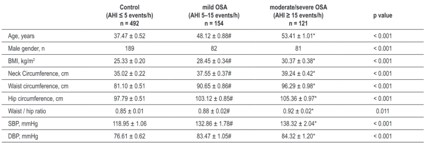
Table 2 – Clinical characteristics of patients with mild, moderate, and severe obstructive sleep apnea (OSA) and controls
Control
(AHI ≤ 5 events/h)
n = 492
mild OSA
(AHI 5–15 events/h) n = 154
moderate/severe OSA (AHI ≥ 15 events/h)
n = 121
p value
Age, years 37.47 ± 0.52 48.12 ± 0.88# 53.41 ± 1.01* < 0.001
Male gender, n 189 82 81 < 0.001
BMI, kg/m2 25.33 ± 0.20 28.45 ± 0.34# 30.37 ± 0.38* < 0.001
Neck Circumference, cm 35.02 ± 0.22 37.55 ± 0.37# 39.24 ± 0.42* < 0.001
Waist circumference, cm 81.10 ± 0.51 90.65 ± 0.86# 96.29 ± 0.98* < 0.001
Hip circumference, cm 97.79 ± 0.51 103.12 ± 0.85# 105.36 ± 0.97* < 0.001
Waist / hip ratio 0.85 ± 0.01 0.88 ± 0.02# 0.92 ± 0.02* 0.011
SBP, mmHg 118.95 ± 1.06 132.86 ± 1.78# 138.32 ± 2.04* < 0.001
DBP, mmHg 76.61 ± 0.62 83.47 ± 1.05# 84.32 ± 1.20* < 0.001
Table 3 – Polysomnographic parameters in patients with mild, moderate, and severe obstructive sleep apnea (OSA) and controls
Control
(AHI ≤ 5 events/h)
(n = 492)
Mild OSA
(AHI 5–15 events/h) (n = 154)
Moderate/severe OSA (AHI ≥ 15 events/h)
(n = 121)
p value
Sleep latency, min 16.20 ± 0.92 18.30 ± 1.57 16.81 ± 1.78 0.517
Total sleep time, min 350.20 ± 3.17 335.56 ± 5.38# 326.48 ± 6.11* 0.001
Sleep eficiency, % 83.60 ± 0.52 79.80 ± 0.89# 78.37 ± 1.01* < 0.001
Stage 1, % 4.30 ± 0.14 4.53 ± 0.24# 5.81 ± 0.27* < 0.001
Stage 2, % 53.90 ± 0.38 54.58 ± 0.64# 57.03 ± 0.73* 0.001
Stage 3, % 22.50 ± 0.33 21.97 ± 0.55# 18.97 ± 0.63* < 0.001
REM phase, % 19.20 ± 0.27 18.92 ± 0.42 18.19 ± 0.52 0.182
Awakening index, events/h 10.90 ± 0.38 16.53 ± 0.64# 27.54 ± 0.73* < 0.001
PLM Index, events/h 0.83 ± 0.27 1.59 ± 0.45 1.30 ± 0.51 0.308
AHI, events/h 1.40 ± 0.28 8.85 ± 0.48# 31.73 ± 0.54* < 0.001
Mean SaO2, % 95.90 ± 0.07 94.41 ± 0.12# 93.55 ± 0.13* < 0.001
SaO2 total time < 90%, min 1.80 ± 0.95 6.77 ± 1.61# 22.10 ± 1.82* < 0.001
Basal SaO2, % 96.50 ± 0.06 95.31 ± 0.10# 94.71 ± 0.11* < 0.001
Minimum SaO2, % 91.20 ± 0.18 85.93 ± 0.30# 81.49 ± 0.34* < 0.001
* Differs from mild OSA and controls; # differs from control (p < 0.01). AHI: Apnea-hypopnea index; REM: rapid eye movement; PLM: periodic leg movement index; SaO2: arterial oxygen saturation.
Table 4 – Distribution of nocturnal atrial and ventricular arrhythmias among patients with obstructive sleep apnea (OSA) - percentage of events
Control
(AHI ≤ 5 events/h)
(n = 492)
Mild OSA (AIH 5–15 events/h)
(n = 154)
Moderate/severe OSA (AHI ≥ 15 events/h)
(n = 121)
p value
General cardiac arrhythmia, % 53.30 77.30# 82.60# < 0.001
Isolated premature ventricular
complex, % 17.30 27.30# 39.70* < 0.001
Ventricular bigeminy, % 0.80 0.00 5.00* < 0.001
Coupled premature ventricular
complex, % 1.00 0.00 5.00* 0.001
Non-sustained ventricular
tachycardia, % 0.20 1.90 0.80 0.06
Isolate or coupled atrial
premature complex, % 43.90 64.30# 73.60# < 0.001
Non-sustained supraventricular
tachycardia, % 6.70 7.10 15.70* 0.005
Chronic and paroxysmal atrial
ibrillation, % 0.20 0.00 1.65* 0.03
Break (sinus breaks > 2.0 seconds and atrioventricular block),%
0.60 1.30 0.80 0.69
is an important factor related to nocturnal cardiac arrhythmia events. Hypoxia as a result of obstructive events is a potent stimulator for the sympathetic nervous system25. Fluctuations
in sympathetic and parasympathetic activity in patients with OSA may predispose them to the development of atrial and ventricular arrhythmias.
Another strong mechanism involved in the pathophysiology of cardiac arrhythmias and OSA is structural heart disease, which could favor the occurrence of cardiac arrhythmia. Oliveira et al26, using three-dimensional echocardiography,
showed that OSA induced an overload in the left atrium, resulting in remodeling. In addition, the effective use of CPAP can improve diastolic function of the left ventricle and the passive emptying of the left atrium27. A structural assessment
of the heart by echocardiography was not performed in this population, which may be considered a limitation of this study.
We did not observe differences in the occurrence of nocturnal cardiac breaks. Harbison et al28 performed Holter
ECG monitoring in 45 patients previously diagnosed with OSA syndrome and observed seven cases of cardiac break nocturnally, which were partially reversed with effective CPAP therapy. However, in randomized clinical trials to evaluate the role of CPAP in cardiac arrhythmias, there was no change after treatment with CPAP29, suggesting that other mechanisms,
not limited to apnea, trigger arrhythmia. The relationship between cardiac breaks, obstructive apnea events, and CPAP is still not fully understood and should be further analyzed in subsequent studies.
The absence of information on the occurrence of cardiac arrhythmia during the day and the variability in the frequency of arrhythmias are also limitations of this study, because the results may not accurately reflect the actual severity of rhythm
(observed by PSG) and the occurrence of cardiac arrhythmia (observed by Holter) in a population-based study can provide new insights into the treatment of arrhythmias as well as highlight the need to assess the sleep of these patients.
Conclusion
Nocturnal cardiac arrhythmias occurred more frequently in patients with obstructive sleep apnea, and the prevalence increased with disease severity. Age, sex and the apnea-hypopnea index were predictors of nocturnal cardiac arrhythmias in this sample.
Author contributions
Conception and design of the research: Cintra FD, Poyares D; Acquisition of data: Cintra FD, Leite RP, Storti LJ; Analysis and interpretation of the data: Cintra FD; Statistical analysis: Poyares D;Obtaining financing: Poyares D, Tufik S; Writing of the manuscript: Cintra FD, Bittencourt LA, Poyares D; Critical revision of the manuscript for intellectual content: Cintra FD, Leite RP, Bittencourt LA, Poyares D, Tufik S, Paola A.
Potential Conflict of Interest
No potential conflict of interest relevant to this article was reported.
Sources of Funding
This study was funded by FAPESP and AFIP.
Study Association
Table 5 – Adjusted logistic model of predictors of the occurrence of nocturnal cardiac arrhythmia
Beta p value Prevalence ratio CI 95%
Male gender 0.40 0.032 1.49 1.04 - 2.16
Age 0.06 < 0.001 1.06 1.04 - 1.08
BMI -0.01 0.80 1.00 0.96 - 1.03
Smoking 0.13 0.51 1.14 0.77 - 1.67
Diabetes 0.90 0.08 2.45 0.90 - 6.70
Hypertension -0.24 0.26 0.79 0.53 - 1.19
AHI 0.04 0.007 1.04 1.01 - 1.07
Total time of oxygen
saturation < 90% -0.01 0.18 0.99 0.98 - 1.00
Awakening index -0.02 0.19 0.98 0.96 - 1.01
Total time awake after
sleep onset 0.00 0.95 1.00 0.99 - 1.01
Total sleep time 0.00 0.15 1.00 1.00 - 1.01
Sleep eficiency -0.02 0.25 0.98 0.94 - 1.02
Constant -0.83 0.62 0.44
1. Kimoff RJ. Sleep fragmentation in obstructive sleep apnea. Sleep. 1996;19(9 Suppl):S61-6.
2. Peled N, Greenberg A, Pillar G, Zinder O, Levi N, Lavie P. Contributions of hypoxia and respiratory disturbance index to sympathetic activation and blood pressure in obstructive sleep apnea syndrome. Am J Hypertens. 1998;11(11 Pt 1):1284-9.
3. Nieto FJ, Young TB, Lind BK, Shahar E, Samet JM, Redline S, et al. Association of sleep-disordered breathing, sleep apnea, and hypertension in a large community-based study. Sleep Heart Health Study. JAMA. 2000;283(14):1829-36. Erratum in: JAMA. 2002;288(16):1985.
4. Peppard PE, Young T, Palta M, Skatrud J. Prospective study of the association between sleep-disordered breathing and hypertension. N Engl J Med. 2000;342(19):1378-84.
5. Bonsignore MR, Esquinas C, Barceló A, Sanchez-de-la-Torre M, Paternó A, Duran-Cantolla J, et al. Metabolic syndrome, insulin resistance and sleepiness in real-life obstructive sleep apnoea. Eur Respir J. 2012;39(5):1136-43.
6. Paulino A, Damy T, Margarit L, Stoïca M, Deswarte G, Khouri L, et al. Prevalence of sleep-disordered breathing in a 316-patient French cohort of sTabela congestive heart failure. Arch Cardiovasc Dis. 2009;102(3):169-75.
7. Marin JM, Carrizo SJ, Vicente E, Agusti AG. Long-term cardiovascular outcomes in men with obstructive sleep apnea-hypopnea with or without treatment with continuous positive airway pressure: an observational study. Lancet. 2005;365(9464):1046-53.
8. Marshall NS, Wong KK, Liu PY, Cullen SR, Knuiman MW, Grunstein RR. Sleep apnea as an independent risk factor for all-cause mortality: the Busselton Health Study. Sleep. 2008;31(8):1079-85.
9. Reynolds EB, Seda G, Ware JC, Vinik AI, Risk MR, Fishback NF. Autonomic function in sleep apnea patients: increased heart rate variability except during REM sleep in obese patients. Sleep Breath. 2007;11(1):53-60.
10. Gilmartin GS, Lynch M, Tamisier R, Weiss JW. Chronic intermittent hypoxia in humans during 28 nights results in blood pressure elevation and increased muscle sympathetic nerve activity. Am J Physiol Heart Circ Physiol. 2010;299(3):H925-31.
11. Pitson DJ, Stradling JR. Autonomic markers of arousal during sleep in patients undergoing investigation for obstructive sleep apnoea, their relationship to EEG arousals, respiratory events and subjective sleepiness. J Sleep Res. 1998;7(1):53-9.
12. Henderson LA, Woo MA, Macey PM, Macey KE, Frysinger RC, Alger JR, et al. Neural responses during Valsalva maneuvers in obstructive sleep apnea syndrome. J Appl Physiol (1985). 2003;94(3):1063-74.
13. Olmetti F, La Rovere MT, Robbi E, Taurino AE, Fanfulla F. Nocturnal cardiac arrhythmia in patients with obstructive sleep apnea. Sleep Med. 2008;9(5):475-80.
14. Kish L. Survey sampling. New York: John Wiley & Sons Inc; 1965.
15. Korn EL, Graubard BI. Analyses of health surveys. New York: John Wiley & Sons Inc, 1999.
16. Santos-Silva R, Tufik S, Conway SG, Taddei JA, Bittencourt LR. São Paulo Epidemiologic Sleep Study: rationale, design, sampling, and procedures. Sleep Med. 2009;10(6):679-85.
17. Rechtschaffen A, Kales A. A manual of standardized terminology: techniques and scoring system for sleep stages of human subjects. Los Angeles: Brain Information Service/ Brain Research Institute; 1968.
18. Iber C, Ancoli-Israel S, Chesson Jr A, Quan S. The AASM manual for the scoring of sleep and associated events: rules, terminology and technical specifications. Westchester: American Academy of Sleep Medicine; 2007.
19. Guilleminault C, Connolly SJ, Winkle RA. Cardiac arrhythmia and conduction disturbances during sleep in 400 patients with sleep apnea syndrome. Am J Cardiol. 1983;52(5):490-4.
20. Olmetti F, La Rovere MT, Robbi E, Taurino AE, Fanfulla F. Nocturnal cardiac arrhythmia in patients with obstructive sleep apnea. Sleep Med. 2008;9(5):475-80.
21. Redline S, Young T. Epidemiology and natural history of obstructive sleep apnea. Ear Nose Throat J. 1993;72(1):20-1, 24-6.
22. Young T, Palta M, Dempsey J, Skatrud J, Weber S, Badr S. The occurrence of sleep-disordered breathing among middle-aged adults. N Engl J Med. 1993;328(17):1230-5.
23. Durán J, Esnaola S, Rubio R, Iztueta A. Obstructive sleep apnea-hypopnea and related clinical features in a population-based sample of subjects aged 30 to 70 yr. Am J Respir Crit Care Med. 2001;163(3 Pt 1):685-9.
24. Ip MS, Lam B, Lauder IJ, Tsang KW, Chung KF, Mok YW, et al. A community study of sleep-disordered breathing in middle-aged Chinese men in Hong Kong. Chest. 2001;119(1):62-9.
25. Somers VK, Mark AL, Zavala DC, Abboud FM. Contrasting effects of hypoxia and hypercapnia on ventilation and sympathetic activity in humans. J Appl Physiol (1985). 1989;67(5):2101-6.
26. Oliveira W, Campos O, Bezerra Lira-Filho E, Cintra FD, Vieira M, Ponchirolli A, et al. Left atrial volume and function in patients with obstructive sleep apnea assessed by real-time three-dimensional echocardiography. J Am Soc Echocardiogr. 2008;21(12):1355-61.
27. Oliveira W, Campos O, Cintra F, Matos L, Vieira ML, Rollim B, et al. Impact of continuous positive airway pressure treatment on left atrial volume and function in patients with obstructive sleep apnoea assessed by real-time three-dimensional echocardiography. Heart. 2009;95(22):1872-8.
28. Harbison J, O’Reilly P, McNicholas WT. Cardiac rhythm disturbances in the obstructive sleep apnea syndrome: effects of nasal continuous positive airway pressure therapy. Chest. 2000;118(3):591-5.
29. Craig S, Pepperell JC, Kohler M, Crosthwaite N, Davies RJ, Stradling JR. Continuous positive airway pressure treatment for obstructive sleep apnoea reduces resting heart rate but does not affect dysrhythmias: a randomized controlled trial. J Sleep Res. 2009;18(3):329-36.