LETTERS
Arq Neuropsiquiatr 2012;70(8):637-644 642
Autoimmune atrophic gastritis presenting
as myelopathy in a young patient
Mielopatia como apresentação de gastrite atrófica autoimune em um doente jovem
Ana Teresa Carvalho Postal1, Sara França1, Dias Costa2, Joana Guimarães1,3
1Neurology Department, Vila Nova Gaia/Espinho Hospital Center, Vila Nova de Gaia, Portugal; 2Neuroradiology Department, São João Hospital Center, Oporto, Portugal;
3Faculty of Medicine, University of Oporto, São João Hospital Center, Oporto, Portugal.
Correspondence: Ana Teresa Carvalho Postal; Alameda Prof. Hernâni Monteiro 4200-319; Oporto - Portugal; E-mail: [email protected]
Conflict of interest: There is no conflict of interest to declare.
Received 30 January 2012; Received in final form 02 March 2012; Accepted 09 March 2012.
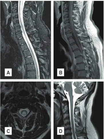
Fig. Spinal cord MRI: (A) extensive swelling of spinal cord, caused by vacuolization of myelin; (B) subtle contrast enhancement of posterior column; (C) cross-section images of spinal cord MRI: bilateral paired areas of hyperintensity affecting dorsal and lateral columns; (D) three months after treatment: significant improvement of lesions, with mild and no gadolinium enhancer T2 hyperintensity extending from bulbo-medular transition until C7 without spinal cord expansion.
A
C
B
D
Non-traumatic causes of extensive hyperintense spinal cord signal on T2-weighted images include a wide-ranging va-riety processes, such as tumors and associated disorders, in-lammation, infection, vasculopathy, congenital disorders or metabolic diseases [namely vitamin B12 deiciency (VBD)].
We present a 36 year-old male patient complaining from progressive sensory loss on both hands and sen-sation of chest and abdominal tightness, lasting for ive weeks. Neurological examination revealed a mild tetrapa-resis, glove and sock hyposthesia, C2 to T11 sensory im-pairment, vibratory anesthesia and proprioceptive errors of lower limbs. Cerebellar tests were normal. Romberg, Babinski and Lhermitte signs were absent. Laboratory tests revealed Hb 14.3 g/dL, mean corpuscular volume 104 fL. Renal and hepatic functions, iron study, copper levels and angiotensin conversion enzyme, and folic acid were nor-mal. Vitamin B12 level was <82 pg/mL (189–883 pg/mL). Serum immunology, virology and bacteriology were nega-tive. CSF study and brain MRI were unremarkable. Cervical spine MRI showed hyperintense spinal cord signal on T2-weighted images afecting mainly dorsal, but also lateral, col-umns between medulla oblongata and T11 levels, with subtle contrast enhancement (Fig A-C). Basing on clinical and lab-oratory examinations, the patient was evaluated for exten-sive myelopathy due to VBD, and it was performed an upper gastrointestinal examination which revealed an atrophic mu-cosa; histological examination showed chronic atrophic gas-tritis and intestinal metaplasia, excluding Helicobacter pylori
infection. Anti-parietal cells were negative, but anti-intrinsic factor antibodies were positive. Treatment was performed with IM cyanocobalamin replacement, 1,000 μg IM daily for a week, then weekly for four weeks and monthly afterwards. hree months later, there was a signiicant clinical recover, only remaining hypoesthesia of hand ingers. MRI performed at this time showed an exuberant improvement (Fig D).
Subacute combined degeneration (SCD) refers to degen-eration of the posterior and sometimes lateral columns of the
spinal cord usually as a result of VBD, which efects may not be appreciated until several years, since there is a signiicant body store of vitamin B121,2. Its absorption occurs via the ileal
LETTERS
provoked by gastric atrophy is the commonest cause of VBD. Autoimmune atrophic gastritis (AAG) is a special type of gas-tric atrophy characterized by serum antibodies antiparietal cells and/or anti-intrinsic factor. AAG was an unexpected di-agnosis since it is a relatively rare disease and the peak age of onset is 60 years, with only 10% of patients being <40 years of age. Myelopathy alone as clinical presentation is also a rare situation, occurring in about 12% of patients1. Concerning on
1. Healton EB, Savage DG, Brust JC, Garrett TJ, Lindenbaum J. Neurologic aspects of cobalamin deficiency. Medicine (Baltimore) 1991;70: 229-245.
2. Stabler SP, Allen RH, Savage DG, Lindenbaum J. Clinical spectrum and diagnosis of cobalamin deficiency. Blood 1990;76:871-881.
3. Shevell MI, Rosenblatt DS. The neurology of cobalamin. Can J Neurol Sci 1992;19:472-486.
4. Hughes JT. Pathology of the spinal cord. 2nd ed. In: Bennington J (Ed). Major problems in pathology. Vol 6. Philadelphia, Pa: Saunders, 1978;192-196.
References
imagiological indings, there was not only the involvement of the posterior column, but also of the lateral column, rarely in-volved in SCD4. Attention must be taken since AAG increases