Cop
yright
© ABE&M t
odos os dir
eit
os r
eser
vados
.
The PI3K signaling pathway mediates
the biological effects of leptin
A via de sinalização intracelular da PI3K medeia os efeitos biológicos da leptina
Jose Donato Jr.1, Renata Frazão1, Carol Fuzeti Elias1
SUMMARY
The activation of the leptin receptor recruits several intracellular signaling pathways, including the phosphatidylinositol 3-kinase (PI3K) pathway. While some of the leptin-induced signaling pathways, such as the JAK2/STAT3 pathway, induce cellular responses primarily through changes in gene expression, the PI3K pathway affects cellular properties more rapidly, through post-translational changes such as protein phosphorylation. Accordingly, several studies have shown that the PI3K pathway is required for the acute effects of leptin, such as a leptin-induced decrease in food intake. Leptin signaling through PI3K also affects the electrophysiological properties of neurons, including changes in their membrane potential and iring rates. In this review, we summarize the recent ad-vances in our understanding of the role played by the PI3K signaling pathway in controlling food intake and energy balance. In particular, we focus on the importance of the PI3K signaling pathway as a mediator of the effects of leptin on hypothalamic neurons. Arq Bras Endocrinol Metab. 2010;54(7):591-602
Keywords
Phosphatidylinositol; phosphatidylinositol 3-kinases; energy balance; insulin; Akt; hypothalamus
SUMÁRIO
A ativação do receptor de leptina recruta diversas vias de sinalização intracelular, entre elas a via da fosfatidilinositol 3-quinase (PI3K). Enquanto algumas dessas vias, como a sinalização pelo JAK2/ STAT3, induzem respostas celulares por meio de mudanças na transcrição gênica, a via da PI3K altera propriedades celulares de forma rápida, via fosforilação de proteínas. Em concordância, estudos mostraram que a via da PI3K é necessária para que a leptina induza seus efeitos agudos, como redução da ingestão alimentar, após administração de leptina. A ativação da PI3K pela lepti-na também afeta as propriedades isiológicas de neurônios, incluindo mudanças no potencial de membrana e no potencial de ação. Nesta revisão, resumimos os recentes avanços na compreen-são do papel desempenhado pela via de sinalização da PI3K no controle da ingestão alimentar e do balanço energético. Discutimos, principalmente, como a via da PI3K é importante para mediar os efeitos da leptina sobre os neurônios hipotalâmicos. Arq Bras Endocrinol Metab. 2010;54(7):591-602
Descritores
Fosfatidilinositol; fosfatidilinositol 3-quinase; balanço energético; insulina; Akt; hipotálamo
1 Division of Hypothalamic Research, Department of Internal Medicine, University of Texas Southwestern Medical Center, Dallas, Texas, United States
Correspondence to:
Jose Donato Jr.
5323 Harry Hines Blvd, Y6.206, Division of Hypothalamic Research, Department of Internal Medicine, University of Texas Southwestern Medical Center,
75390-9077 – Dallas, Texas, United States
Received on Aug/18/2009 Accepted on Sept/28/2010
IntRODUctIOn
P
hosphatidylinositol 3-kinases (PI3Ks) are heterodi-meric complexes composed of regulatory and cata-lytic subunits that recruit lipids as second messengers and control a wide variety of cellular functions, such as survival, growth, metabolism, and chemotaxis. The di-verse biological functions exerted by the PI3K signaling pathway have attracted attention because of the thera-peutic potential of PI3K-targeted drugs. Pharmacologi-cal compounds that target speciic PI3K subunits mightbe used for the prevention and/or treatment of inlam-mation and autoimmune diseases, hypertension, throm-bosis, cardiac dysfunctions, and cancer (1-3).
Cop
yright
© ABE&M t
odos os dir
eit
os r
eser
vados
.
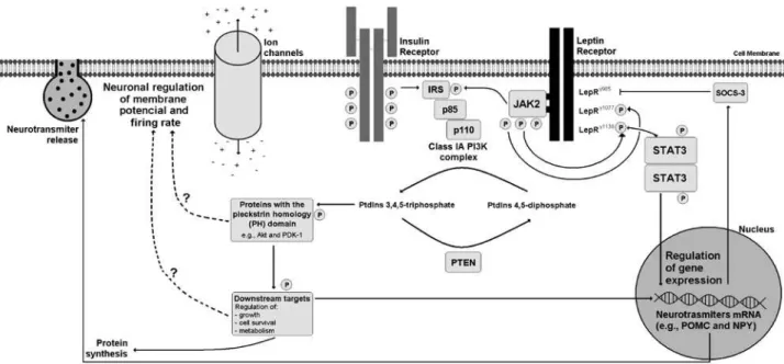
Figure 1. Scheme that illustrates intracellular signaling pathways recruited by leptin, particularly the PI3K pathway. Note that activation of leptin receptor (LepR) induces the subsequent phosphorylation and recruitment of Janus tyrosine kinase -2 (JAK2). JAK2 phosphorylates the LepRy1077 and LepRy1138 residues, and the insulin receptor substrates (IRS). The phosphorylation of LepRy1138 residue will recruit the signal transducer and activator of transcription -3 (STAT3) that under phosphorylation it dimerizes and is transported to the nucleus, controlling the transcription of target genes, including the suppressor of cytokine signaling-3 (SOCS-3). By its turn, SOCS-3 binds to the LepRy985 residue, inhibiting the LepR signaling. The insulin receptor also phosphorylates the IRS, which binds to the p85 regulatory subunits of Class IA PI3K, inducing the action of the PI3K complex, also composed of p110 catalytic subunits (α, β or δ). When activated, the Class IA PI3Ks phosphorylates the phosphatidylinositol (PtdIns) 4,5-diphosphate in the 3-hydroxyl group of the inositol ring, generating PtdIns 3,4,5-triphosphate. The activity of PI3Ks is counterbalanced by the enzyme phosphatase and tensin homologue (PTEN). Proteins that contain the pleckstrin homology (PH) domain are regulated by PtdIns 3,4,5-triphosphate and they are responsible to coordinate and convey the PI3K-dependent intracellular effects induced by leptin and insulin. These effects include the regulation of growth, cell survival, metabolism and chemotaxis through downstream targets. Regarding the effects of PI3K signaling in the regulation of energy balance, the induction of PI3K can affect the electrophysiological property of leptin-sensitive neurons, e.g., the membrane potential and fire rate, influencing the release of neurotransmitters. Besides, gene expression and protein synthesis are also regulated by downstream components recruited by PI3K signaling pathway. Interestingly, the mechanisms by which PI3K regulates ion channels properties are still unknown (dotted arrows).
The PI3K signaling pathway is induced by several growth factors and other hormones. For instance, ac-tivation of insulin receptor causes phosphorylation of tyrosine residues in insulin receptor substrate -1 and -2 (IRS-1 and IRS-2). This phosphorylation induces a ra-pid association between IRS-1 or IRS-2 and PI3Ks, cau-sing the subsequent activation of the PI3K complex (4). The PI3K-induced phosphorylation of PtdIns activates downstream targets, such as Akt (also known as protein kinase B or PKB) and phosphoinositide-dependent ki-nase-1 (PDK-1), which, in turn, convey and coordinate most of the intracellular effects induced by insulin.
More recently, the PI3K signaling pathway was sho-wn to play a role in the regulation of food intake and energy balance through hypothalamic neurons. Resem-bling many peripheral cells, neurons also respond to hormonal signals through the activation of intracellular pathways that regulate membrane potential, iring rate,
the synthesis and release of neurotransmitters, and gene expression. Insulin and leptin are important hormones that act in speciic populations of hypothalamic neurons, which ultimately control glucose homeostasis, food in-take, energy expenditure and sympathetic activity.
In this review, we summarize recent advances in the understanding of the role of the PI3K signaling pa-thway in the control of food intake and energy balance. In particular, we focus on the importance of the PI3K pathway as a mediator of leptin’s effects in hypothala-mic neurons.
LEPtIn AnD EnERGY BALAncE
Cop
yright
© ABE&M t
odos os dir
eit
os r
eser
vados
.
hyperphagic obesity, diabetes mellitus, and neuroendo-crine dysfunctions in rodents and humans (7,8). These monogenic mutations were initially described in mice (ob/ob, mutation in the leptin gene and db/db, mu-tation in the LepR gene) and were later shown to be recapitulated in humans. Importantly, leptin treatment in leptin-deicient humans or mice reduces food intake and body weight and restores their metabolic and neu-roendocrine functions (9-11).
Innumerous studies have shown that the hypotha-lamus is the primary target of leptin in regulating ener-gy balance, glucose homeostasis, and neuroendocrine function (12). The availability of leptin favors a re-duction in food intake and body weight. On the other hand, fasting, a condition of low leptin levels, causes an opposite effect that can be partially or completely rever-ted by leptin replacement (5,6,13). For example, mice fasted for 48 h show increased expression of the ore-xigenic neuropeptide Y (NPY) and a reduction in the expression of pro-opiomelanocortin (POMC), which is a precursor of the anorectic peptide alpha melanocyte-stimulating hormone (α-MSH). However, intraperito-neal (i.p.) treatment of leptin during fasting prevents the changes in the expression of POMC and NPY in the arcuate nucleus (ARH), an important hypothalamic area affected by leptin (13,14).
Alternative splicing of the LepR mRNA and/or post-translational processing generates at least ive isoforms of LepR (LepRa-e). Of the isoforms, LepRb (ObRb or long isoform) is considered the functional isoform, as upon leptin binding it induces a series of in-tracellular signaling cascades (15,16). LepRe does not carry a transmembrane domain and circulates as a lep-tin binding protein. The isoforms LepRa, LepRc, and LepRd have a short intracellular domain, compared to the LepRb isoform. As a consequence of incomplete intracellular domains, LepRa, LepRc, and LepRd are unable to activate intracellular pathways normally re-cruited by LepRb signaling (17). Because the focus of the present review is to discuss intracellular pathways recruited by leptin signaling, we will deine LepRb as LepR.
LepR is a member of the class I cytokine receptors. Upon leptin binding, LepR undergoes dimerization that culminates in a cascade of intracellular events. In most cases, this cascade is triggered by the phos-phorylation of a Janus tyrosine kinase (JAK) family member, known as JAK2. JAK2 is constitutively at-tached to LepR and becomes phosphorylated and
ac-tivated when leptin binds to its receptor (17). Three tyrosine resides in the intracellular domain of LepR, named LepRy985, LepRy1077, and LepRy1138, can become
phosphorylated after leptin signaling. Phosphorylation of LepRy985 reduces LepR signaling; therefore, this
residue is considered a site for negative feedback. On the other hand, the phosphorylation of the LepRy1077
and LepRy1138 residues is necessary for the subsequent
phosphorylation and recruitment of signal transducers and activators of transcription 5 and 3 (STAT5 and STAT3, respectively) (17).
The best-described signaling pathway recruited by leptin involves the coordinated activation of JAK2/ STAT3. STAT3 is a transcriptional factor that, upon phosphorylation, dimerizes and is transported to the nucleus, where it controls the transcription of target genes (Figure 1). The phosphorylated form of STAT3 (pSTAT3) has been used as a marker of leptin-responsi-ve neurons, whether the cells are activated or inhibited by leptin (18-20). One of the genes rapidly induced by leptin and regulated by pSTAT3 is the suppressor of cytokine signaling-3 (SOCS-3) (21). SOCS-3 is a family member of cytokine-inducible signaling inhi-bitors. It causes inhibition of LepR signaling by bin-ding to LepRy985 and blocking JAK2 activity (Figure 1).
Changes in SOCS-3 expression have been related to conditions of leptin resistance, like those observed du-ring diet-induced obesity. Accordingly, the hypothala-mic expression of SOCS-3 is increased in obese animals (21). Besides, the genetic deletion of SOCS-3 in spe-ciic populations of hypothalamic neurons increases the leptin sensitivity and reduces the susceptibility to diet-induced obesity (22).
To directly test the importance of the LepR/JAK2/ STAT3 signaling pathway on the biological effects of leptin, Bates and cols. (18) generated genetically mo-diied mice in which the LepRy1138 was substituted with
Cop
yright
© ABE&M t
odos os dir
eit
os r
eser
vados
.
In vitro studies also demonstrated that fast respon-ses, presumably too rapid to be mediated by changes in gene expression, are evoked by acute application of lep-tin in energy homeostasis-related neurons (23-26). For instance, Spanswick and cols. (24) demonstrated that leptin applied to hypothalamic slices decreases the i-ring rate of action potentials and the input resistance of glucose-responsive neurons located in the ARH and in the ventromedial nucleus of the hypothalamus (VMH). These effects were accompanied by a slow progressive hyperpolarization to a new equilibrium, 5 to 15 min after application. Importantly, these effects were obser-ved in wild-type animal models, but not in the obese Zucker rats, which have a mutant LepR isoform. The activation of ATP-sensitive potassium channel (KATP) was evidenced by the application of tolbutamide, a po-tassium channel blocker, which reversed the actions of leptin (24). Later, electrophysiological studies demons-trated that leptin mainly depolarized POMC neurons in 2 to 10 min, in a dose-response manner (27). The depolarization of POMC neurons was caused by a small inward current that reversed at about -20 mV, sugges-ting the involvement of a nonspeciic cation channel. In addition, leptin treatment decreased the frequency of GABA-mediated inhibitory postsynaptic currents by 25% in 30% of the POMC cells, indicating that it may act presynaptically to reduce GABA release, which was conirmed by the application of tetrodotoxin (27). Therefore, these results indirectly imply that other in-tracellular pathways faster than JAK2/STAT3 signaling pathway, which requires changes in gene expression to convey its signal, are responsible for the acute leptin-induced changes in neuronal activity.
PI3K SIGnALInG IS REQUIRED FOR tHE AcUtE
EFFEctS OF LEPtIn In FOOD IntAKE
Initial reports, showing that the activity of the PI3K signaling pathway in the brain is important for the re-gulation of food intake, employed pharmacologic inhi-bitors of PI3K, including wortmannin and LY294002. In a classic study, wortmannin or LY294002 was ad-ministered intracerebroventricularly (i.c.v.) to Wistar rats (28). These authors observed that i.c.v. injection of LY294002 did not change food intake or body weight after 4 h or 24 h of treatment. However, pretreatment with either wortmannin or LY294002 was effective in blunting the anorectic effect of i.c.v. injection of leptin. As evidence that LepR signaling recruits PI3K in
hypo-thalamic neurons, these authors also showed an increa-se in PI3K activity associated with IRS-2 at 30 min after i.p. injection of leptin (28). These results revealed that PI3K in the brain is required for the short-term effects of leptin on food intake regulation.
Additional studies have conirmed and extended these indings. Of note, it was shown that leptin-in-duced activation of PI3K in the hypothalamus recruits phosphodiesterase 3B (PDE3). PDE3 counterbalances the activity of adenylyl cyclases, which are responsible for the synthesis of the second messenger cAMP, whe-reas PDE3 induces cAMP clearance (29). Using i.c.v. administration of a PDE3 inhibitor, cilostamide, the re-duction in food intake and body weight observed after leptin treatment was prevented by the inhibition of the PI3K-PDE3 pathway.
As previously mentioned, leptin administration inhi-bits the expression of the orexigenic NPY. Leptin- res-ponsive NPY cells are found in the ARH and coexpress agouti-related peptide (AgRP) (5-7). The involvement of PI3K in this group of cells was investigated in rats subjected to a 52-h fast (30). The fasted rats showed increased NPY and AgRP mRNA levels, compared to ad libitum fed animals. As predicted, i.c.v. administra-tion of leptin over 12-h intervals blocked the fasting-induced increase in NPY and AgRP expression. Howe-ver, pretreatment with LY294002 blunted the effects of leptin in the expression of NPY and AgRP.
PI3K IntEGRAtES tHE EFFEctS OF LEPtIn AnD
InSULIn In tHE REGULAtIOn OF FOOD IntAKE
Insulin also acts in the brain to regulate food intake. For example, administration of insulin in rats causes a signi-icant reduction in food intake and body weight (31). This effect was completely prevented by the administra-tion of PI3K inhibitors, wortmannin and LY294002, one hour before insulin treatment (31). Furthermore, insulin treatment induces phosphorylation of IRS-1, IRS-2, and Akt in the hypothalamus and increases the immunoreactivity of PtdIns 3,4,5-triphosphate specii-cally in the ARH (31). These results suggest that PI3K is a common intracellular signaling pathway required for the hypothalamic effects of either leptin or insulin in food intake regulation.
Cop
yright
© ABE&M t
odos os dir
eit
os r
eser
vados
.
In this regard, Xu and cols. (25) generated a mouse model, which expresses the enhanced green luores-cent protein (EGFP) fused to the pleckstrin homology (PH) domain from Grp1 [EGFP-PH(Gpr1)]. Follo-wing PI3K generation of PtdIns 3,4,5-triphosphate, the EGFP-PH(Gpr1) complex translocates to the cell membrane, allowing the visualization of neurons in which the PI3K signaling pathway is activated (25). Using this reporter mouse, these authors showed that both leptin and insulin activate the PI3K pathway in POMC neurons. But, while insulin induces the activa-tion of PI3K in AgRP neurons, leptin shows an oppo-site effect. Interestingly, the action of insulin and leptin in POMC neurons, and of insulin in AgRP neurons, was shown to be direct in those cells. However, the inhibitory effect of leptin on AgRP neurons was blo-cked by inhibitors of synaptic transmission. These re-sults indicate that leptin recruits another population of neurons to affect PI3K activity in AgRP neurons (25).
PI3K SIGnALInG MEDIAtES LEPtIn’S EFFEctS
On GLUcOSE HOMEOStASIS
Experimental evidence also indicates that the PI3K signaling pathway in hypothalamic neurons mediates leptin’s effects on glucose homeostasis. Using stere-otaxic techniques, leptin receptor-deicient Koletsky (fak/fak) rats received adenoviral gene therapy to
in-duce the expression of LepR in the ARH (32). The virus-induced LepR signaling in mediobasal hypotha-lamic neurons markedly increased the insulin tolerance of the fak/fak rats. On the other hand, i.c.v.
adminis-tration of LY294002 attenuated the improvement in insulin tolerance of rats submitted to the adenoviral therapy, suggesting that the brain-mediated effects of leptin on glucose homeostasis involve induction of the PI3K signaling pathway. Accordingly, virus-induced expression of a constitutively active Akt in the hypo-thalamus recapitulated the insulin-sensitizing effects of adenoviral-induced LepR gene expression (32). Ho-wever, it is worth mentioning that this approach may have induced LepR expression in cells that normally do not express this receptor. Additionally, the degree of gene expression achieved is probably not similar to the physiological levels observed in wild-type rats. There-fore, although these results highlight a potential role of leptin-induced PI3K signaling in the hypothalamus to control glucose homeostasis, additional studies using alternative methods are necessary to establish the
phy-siologic relevance of PI3K signaling pathways in leptin-induced regulation of insulin sensitivity.
More recently, it was demonstrated that the effects of i.c.v. injection of leptin on improving insulin sen-sitivity in skeletal muscle is also dependent on PI3K signaling (33). Administration of LY294002 blocked leptin’s effect on improving glucose tolerance and on the phosphorylation of AMP-activated protein kinase (AMPK)/acetyl-CoA carboxylase pathway in the soleus muscle of rats. Therefore, the leptin-induced activation of the PI3K signaling pathway in the hypothalamus se-ems to modulate energy sensing pathways, e.g., AMPK signaling, in peripheral tissues, and ultimately control glucose homeostasis.
tHE PI3K SUBUnItS
One challenge in designing studies to investigate the physiologic relevance of PI3K signaling using geneti-cally modiied mouse models is that the PI3K complex is composed of several subunits encoded by different genes. So far, only a subset of the PI3K subunits has been targets of genetic manipulation to investigate the role played by PI3K signaling in the regulation of ener-gy balance. Three classes of PI3K have been described: Class I, Class II and Class III. Each class shows diffe-rent molecular structures and lipid substrate preferen-ces. Class I PI3Ks have great afinity to phosphorylate PtdIns 4,5-diphosphate into PtdIns 3,4,5-triphosphate. Class II PI3Ks phosphorylate PtdIns 4-phosphate into PtdIns 3,4-diphosphate. Finally, Class III PI3Ks phos-phorylate PtdIns into PtdIns 3-phosphate. The downs-tream targets change according to the domain activated by different 3-phosphoisositides. For example, PtdIns 3,4,5-triphosphate and PtdIns 3,4-diphosphate recog-nize, bind, and regulate proteins that contain the PH domain (Figure 1), whereas proteins that contain the phox homology (PX) domain and the FYVE domain are regulated by PtdIns 3-phosphate (34).
Cop
yright
© ABE&M t
odos os dir
eit
os r
eser
vados
.
the gene Pik3cd). The regulatory subunits consist of p85α, p55α, p50α (all three encoded by the gene Pi-k3r1), p85β (encoded by the gene Pik3r2), and p55γ (encoded by the gene Pik3r3). The Class IB PI3Ks are composed of the p110γ catalytic subunit (encoded by the gene Pik3cg) and the p101 regulatory subunit (en-coded by the gene Pik3r5). Class IA PI3Ks are recrui-ted by receptors that display tyrosine kinase activity and by Ras, whereas the Class IB PI3Ks are downstream of G protein-coupled receptors (GPCRs) and Ras (34). The insulin receptor has intrinsic tyrosine kinase acti-vity and, as mentioned, LepR signaling recruits JAK2. Therefore, it is likely that both hormones recruit prima-rily the Class IA PI3Ks. Nonetheless, it is not possible to rule out an involvement of leptin and insulin with other PI3K classes. The leptin- and insulin-induced ac-tivation of Class IA PI3Ks involves the phosphorylation of IRS-1 or IRS-2, which bind to the regulatory PI3K subunits and induce the activation of the PI3K complex (Figure 1).
The disruption of either catalytic or regulatory su-bunits can interfere with PI3K activity (Table 1). Ho-wever, the regulation of PI3K is very complex, and the consequences of manipulations in speciic subunits may generate unexpected phenotypes. One good example is the mouse models carrying genetic deletions of the p85α or p85β regulatory subunits. Despite the fact that PI3K is a signaling pathway important for insulin to regulate glucose homeostasis, the genetic disruption of genes that encode the p85 regulatory subunits in-creases insulin sensitivity and causes hypoglycemia and hypoinsulinemia (34-39).
To investigate the role of PI3K signaling in POMC neurons, Hill and cols. (26,40) generated a conditio-nal knockout mouse in which the gene encoding the p85α, p55α and p50α regulatory subunits was selec-tively disrupted in POMC cells. These authors showed that despite the fact that POMC-speciic Pik3r1 dele-tion did not change the leptin-induced depolarizadele-tion and increased iring rate in these cells (26), the female mice were resistant to a diet-induced obesity (40). Ne-vertheless, no protection against obesity induced by a high-fat diet was observed in male mice carrying the same mutation (40).
Hill and cols. (26) also crossed the POMC-speciic Pik3r1 knockout mice with animals that carry a global deletion in the gene that encodes the p85β regulatory subunit, generating mice lacking p85α and p85β Class IA PI3K regulatory subunits in POMC cells. These
au-thors observed that, in intact POMC neurons, leptin induces a depolarization that can be veriied by incre-ases in iring rate and inward current, as well as by a decrease in the whole-cell input resistance. However, in mice lacking the PI3K regulatory subunits in POMC cells, leptin failed to change the membrane potential and to induce inward current in those neurons (Table 2). Furthermore, the deletion of Pik3r1 and Pik3r2 genes in POMC cells prevented the reduction in food intake and body weight caused by acute i.c.v. injection of lep-tin (26). These data suggest that leplep-tin directly excites POMC neurons via a PI3K-dependent mechanism and that deletion of both Pik3r1 and Pik3r2 is required to completely disrupt PI3K activity in POMC neurons. Nonetheless, no changes were observed in the long-term regulation of body weight and metabolism (26). These results also suggest that PI3K is important to mediate the acute effects of leptin, but other signa-ling pathways, possibly JAK2/STAT3, are necessary for the long-term regulation of energy homeostasis. It is important to mention that the use of animals car-rying mutations in PI3K regulatory subunits in studies aiming to evaluate the role of PI3K in energy home-ostasis may produce ambiguous results (Table 1). As demonstrated before, mouse mutants of p85α or p85β subunits have a reduced body size and increased insulin sensitivity (34,35,39).
tHE cAtALYtIc SUBUnItS OF cLASS IA PI3Ks
A variety of genetic mouse models with deletions in the catalytic subunits of Class IA PI3Ks has been produced (Table 1). Mice carrying a disruption in the gene that encodes the p110δ catalytic subunit are viable and show no changes in the regulation of energy balance, but they present several immunological problems and abnormali-ties in B cells, T cells, and neutrophils (41). This pheno-type is in accordance with the sites of p110δ expression in the body. While p110α and p110β are ubiquitously distributed among all tissues, the p110δ mRNA is ex-pressed predominantly in leukocytes (42). Thus, consi-dering the brain as the primary target of leptin in energy homeostasis, the role played by the p110δ PI3K catalytic subunit in leptin signaling is likely negligible.
acti-Cop
yright
© ABE&M t
odos os dir
eit
os r
eser
vados
.
vity (p110αko mice). These mice showed reduced body
weight and body length, glucose and insulin intoleran-ce, higher levels of insulin and leptin, and an increased food intake and body fat mass (45). Using different approaches, other groups also showed that the p110α PI3K catalytic subunit is essential for proper growth factor signaling (46). Thus, the p110α PI3K catalytic subunit seems to be critical for metabolism regulation and growth (Table 1).
Recently, two studies speciically focused on the physiological role played by PI3K catalytic subunits in speciic populations of hypothalamic neurons (40,47). In these studies, the p110α subunit was selectively de-leted from POMC cells or from cells expressing the ste-roidogenic factor 1 (SF-1). In the brain, the expression of SF-1 is restricted to neurons located in the VMH, including those that express LepR (47). Deletion of p110α in POMC neurons, but not in SF-1 cells, increa-sed the body weight in mice consuming a regular chow diet. However, the deletion of p110α in either POMC or SF-1 cells increased the susceptibility to diet-induced obesity. The food intake remained unchanged, where-as decrewhere-ased energy expenditure wwhere-as observed in both conditional knockout models. Also, the p110α deletion in POMC neurons reduced insulin sensitivity without affecting the glucose tolerance of these mice (40). Similarly to the results observed after deletion of p85α
and p85β in POMC cells, the disruption of p110α in the VMH decreased the acute metabolic responses ob-served after i.c.v. leptin administration (47). Overall, these indings indicate that the activity of p110α in leptin responsive neurons is required for the regulation of energy balance and glucose homeostasis (Table 1). Furthermore, these studies demonstrated that body weight changes in mice carrying a conditional deletion of p110α were primarily caused by changes in energy expenditure, instead of food intake. In particular, Xu and cols. (47) showed that the lack of p110α in SF-1 cells blunted the acute increase in energy expenditure, usually observed after exposure to a high-fat diet. Thus, PI3K signaling in the hypothalamus potentially modi-ies the energy balance through the regulation of either food intake and/or energy expenditure.
Although the aforementioned information strongly indicates that the p110α catalytic subunit is essential for the regulation of energy metabolism, there is still a debate about the role played by other PI3K catalytic subunits. For example, using pharmacological inhibi-tors speciic for each PI3K catalytic subunit, Knight and
cols. (2) showed that only the p110α subunit is requi-red for the insulin-induced PI3K activation in culturequi-red cells, whereas the other catalytic subunits are dispensa-ble for the same effect. Nonetheless, p110β and p110δ maintained a basal level of PtdIns 3,4,5-triphosphate, setting a phenotypic threshold for p110α activity in myotubes, but not in adipocytes in cell culture (2). Also, experiments in vivo using speciic pharmacologi-cal inhibitors showed that inhibition of p110α, but not p110β, blocks the acute effects of insulin (2). However, another report found that although p110α is required for insulin-stimulated phosphorylation of Akt in several cultured cell lines, the p110β and p110δ subunits are also necessary for insulin signaling in HepG2 hepatoma cells (48). In accordance, Jia and cols. (49) produced a conditional knockout mouse, in which the p110β subunit was ablated speciically from the liver. These mice displayed impaired insulin sensitivity and glucose homeostasis, but no changes in insulin-induced phos-phorylation of Akt in the liver (49). In another study, the role played by the p110β subunit was studied in mutant mice that express a catalytically inactive Pik3cb gene (50). These mice survived to adulthood, but they displayed insulin resistance and reduced body growth, suggesting that the p110β PI3K Class IA subunit is also involved in growth and metabolism (50).
Cop
yright
© ABE&M t
odos os dir
eit
os r
eser
vados
.
In a recent study, the authors generated a mouse model carrying a conditional deletion of p110α or p110β speciically from cells that express POMC or AgRP (52). As opposed to what was previously sug-gested, these authors showed that the p110β subunit exerts a dominant role over p110α in the regulation of energy homeostasis. They showed that p110β inacti-vation in POMC neurons induces an increase in food intake and fat mass in mice exposed to a regular chow diet and to a high fat diet. In contrast, the same stu-dy showed that p110α deletion from POMC cells in-creased the fat mass only in mice consuming a high fat diet. In AgRP neurons, the deletion of p110β, but not p110α, induced a decrease in body weight, fat mass and food intake. This study also emphasized the importance of the p110β subunit for the electro-physiological properties of these neurons, although some controversial points were presented. For instan-ce, POMC-speciic deletion of p110α (POMCp110α null) created a more hyperpolarized resting membrane potential, compared to intact POMC neurons (52). Apparently, these changes did not interfere with lep-tin activity, since leplep-tin depolarized POMCp110αnull neurons and increased their spike iring frequency, as observed in intact POMC neurons. However, while 30% of intact POMC cells were responsive to leptin, 53% of POMC p110α null neurons depolarized in response to leptin administration. Therefore, whe-ther deletion of p110α subunit from POMC neurons causes any effect on leptin’s action is still controver-sial. Besides, the deletion of the p110β subunit from POMC neurons (POMC p110β null) blocked the de-polarizing effects of leptin in these cells. Intriguingly, 40% of the POMC p110β null neurons were hyper-polarized by leptin (resting membrane potential -50 ± 1 mV, responsive cell hyperpolarized about -6 ± 1.7 mV). Additionally, insulin hyperpolarizes a subset of POMC neurons, an effect that is blocked by selective deletion of either p110 subunit from these neurons. This indicates that both p110α and p110β subunits may contribute to the electrophysiological properties of insulin in POMC neurons (52).
Another controversial issue presented by Al-Qas-sab and cols. (52) is that leptin did not evoke any res-ponse in AgRP neurons, including cells from control or from conditional knockout mice, even though the literature clearly shows the involvement of leptin on NPY/AgRP neurons (5,6,25). The small number of neurons assessed (n = 7) might explain the lack of res-ponse in these cells. In contrast, the authors
demons-trated that insulin depolarized approximately 30% of the AgRP neurons, while insulin hyperpolarized both AgRPp110αnull and AgRPp110βnull neurons (52). Hence, although this study showed that the deletion of p110α caused fewer changes in the regulation of long-term energy homeostasis compared to p110β, both subunits seem to play a role in the lept and in-sulin-evoked electrophysiological responses in POMC and AgRP neurons.
tARGEtInG PtEn: AnOtHER WAY tO StUDY tHE
PI3K PAtHWAY
As mentioned before, while PI3Ks phosphorylate PtdIns at the 3-hydroxyl group of the inositol ring, PTEN is responsible for its dephosphorylation. Thus, changes in the activity of PTEN are expected to dra-matically affect PI3K signaling. Following this idea, Plum and cols. (53) generated a conditional kno-ckout mouse lacking the Pten gene in POMC cells (PPKO mice). These mice exhibited an enhancement of PtdIns signaling in POMC cells. The PPKO mice exhibited hyperphagia and developed a sexually di-morphic diet-sensitive obese phenotype, accompanied by the resistance to leptin’s activity in vivo, despite leptin-stimulated STAT3 phosphorylation in POMC neurons being unaltered (Table 1). PPKO mice exhi-bited a marked hyperpolarization and reduced basal iring rate in POMC neurons resulting from increased KATP activity. Leptin induced a small depolarization in PPKO POMC neurons from -58 ± 3 mV to -54 ± 2 mV, but this effect was not suficient to elicit electrical activity (Table 2). However, the reversal of the incre-ased KATP current in PPKO neurons by tolbutamide restored the ability of leptin to increase iring in the-se cells. In addition, LY294002 normalized both the amplitude of KATP conductance and the iring rate of the PPKO POMC neurons. Importantly, blocking KATP channels by i.c.v. administration of tolbutami-de reversed the hyperphagic phenotype observed in PPKO mice. These indings suggest that PI3K-depen-dent regulation of KATP channels in POMC neurons plays a central role in the regulation of energy home-ostasis (53).
In another study from the same group, PTEN was disrupted in LepR expressing cells (PtenΔObRb mice).
PtenΔObRb mice showed enhanced PI3K activity, a
Cop
yright
© ABE&M t
odos os dir
eit
os r
eser
vados
.
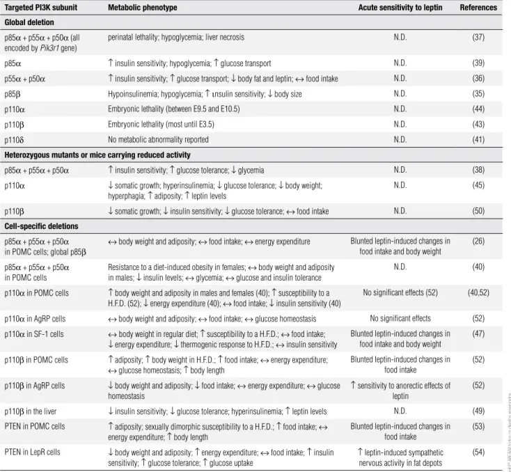
table 1. List of studies that investigated the physiological role played by different Class IA PI3K subunits and by PTEN in the regulation of energy homeostasis
targeted PI3K subunit Metabolic phenotype Acute sensitivity to leptin References
Global deletion
p85α + p55α + p50α (all
encoded by Pik3r1 gene)
perinatal lethality; hypoglycemia; liver necrosis N.D. (37)
p85α ↑ insulin sensitivity; hypoglycemia; ↑ glucose transport N.D. (39)
p55α + p50α ↑ insulin sensitivity; ↑ glucose transport; ↓ body fat and leptin; ↔ food intake N.D. (36)
p85β Hypoinsulinemia; hypoglycemia; ↑ ιnsulin sensitivity; ↓ body size N.D. (35)
p110α Embryonic lethality (between E9.5 and E10.5) N.D. (44)
p110β Embryonic lethality (most until E3.5) N.D. (43)
p110δ No metabolic abnormality reported N.D. (41)
Heterozygous mutants or mice carrying reduced activity
p85α + p55α + p50α ↑ insulin sensitivity; ↑ glucose tolerance; ↓ glycemia N.D. (38)
p110α ↓ somatic growth; hyperinsulinemia; ↓ glucose tolerance; ↓ body weight;
hyperphagia; ↑ adiposity; ↑ leptin levels
N.D. (45)
p110β ↓ somatic growth; ↓ insulin sensitivity; ↓ glucose tolerance; ↔ food intake N.D. (50)
cell-speciic deletions
p85α + p55α + p50α
in POMC cells; global p85β
↔ body weight and adiposity; ↔ food intake; ↔ energy expenditure Blunted leptin-induced changes in
food intake and body weight
(26)
p85α + p55α + p50α
in POMC cells
Resistance to a diet-induced obesity in females; ↔ body weight and adiposity
in males; ↓ insulin levels; ↔ glycemia; ↔ glucose and insulin tolerance
N.D. (40)
p110α in POMC cells ↑ body weight and adiposity in males and females (40); ↑ susceptibility to a
H.F.D. (52); ↓ energy expenditure (40); ↔ food intake; ↓ insulin sensitivity (40)
No significant effects (52) (40,52)
p110α in AgRP cells ↔ body weight and adiposity; ↔ food intake; ↔ glucose homeostasis No significant effects (52)
p110α in SF-1 cells ↔ body weight in regular diet; ↑ susceptibility to a H.F.D.; ↔ food intake;
↓ energy expenditure; ↓ thermogenic response to H.F.D.; ↔ insulin sensitivity
Blunted leptin-induced changes in food intake and body weight
(47)
p110β in POMC cells ↑ adiposity; ↑ body weight in H.F.D.; ↑ food intake; ↔ energy expenditure;
↔ glucose homeostasis; ↑ body length
Blunted leptin-induced changes in food intake
(52)
p110β in AgRP cells ↓ body weight and adiposity; ↓ food intake; ↔ energy expenditure; ↔ glucose
homeostasis
↑ sensitivity to anorectic effects of
leptin
(52)
p110β in the liver ↓ insulin sensitivity; ↓ glucose tolerance; hyperinsulinemia; ↑ leptin levels N.D. (49)
PTEN in POMC cells ↑ adiposity; sexually dimorphic susceptibility to a H.F.D.; ↑ food intake; ↔
energy expenditure; ↑ body length
Blunted leptin-induced changes in food intake
(53)
PTEN in LepR cells ↓ body weight and adiposity; ↑ energy expenditure; ↔ food intake; ↑ insulin
sensitivity; ↑ glucose tolerance; ↑ glucose uptake
↑ leptin-induced sympathetic
nervous activity in fat depots
(54)
N.D.: not determined.
adiposity as a result of increased energy expenditure (54). Part of the changes in energy expenditure in the PtenΔObRb mice was caused by white adipose
tis-sue (WAT) transdifferentiation. The perigonadal WAT of PtenΔObRb mice exhibited markers of brown adipose
tissue, e.g., increased mitochondrial content and un-coupling protein-1 expression. Interestingly, leptin was required for all these effects because the condi-tional disruption of PTEN in leptin-deicient mice did not affect their metabolism or WAT morphophysio-logy (54).
Cop
yright
© ABE&M t
odos os dir
eit
os r
eser
vados
.
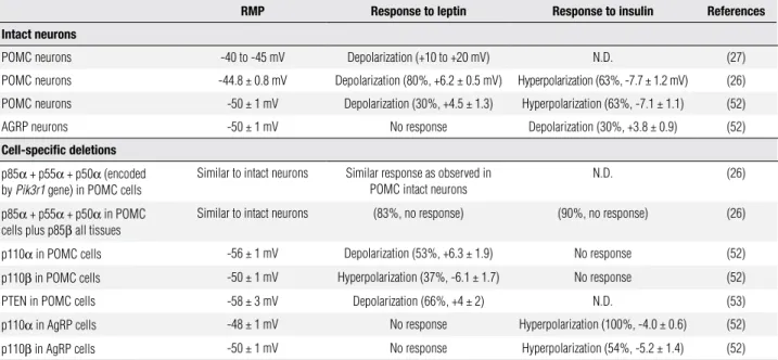
table 2. Electrophysiological properties of intact neurons and of neurons with specific Class IA PI3K subunit deletions
RMP Response to leptin Response to insulin References
Intact neurons
POMC neurons -40 to -45 mV Depolarization (+10 to +20 mV) N.D. (27)
POMC neurons -44.8 ± 0.8 mV Depolarization (80%, +6.2 ± 0.5 mV) Hyperpolarization (63%, -7.7 ± 1.2 mV) (26)
POMC neurons -50 ± 1 mV Depolarization (30%, +4.5 ± 1.3) Hyperpolarization (63%, -7.1 ± 1.1) (52)
AGRP neurons -50 ± 1 mV No response Depolarization (30%, +3.8 ± 0.9) (52)
cell-speciic deletions
p85α + p55α + p50α (encoded
by Pik3r1 gene) in POMC cells
Similar to intact neurons Similar response as observed in
POMC intact neurons
N.D. (26)
p85α + p55α + p50α in POMC
cells plus p85β all tissues
Similar to intact neurons (83%, no response) (90%, no response) (26)
p110α in POMC cells -56 ± 1 mV Depolarization (53%, +6.3 ± 1.9) No response (52)
p110β in POMC cells -50 ± 1 mV Hyperpolarization (37%, -6.1 ± 1.7) No response (52)
PTEN in POMC cells -58 ± 3 mV Depolarization (66%, +4 ± 2) N.D. (53)
p110α in AgRP cells -48 ± 1 mV No response Hyperpolarization (100%, -4.0 ± 0.6) (52)
p110β in AgRP cells -50 ± 1 mV No response Hyperpolarization (54%, -5.2 ± 1.4) (52)
The percentage represents the proportion of neurons that responded to a specific drug application. RMP, resting membrane potential. N.D., not determined.
PI3K: A POtEntIAL LInK BEtWEEn MEtABOLISM
AnD cAncER
Obesity, which is a condition of hyperleptinemia (7), has a strong correlation with the onset of certain can-cers. According to a longitudinal study that evaluated more than 900,000 American adults, overweight or obesity accounted for 14% and 20% of all cancer deaths in men and women, respectively, after a follow-up pe-riod of 16 years (57). Interestingly, a high association between the development of various types of cancer and abnormalities in the PI3K pathway has also been reported (45,46,49,58). For instance, somatic missense mutations in the Pik3ca gene occur at a high frequency in many types of human cancers (3). The relationship between PI3K and the development of cancers indica-tes that drugs that affect the activity of PI3K can be a promising anti-cancer therapy (1-3). Thus, it is possi-ble that the PI3K signaling pathway is an important link between obesity, leptin and increased risk of cancer (59). Nonetheless, other intracellular pathways, inclu-ding JAK/STAT3 and MAP kinase, may also be invol-ved in leptin-induced development of certain types of cancer (60).
cOncLUSIOnS AnD FUtURE PERSPEctIVES
In the present review, we discussed the evidence offe-red by recent indings that the PI3K signaling pathway is required for leptin-induced regulation of energy
ho-meostasis. Manipulation of the PI3K signaling pathway in speciic populations of leptin-sensitive neurons cau-ses different biological effects, possibly related to the role played by particular neurons in leptin physiology. Although the role played by PI3K signaling in each deined population of leptin-sensitive neurons is of fundamental importance, potential drugs that target PI3K subunits are likely to produce a systemic effect. Therefore, broader strategies to study leptin and PI3K will expand our knowledge on the whole-body effects of PI3K in the regulation of energy homeostasis. Mo-reover, the various subunits of PI3K appear to differen-tially regulate energy homeostasis, but their exact role and their physiological relevance still need to be further assessed in more detail. Studies that directly assess the possible involvement of Class IB, Class II and Class III PI3Ks in leptin’s physiology will be informative. Fi-nally, the PI3K pathway seems to be a very good can-didate for regulating the rapid (acute) effects of leptin, whereas the JAK2/STAT3 pathway plays a dominant role in the long-term regulation of energy homeostasis. However, changing the acute cellular response to lep-tin may create a chronic condition that will ultimately affect the long-term regulation of energy homeostasis. Understanding how these conditions develop and how PI3K interacts with other intracellular pathways repre-sents important challenges for future studies.
manus-Cop
yright
© ABE&M t
odos os dir
eit
os r
eser
vados
.
cript. CFE is the Distinguished Scholar in Medical Research (UTSW, Dallas, TX). This work was supported by NIH grant R01HD061539, by the National Council for Scientiic and Te-chnological Development (CNPq-Brazil) 201804/2008-5 (to R.F.), and by the President’s Council Award and the Regent’s Research Award to CFE (UTSW, Dallas – TX).
Disclosure: no potential conlict of interest relevant to this article was reported.
REFEREncES
1. Ward S, Sotsios Y, Dowden J, Bruce I, Finan P. Therapeutic po-tential of phosphoinositide 3-kinase inhibitors. Chem Biol. 2003;10(3):207-13.
2. Knight ZA, Gonzalez B, Feldman ME, Zunder ER, Goldenberg DD, Williams O, et al. A pharmacological map of the PI3-K fa-mily deines a role for p110[alpha] in insulin signaling. Cell. 2006;125(4):733-47.
3. Jia S, Roberts TM, Zhao JJ. Should individual PI3 kinase isoforms be targeted in cancer? Curr Opin Cell Biol. 2009;21(2):199-208. 4. Myers MG Jr, Backer JM, Sun XJ, Shoelson S, Hu P, Schlessinger J,
et al. IRS-1 activates phosphatidylinositol 3’-kinase by associa-ting with src homology 2 domains of p85. Proc Natl Acad Sci U S A. 1992;89(21):10350-4.
5. Williams KW, Scott MM, Elmquist JK. From observation to expe-rimentation: leptin action in the mediobasal hypothalamus. Am J Clin Nutr. 2009;89(3):985S-90S.
6. Schwartz MW. Central nervous system regulation of food intake. Obesity (Silver Spring). 2006;14 Suppl 1:1S-8S.
7. Simpson KA, Martin NM, Bloom SR. Hypothalamic regulation of food intake and clinical therapeutic applications. Arq Bras Endo-crinol Metabol. 2009;53(2):120-8.
8. Ribeiro SM, dos Santos ZA, da Silva RJ, Louzada E, Donato J Jr, Tirapegui J. [Leptin: aspects on energetic balance, physical exercise and athletic amenorhea]. Arq Bras Endocrinol Metabol. 2007;51(1):11-24.
9. Halaas JL, Gajiwala KS, Maffei M, Cohen SL, Chait BT, Rabinowitz D, et al. Weight-reducing effects of the plasma protein encoded by the obese gene. Science. 1995;269(5223):543-6.
10. Farooqi IS, Matarese G, Lord GM, Keogh JM, Lawrence E, Agwu C, et al. Beneicial effects of leptin on obesity, T cell hyporespon-siveness, and neuroendocrine/metabolic dysfunction of human congenital leptin deiciency. J Clin Invest. 2002;110(8):1093-103. 11. Licinio J, Caglayan S, Ozata M, Yildiz BO, de Miranda PB, O’Kirwan F,
et al. Phenotypic effects of leptin replacement on morbid obe-sity, diabetes mellitus, hypogonadism, and behavior in leptin-deicient adults. Proc Natl Acad Sci U S A. 2004;101(13):4531-6. 12. de Luca C, Kowalski TJ, Zhang Y, Elmquist JK, Lee C, Kilimann
MW, et al. Complete rescue of obesity, diabetes, and infertility in db/db mice by neuron-speciic LEPR-B transgenes. J Clin Invest. 2005;115(12):3484-93.
13. Ahima RS, Prabakaran D, Mantzoros C, Qu D, Lowell B, Maratos-Flier E, et al. Role of leptin in the neuroendocrine response to fasting. Nature. 1996;382(6588):250-2.
14. Schwartz MW, Seeley RJ, Woods SC, Weigle DS, Campield LA, Burn P, et al. Leptin increases hypothalamic pro-opiomelanocor-tin mRNA expression in the rostral arcuate nucleus. Diabetes. 1997;46(12):2119-23.
15. Tartaglia LA, Dembski M, Weng X, Deng N, Culpepper J, Devos R, et al. Identiication and expression cloning of a leptin receptor, OB-R. Cell. 1995;83(7):1263-71.
16. Chua SC, Koutras IK, Han L, Liu S-M, Kay J, Young SJ, et al. Fine structure of the murine leptin receptor gene: splice site suppres-sion is required to form two alternatively spliced transcripts. Ge-nomics. 1997;45(2):264-70.
17. Myers MG Jr. Leptin receptor signaling and the regulation of mammalian physiology. Recent Prog Horm Res. 2004;59:287-304. 18. Bates SH, Stearns WH, Dundon TA, Schubert M, Tso AW, Wang Y,
et al. STAT3 signalling is required for leptin regulation of energy balance but not reproduction. Nature. 2003;421(6925):856-9. 19. Scott MM, Lachey JL, Sternson SM, Lee CE, Elias CF, Friedman
JM, et al. Leptin targets in the mouse brain. J Comp Neurol. 2009;514(5):518-32.
20. Donato J Jr, Silva RJ, Sita LV, Lee S, Lee C, Lacchini S, et al. The ventral premammillary nucleus links fasting-induced changes in leptin levels and coordinated luteinizing hormone secretion. J Neurosci. 2009;29(16):5240-50.
21. Bjorbaek C, Elmquist JK, Frantz JD, Shoelson SE, Flier JS. Identi-ication of SOCS-3 as a potential mediator of central leptin resis-tance. Molecular Cell. 1998;1(4):619-25.
22. Howard JK, Flier JS. Attenuation of leptin and insulin signaling by SOCS proteins. Trends Endocrinol Metab. 2006;17(9):365-71. 23. Glaum SR, Hara M, Bindokas VP, Lee CC, Polonsky KS, Bell GI,
et al. Leptin, the obese gene product, rapidly modulates sy-naptic transmission in the hypothalamus. Mol Pharmacol. 1996;50(2):230-5.
24. Spanswick D, Smith MA, Groppi VE, Logan SD, Ashford ML. Lep-tin inhibits hypothalamic neurons by activation of ATP-sensitive potassium channels. Nature. 1997;390(6659):521-5.
25. Xu AW, Kaelin CB, Takeda K, Akira S, Schwartz MW, Barsh GS. PI3K integrates the action of insulin and leptin on hypothalamic neurons. J Clin Invest. 2005;115(4):951-8.
26. Hill JW, Williams KW, Ye C, Luo J, Balthasar N, Coppari R, et al. Acute effects of leptin require PI3K signaling in hypotha-lamic proopiomelanocortin neurons in mice. J Clin Invest. 2008;118(5):1796-805.
27. Cowley MA, Smart JL, Rubinstein M, Cerdan MG, Diano S, Horvath TL, et al. Leptin activates anorexigenic POMC neu-rons through a neural network in the arcuate nucleus. Nature. 2001;411(6836):480-4.
28. Niswender KD, Morton GJ, Stearns WH, Rhodes CJ, Myers MG Jr, Schwartz MW. Intracellular signalling. Key enzyme in leptin-induced anorexia. Nature. 2001;413(6858):794-5.
29. Zhao AZ, Huan JN, Gupta S, Pal R, Sahu A. A phosphatidylinositol 3-kinase phosphodiesterase 3B-cyclic AMP pathway in hypotha-lamic action of leptin on feeding. Nat Neurosci. 2002;5(8):727-8. 30. Morrison CD, Morton GJ, Niswender KD, Gelling RW, Schwartz
MW. Leptin inhibits hypothalamic Npy and Agrp gene expression via a mechanism that requires phosphatidylinositol 3-OH-kinase signaling. Am J Physiol Endocrinol Metab. 2005;289(6):E1051-7. 31. Niswender KD, Morrison CD, Clegg DJ, Olson R, Baskin DG,
Myers MG Jr, et al. Insulin activation of phosphatidylinositol 3-kinase in the hypothalamic arcuate nucleus: a key mediator of insulin-induced anorexia. Diabetes. 2003;52(2):227-31.
32. Morton GJ, Gelling RW, Niswender KD, Morrison CD, Rhodes CJ, Schwartz MW. Leptin regulates insulin sensitivity via phospha-tidylinositol-3-OH kinase signaling in mediobasal hypothalamic neurons. Cell Metabolism. 2005;2(6):411-20.
33. Roman EAFR, Reis D, Romanatto T, Maimoni D, Ferreira EA, San-tos GA, et al. Central leptin action improves skeletal muscle AKT, AMPK, and PGC1[alpha] activation by hypothalamic PI3K-depen-dent mechanism. Mol Cell Endocrinol. 2010;314(1):62-9.
Cop
yright
© ABE&M t
odos os dir
eit
os r
eser
vados
.
35. Ueki K, Yballe CM, Brachmann SM, Vicent D, Watt JM, Kahn CR, et al. Increased insulin sensitivity in mice lacking p85beta su-bunit of phosphoinositide 3-kinase. Proc Natl Acad Sci U S A. 2002;99(1):419-24.
36. Chen D, Mauvais-Jarvis F, Bluher M, Fisher SJ, Jozsi A, Goodyear LJ, et al. p50alpha/p55alpha phosphoinositide 3-kinase kno-ckout mice exhibit enhanced insulin sensitivity. Mol Cell Biol. 2004;24(1):320-9.
37. Fruman DA, Mauvais-Jarvis F, Pollard DA, Yballe CM, Brazil D, Bronson RT, et al. Hypoglycaemia, liver necrosis and perinatal death in mice lacking all isoforms of phosphoinositide 3-kinase p85 alpha. Nat Genet. 2000;26(3):379-82.
38. Mauvais-Jarvis F, Ueki K, Fruman DA, Hirshman MF, Sakamoto K, Goodyear LJ, et al. Reduced expression of the murine p85alpha subunit of phosphoinositide 3-kinase improves insulin signaling and ameliorates diabetes. J Clin Invest. 2002;109(1):141-9. 39. Terauchi Y, Tsuji Y, Satoh S, Minoura H, Murakami K, Okuno A,
et al. Increased insulin sensitivity and hypoglycaemia in mice lacking the p85 alpha subunit of phosphoinositide 3-kinase. Nat Genet. 1999;21(2):230-5.
40. Hill JW, Xu Y, Preitner F, Fukuda M, Cho Y-R, Luo J, et al. Phospha-tidyl inositol 3-kinase signaling in hypothalamic proopiomelano-cortin neurons contributes to the regulation of glucose homeos-tasis. Endocrinology. 2009;150(11):4874-82.
41. Okkenhaug K, Bilancio A, Farjot G, Priddle H, Sancho S, Peskett E, et al. Impaired B and T cell antigen receptor signaling in p110delta PI 3-kinase mutant mice. Science. 2002;297(5583):1031-4. 42. Vanhaesebroeck B, Welham MJ, Kotani K, Stein R, Warne PH,
Zve-lebil MJ, et al. p110δ, a novel phosphoinositide 3-kinase in leu-kocytes. Proc Natl Acad Sci U S A. 1997;94(9):4330-5.
43. Bi L, Okabe I, Bernard DJ, Nussbaum RL. Early embryonic lethali-ty in mice deicient in the p110beta catalytic subunit of PI 3-kina-se. Mamm Genome. 2002;13(3):169-72.
44. Bi L, Okabe I, Bernard DJ, Wynshaw-Boris A, Nussbaum RL. Pro-liferative defect and embryonic lethality in mice homozygous for a deletion in the p110alpha subunit of phosphoinositide 3-kinase. J Biol Chem. 1999;274(16):10963-8.
45. Foukas LC, Claret M, Pearce W, Okkenhaug K, Meek S, Peskett E, et al. Critical role for the p110alpha phosphoinositide-3-OH kinase in growth and metabolic regulation. Nature. 2006;441(7091):366-70. 46. Zhao JJ, Cheng H, Jia S, Wang L, Gjoerup OV, Mikami A, et al.
The p110α isoform of PI3K is essential for proper growth factor signaling and oncogenic transformation. Proc Natl Acad Sci U S A. 2006;103(44):16296-300.
47. Xu Y, Hill JW, Fukuda M, Gautron L, Sohn J-W, Kim K-W, et al. PI3K Signaling in the ventromedial hypothalamic nucleus is required for normal energy homeostasis. Cell Metab. 2010;12(1):88-95.
48. Chaussade C, Rewcastle GW, Kendall JD, Denny WA, Cho K, Gron-ning LM, et al. Evidence for functional redundancy of class IA PI3K isoforms in insulin signalling. Biochem J. 2007;404(3):449-58. 49. Jia S, Liu Z, Zhang S, Liu P, Zhang L, Lee SH, et al. Essential roles
of PI(3)K-p110β in cell growth, metabolism and tumorigenesis. Nature. 2008;454(7205):776-9.
50. Ciraolo E, Iezzi M, Marone R, Marengo S, Curcio C, Costa C, et al. Phosphoinositide 3-kinase p110beta activity: key role in me-tabolism and mammary gland cancer but not development. Sci Signal. 2008;1(36):ra3.
51. Tups A, Anderson GM, Rizwan M, Augustine RA, Chaussade C, Shepherd PR, et al. Both p110alpha and p110beta isoforms of phos-phatidylinositol 3-OH-kinase are required for insulin signalling in the hypothalamus. J Neuroendocrinol. 2010;22(6):534-42. 52. Al-Qassab H, Smith MA, Irvine EE, Guillermet-Guibert J, Claret M,
Choudhury AI, et al. Dominant role of the p110beta isoform of PI3K over p110alpha in energy homeostasis regulation by POMC and AgRP Neurons Cell Metab. 2009;10(5):343-54.
53. Plum L, Ma X, Hampel B, Balthasar N, Coppari R, Münzberg H, et al. Enhanced PIP3 signaling in POMC neurons causes KATP chan-nel activation and leads to diet-sensitive obesity. J Clin Invest. 2006;116(7):1886-901.
54. Plum L, Rother E, Münzberg H, Wunderlich FT, Morgan DA, Ham-pel B, et al. Enhanced leptin-stimulated Pi3k activation in the CNS promotes white adipose tissue transdifferentiation. Cell Metabo-lism. 2007;6(6):431-45.
55. Gildea JJ, Herlevsen M, Harding MA, Gulding KM, Moska-luk CA, Frierson HF, et al. PTEN can inhibit in vitro organotypic and in vivo orthotopic invasion of human bladder cancer cells even in the absence of its lipid phosphatase activity. Oncogene. 2004;23(40):6788-97.
56. Gu J, Tamura M, Yamada KM. Tumor suppressor PTEN inhibits integrin- and growth factor-mediated mitogen-activated protein (MAP) kinase signaling pathways. J Cell Biol. 1998;143(5):1375-83. 57. Calle EE, Rodriguez C, Walker-Thurmond K, Thun MJ. Overweight,
obesity, and mortality from cancer in a prospectively studied co-hort of U.S. adults. N Engl J Med. 2003;348(17):1625-38. 58. Osorio-Costa F, Rocha GZ, Dias MM, Carvalheira JB.
Epidemio-logical and molecular mechanisms aspects linking obesity and cancer. Arq Bras Endocrinol Metabol. 2009;53(2):213-26. 59. Huang X-F, Chen J-Z. Obesity, the PI3K/Akt signal pathway and
colon cancer. Obesity Reviews. 2009;10(6):610-6.