Joana Rita Batuca
HUMORAL RESPONSE TOWARDS
HIGH DENSITY LIPOPROTEIN: A
NEW MECHANISM FOR
ATHEROGENESIS
Dissertation presented to obtain the PhD degree in Ciências da Vida - Especialidade Farmacologia in the Faculdade de Ciências Médicas da Universidade Nova de Lisboa
Experimental work conducted in the Departamento de Farmacologia - Centro de Estudo de Doenças Crónicas (CEDOC), Faculdade de Ciências Médicas da Universidade Nova de Lisboa,
This work was financially supported by:
Thinking is more interesting than knowing, but less interesting than looking
The scientific content of the present thesis has been included in the publication of
the following international scientific periodicals with referees:
- Batuca JR, Ames PRJ, Isenberg DA and Delgado Alves J. Antibodies towards high-density
lipoprotein components inhibit paraoxonase activity in patients with Systemic Lupus Erythematosus. Ann N Y Acad Sci. 2007;1108 (1):137–146.
- Batuca JR, Ames PRJ, Amaral MC, Favas C, Isenberg DA and Delgado Alves J.
Anti-atherogenic and anti-inflammatory properties of high-density lipoprotein are affected by specific antibodies in systemic lupus erythematosus. Rheumatology (Oxford). 2009;48(1):26-31.
- Batuca J & Delgado Alves J. C-reactive protein in systemic lupus erythematosus. Autoimmunity. 2009 May;42(4):282-5
- Batuca JR, Amaral MC and Delgado Alves J. Humoral mechanism of atherogenesis. Ann N Y
Acad Sci. 2009;1173:401-408.
- Batuca JR, Gomes AL, Amaral MC, Favas C, Justino GC, Dias S and Delgado Alves J.
Antibodies against high density lipoprotein components a new risk factor for atherosclerosis (submitted)
- Batuca JR, Amaral MC, Favas C and Delgado Alves J. Antibodies toward high-density
lipoprotein components in patients with type 2 diabetes (submitted)
- Batuca JR, Amaral MC, Favas C and Delgado Alves J. Nicotinic acid increases anti-ApoA-I
antibodies (submitted)
The work presented in this thesis was awarded with the - Prémio NEDAI de Investigação em Auto-imunidade from the Portuguese Society of Internal Medicine in 2010.
i
ACKNOWLEDGEMENTS
The first thankfulness goes to my supervisor Prof. Doutor José Delgado Alves, for his consistent guidance into the scientific way of thinking, encouragement and support. Above of all, I greatly appreciate his truly enthusiastic approach to science, for giving me the opportunity to be his "Portuguese biochemist " and for be the person with whom I most liked to share good results after a long day in the lab.
I also wish to express my heartfelt gratitude to Prof. Doutora Emília Monteiro head of Pharmacology Department for giving me the opportunity to work in her laboratory. I could ever forget that she was my first mentor and continue to be very helpful to my growth as a scientist. I greatly appreciate the trust she have placed in me over the years and her dedication to her work.
I remain greatly indebted to all the clinicians and basic researchers that in somehow helped me throughout this project. My grateful thanks to Drª Marta Amaral, Drª Catarina Favas, Prof. Doutor David Isenberg, Doutor Paul Ames, Doutor Luis Lopez for their support and assistance with recruitment of subjects for the study, for allowing me to use the clinical trials in my thesis and for all those interesting discussions during the development of this project. I am also very grateful to Doutor Constantin Fesel, Doutor Sergio Dias, Doutor Michel Kranendonk, Doutora Ana Gomes, Doutor Gonçalo Justino for being so well received in their laboratories, for all the constructive inputs in scientific discussions and fruitful collaboration between our research groups.
ii
I would also like to acknowledge the institutional support given by the Science Medical Faculty and the CEDOC for providing the research facilities and for funding my salary, which have made this work possible.
My sincere thanks to all foundations and companies that provide the financially supported for this study.
Finally grateful thanks to:
My parents Manuel and Teresa, my sisters Marta and Inês, for their steadfast love, for always believing in me and encouraging me, for a firm foundation and through their example shown me the importance of honest and hard-work to achieve any goal.
My dear husband, Marco, for being there with limitless love and for giving me all the support I could ever have wished for. And to the greatest gift of all, our son, António, who changed everything, but with just a smile or a "gosto de ti mamã" make me believe that we'll get everything.
My remaining family and friends for their continued love, for filling my life with memorable fun moments and for putting up with me when I kept going on about my research.
iii
TABLE OF CONTENTS
ACKNOWLEDGEMENTS _______________________________________________________ i
TABLE OF CONTENTS _______________________________________________________ iii
LIST OF FIGURES ___________________________________________________________ vii
LIST OF TABLES ____________________________________________________________ xi
LIST OF ABBREVIATIONS ____________________________________________________ xiii
ABSTRACT ________________________________________________________________xix
RESUMO ________________________________________________________________xxiii
1. INTRODUCTION _________________________________________________________ 1
1.1 Atherosclerosis: a global overview ___________________________________________ 3
1.1.1 Hypercholesterolemia ... 4
1.1.2 Diet ... 4
1.1.3 Impaired glucose metabolism ... 4
1.1.4 Hypertension ... 5
1.1.5 Smoking ... 5
1.1.6 Lack of exercise ... 5
1.1.7 Hyperhomocysteinemia ... 5
1.1.8 Infections ... 6
1.1.9 Genetic factors ... 6
1.2 Lipids and Plasma Lipoproteins in Atherogenesis ________________________________ 8 1.2.1 Lipoprotein structure and composition ... 8
1.2.2 Lipoprotein metabolism ... 9
1.2.3 Lipoproteins and cardiovascular disease (CVD)... 13
1.3 High Density Lipoprotein (HDL) _____________________________________________ 15 1.3.1 Structure (heterogeneity) and composition ... 15
1.3.2 Atheroprotective functions of HDL... 27
1.3.3 Functionally defective HDL ... 40
1.3.4 Epidemiology of cardiovascular risk in relation to low HDL-C ... 43
1.3.5 Pharmacologic modulation of HDL-C... 44
1.4 Immunity in Atherogenesis ________________________________________________ 57 1.4.1 Overview ... 57
1.4.2 General innate immune response ... 57
iv
1.4.4 The immune response in atherogenesis ... 60
1.4.5 Immune response in atherogenesis and HDL ... 69
1.5 Aims of the thesis ________________________________________________________ 72 2. METHODS ____________________________________________________________ 73 2.1 Immunologic-related methods _____________________________________________ 75 2.1.1 Enzyme-Linked Immunoabsorbent Assays (ELISAs) ... 75
2.1.2 Immunoturbidimetric immunoassay ... 79
2.1.3 Antibody isolation ... 79
2.2 Biochemistry-related methods _____________________________________________ 81 2.2.1 Paraoxonase 1 (PON1) activity ... 81
2.2.2 Total anti-oxidant capacity (TAC) ... 81
2.2.3 Nitric oxide metabolites ... 82
2.2.4 Total HDL cholesterol, HDL2 and HDL3 ... 82
2.3 Functional assays in vitro __________________________________________________ 83 2.3.1 Inhibition of paraoxonase 1 (PON1) activity by the antibodies isolated from patient’s serum 83 2.3.2 In vitro exposure of human umbilical vein endothelial cells (HUVECs) to aHDL antibodies 83 2.4 Statistical analysis _______________________________________________________ 84 3. HUMORAL RESPONSE TOWARDS HDL COMPONENTS AND ITS ASSOCIATION WITH MODIFICATIONS OF THE ANTI-ATHEROGENIC PROPERTIES OF HDL __________________ 85 3.1 aHDL and aApoA-I antibodies in systemic lupus erythematosus (SLE) _______________ 87 3.2 Anti-HDL and anti-ApoA-I antibodies in non auto-immune patients with atherosclerosis-related clinical events ___________________________________________________________ 97 3.2.1 Coronary artery disease (CAD) and ischemic stroke (IS) ... 97
3.2.2 Type 2 diabetes ... 108
4. BIOLOGIC ACTIVITY AND PATHOGENIC POTENTIAL OF ANTIBODIES TOWARDS THE HDL COMPLEX ________________________________________________________________ 119 4.1 Introduction ___________________________________________________________ 121 4.2 Aim __________________________________________________________________ 121 4.3 Patients and Methods ___________________________________________________ 121 4.4 Isolation of aHDL and aApoA-I antibodies from patients with SLE and in vitro inhibition of PON1 activity _________________________________________________________________ 122 4.4.1 Results ... 122
4.4.2 Discussion ... 123
v
4.5.2 Discussion ... 125
4.6 In vitro inhibition of PON1 activity by aHDL antibodies _________________________ 125 4.6.1 Results ... 125
4.6.2 Discussion ... 125
4.7 In vitro exposure of human umbilical vein endothelial cells (HUVECs) to aHDL antibodies 126 4.7.1 Results ... 126
4.7.2 Discussion ... 128
5. PHARMACOLOGIC MANIPULATION OF HDL-C AND aHDL ANTIBODIES TITRES _____ 131 5.1 Exploratory, double blind placebo controlled, randomized, single cross-over study to evaluate the potential anti-oxidant activity of Niaspan® (EXPLORE) ______________________ 133 5.1.1 Introduction ... 133
5.1.2 Aim ... 134
5.1.3 Patients and methods ... 134
5.1.4 Results ... 137
5.1.5 Discussion ... 142
5.2 Prevention of atherosclerosis with atorvastatin in patients with systemic lupus erythematosus (SLE): a pilot study ________________________________________________ 144 5.2.1 Introduction ... 144
5.2.2 Aim ... 145
5.2.3 Patients and methods ... 145
5.2.4 Results ... 146
5.2.5 Discussion ... 148
5.3 Influence of Rosuvastatin on the oxidative modification of LDL in type 2 diabetes ____ 149 5.3.1 Introduction ... 149
5.3.2 Aim ... 150
5.3.3 Patients and methods ... 150
5.3.4 Results ... 152
5.3.5 Discussion ... 154
6. OVERALL DISCUSSION AND PROPOSALS FOR FUTURE DIRECTIONS ______________ 157
vii
LIST OF FIGURES
Figure 1. Schematic presentation of the major lipoproteins in normal human plasma. ... 9
Figure 2. Overview of lipoprotein metabolism ... 10
Figure 3. High density lipoproteins (HDL) heterogeneity... 17
Figure 4. Nomenclature of the high density lipoproteins (HDL) subclasses determined by different methods. ... 18
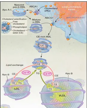
Figure 5. Cholesterol ester transfer protein (CETP)-mediated lipid exchange between lipoproteins. . 26
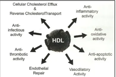
Figure 6. Summary of the key anti-atherogenic properties attributed to high density lipoprotein (HDL). ... 28
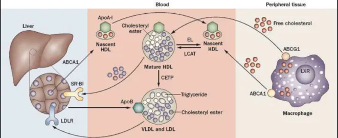
Figure 7. HDL metabolism and reverse cholesterol transport (RCT) ... 29
Figure 8. Major pathways involved in vasodilatory activity of HDL. ... 34
Figure 9. Contribution of platelets to the initiation and the progression of atherosclerosis ... 37
Figure 10. Intracellular signalling cascate triggered by the interaction of HDL with the platelet receptors SR-B1 and ApoER2. ... 38
Figure 11. HDL and glucose homeostasis. ... 39
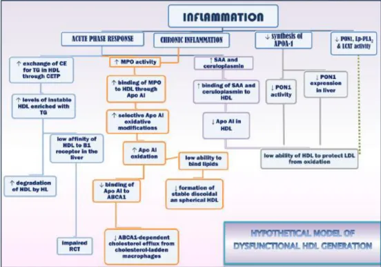
Figure 12. Proposed model for generating dysfunctional HDL. ... 41
Figure 13. Coronary artery disease (CAD) risk predicted by high density lipoprotein cholesterol (HDL-C) and low density lipoprotein cholesterol (LDL-(HDL-C) in the Framingham Heart Study. ... 43
Figure 14. Residual cardiovascular risk despite statin treatment. ... 45
Figure 15. Percentage reductions in serum LDL cholesterol levels according to statin and daily dose. ... 46
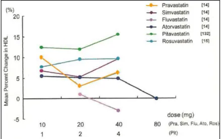
Figure 16. Effect of each statin on HDL-C levels. ... 46
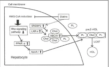
Figure 17. Mechanism of up-regulation of ApoA-I production by statins and effect on ABCA1. ... 47
Figure 18. Cholesterol biosynthetic pathway ... 48
Figure 19. Mechanism of action of fibrates. ... 49
Figure 20. Mechanisms of nicotinic acid-induced changes in lipolysis. ... 52
Figure 21. Emerging HDL-based therapies. ... 53
Figure 22. Interaction between innate and adaptative immunity. ... 59
Figure 23. T cell-dependent B cell activation. ... 60
Figure 24. Activation of innate immune response in atherosclerotic lesion. ... 61
Figure 25. Activation of T cells in atherosclerotic lesions. ... 63
Figure 26. Naive T-cell differentiation into specific T helper (Th) substes involved in atherosclerosis and its complex interactions. ... 64
Figure 27. Role of HDL and ApoA-I in regulating immune responses. ... 71
Figure 28: Levels of IgG aHDL (A) and IgG aApoA-I antibodies (B) in healthy controls (CTRL) and patients with systemic lupus erythematosus (SLE).. ... 90
Figure 29: Correlation (Spearman’s rank test) between PON1 activity (U/L) and IgG aHDL levels (A) and IgG aApoA-I levels (B) in patients with systemic lupus erythematosus (SLE). ... 91
viii
Figure 31. Relationship between the titres of IgG aHDL, aApoA-I antibodies and PON1 activity with SLICC/ACR damage index (A) and disease activity (BILAG score) (B) in patients with systemic lupus erythematosus (SLE). ... 93 Figure 32. Relationship between the NO2-+NO3- (A) and VCAM-1 (B, C) levels with damage
(SLICC/ACR DI) and disease activity (BILAG score) in patients with systemic lupus erythematosus (SLE). ... 94 Figure 33. Levels of IgG aHDL (A), aApoA-I (B) and aPON1 (C) antibodies in healthy controls (CTRL) and patients with atherosclerosis-associated clinical events: coronary artery disease (CAD) and ischemic stroke (IS). ... 101 Figure 34. Relationship between the titres of IgG aHDL and IgG aApo A-I (A) and aPON1 antibodies (B) in both group of patients with atherosclerosis-associated clinical events: coronary artery disease (CAD) and ischemic stroke (IS). ... 102 Figure 35. Correlation (Spearman’s rank test) between PON1 activity (U/L) and IgG aApoA-I (A) and aPON1 antibodies (B) in both groups of patients with atherosclerosis-associated clinical events coronary artery disease (CAD) and ischemic stroke (IS). ... 103 Figure 36. Correlation (Spearman’s rank test) between NO3-levels and IgG aHDL (A), aApoA-I (B),
ix Figure 51. Serum nitric oxide metabolites (NO2- + NO3-) acording treatment groups at baseline and at
xi
LIST OF TABLES
Table 1. Composition of the major lipoprotein complexes. ... 9 Table 2. Proteins detected in high density lipoproteins (HDL) by mass spectrometry. ... 15 Table 3. Classification of high density lipoproteins (HDL) by physical properties. ... 18 Table 4. Clinical evidence for ApoA-I/ HDL infusion. ... 55
Table 5. Several ligands for the different pattern-recognition receptors (PRRs), scavenger receptors (SRs) and toll-like receptors (TLRs). ... 61
Table 6. Demographic characteristics and clinical and serological data of healthy controls (CTRL) and patients with systemic lupus erythematosus (SLE).. ... 90
Table 7: Biological variables (oxidation and endothelial dysfunction markers) measured in healthy controls (CTRL) and patients with systemic lupus erythematosus (SLE). ... 91
Table 8: Demographic characteristics and clinical and serological data of healthy controls (CTRL) and patients with atherosclerosis-associated clinical events: coronary artery disease (CAD) and ischemic stroke (IS). ... 100
Table 9: Antibodies titres measured in healthy controls (CTRL) and in patients with atherosclerosis-associated clinical events coronary artery disease (CAD) and ischemic stroke (IS). ... 100
Table 10: Biological variables (oxidation and inflammation markers) measured in healthy controls (CTRL) and in patients with atherosclerosis-associated clinical events: coronary artery disease (CAD) and ischemic stroke (IS). ... 104
Table 11. Demographic characteristics and clinical and serological data of healthy controls (CTRL) and patients with type 2 diabetes. ... 111
Table 12. Antibodies titres measured in healthy controls (CTRL) and in patients with type 2 diabetes. ... 112
Table 13. Biological variables (oxidation and inflammation markers) measured in healthy controls (CTRL) and in patients with type 2 diabetes. ... 114
Table 14. Baseline demographic characteristics and clinical and serological data of two groups ... 138
Table 15. Demographic characteristics, clinical and laboratory data of patients with systemic lupus erythematosus (SLE) randomized to placebo and atorvastatin group. ... 147
Table 16. Biological variables mesuared of patients with systemic lupus erythematosus (SLE) randomized to placebo and atorvastatin group... 148
Table 17. Demographic characteristics, clinical and serological data of patients with type 2 diabetes randomized to receive or not rosuvastatin. ... 153
Table 18. Effect of six weeks treatment with rosuvastatin in serological data of patients with type 2 diabetes ... 153
xiii
LIST OF ABBREVIATIONS
aa - amino acid
aApoA-I - anti-apolipoprotein A-I
aApoA-II - anti-apolipoprotein A-II
aApoC-I - anti-apolipoprotein C-I
ABCA1 - adenosine triphosphate-binding cassette transporter A1
ABCG1/G4 - adenosine triphosphate-binding cassette transporter G1/G4
AC - adenylate cyclase
aCL - anti-cardiolipin
ACAT - acyl-CoA:cholesterol acyltransferase
ACE - angiotensin-converting enzyme
ACR - American College of Rheumatology
ADMA - asymmetric dimethylarginine
AGEs - advanced glycation end products
aHDL - anti-high density lipoprotein
AI-BP - apolipoprotein A-I binding protein
ALT - alanine transaminase
AMPK - 5' adenosine monophosphate-activated protein kinase
AP - alkaline phosphatase
AP-1 - activator protein-1
APCs - antigen presenting cells
APS - antiphospholipid syndrome
Apo - apolipoprotein
aPON1 - anti-paraoxonase1
ARB - angiotensin receptor blocker
AST - aspartate aminotransferase
ATGL - adipose triacylglycerol lipase
BAFF - B cell activating factor
β2GP1 - beta 2 glicoprotein 1
BCRs - B cell antigen receptor
BIC - bicarbonate
BILAG - British Isles Lupus Assessment Group
BSA - bovine serum albumin
xiv
CAD - coronary artery disease
cAMP - cyclic adenosine monophosphate
CCL - chemokine C-C motif ligand
CCR2 - chemokine receptor type 2
CE - cholesteryl ester
CETP - cholesteryl ester transfer protein
CHD - coronary heart disease
cGMP - cyclic guanosine monophosphate
COX - cyclooxygenase
CREBP - cAMP-response element binding protein
CRP - C-reactive protein
CTRL - healthy controls
CVD - cardiovascular disease
CX3CR1 - chemokine C-X3-C motif ligand 1
DBP - diastolic blood pressure
DCs - dendritic cells
DNA - deoxyribonucleic acid
EGFR - epidermal growth factor receptor
EL - endothelial lipase
ELISA - enzyme-linked immunoabsorbent assay
EPCs - endothelial progenitor cells
eNOS - endothelial nitric oxide synthase
ET-1 - endothelin-1
FC - free cholesterol
FFA - free fatty acid
FH - familial hypercholesterolemia
FMD - flow-mediated dilation
FPG - fasting plasma glucose
FXR - farnesoid X-activated receptor
GPx3 - glutathione peroxidases isoenzyme 3
HbA1c - glycosylated haemoglobin
HBP - high density lipoprotein binding protein
Hcy - Homocysteine
HDL - high-density lipoprotein
xv 12-HETE - 12-hydroxyeicosatetraenoic acid
HL - hepatic lipase
HMG-CoA reductase - 3-hydroxy-3- methylglutaryl coenzyme A reductase
HOMA - homeostatic model assessment
hs-CRP - high sensitivity - C-reactive protein
HSL - hormone-sensitive lipase
HSP - heat shock proteins
HUVECs - human umbilical vein endothelial cells
ICAM-1 - intercellular adhesion molecule 1
IDL - intermediate-density lipoprotein
IgG - immunoglobulin G
IgM - immunoglobulin M
IL - interleukin
IMT - intima-media thickness
INF-γ - interferon-gamma
iNOS - inductible nitric oxide synthase
IS - ischemic stroke
KO - knockout
LCAT - lecithin:cholesterol acyltransferase
LDL - low-density lipoprotein
LDL-C - low-density lipoprotein cholesterol
LOX-1 - lectin-like oxidized LDL receptor 1
Lp(a) - lipoprotein(a)
LPL - lipoprotein lipase
Lp-PLA2 - lipoprotein-associated phospholipase A2
LPS - lipopolysaccharide
LTB4 - leukotriene B4
LXR - liver X receptor
MAPK - mitogen-activated protein kinase
MCP-1 - monocyte chemoattractant protein
MDA - malondialdehyde
MHC - major histocompatibility complex
MI - myocardial infarction
MPO - myeloperoxidase
xvi
MyD88 - myeloid differentiation factor 88
NAD - nicotinamide adenine dinucleotide
NADP - nicotinamide adenine dinucleotide phosphate
NADPH - nicotinamide adenine dinucleotide reduced form
NF-κB - nuclear factor kappa-light-chain-enhancer of activated B cells
NMD - nitroglycerin-mediated endothelium-independent dilation
NK - natural killers NO• - nitric oxide NO2- - nitrite
NO3- - nitrate
NOS - nitric oxide synthase
3-NT - 3-nitrotyrosine
O2•- - superoxide anion
ONOO- - peroxynitrite
OP - organophosphate
oxLDL - oxidized low-density lipoprotein
PAF-AH - platelet-activating factor acetyl hydrolase
PAI-1 - plasminogen activator inhibitor-1
PAMPs - pathogen associated molecular patterns
PAPS - primaty antiphospholipid syndrome
PBS - phosphate buffered saline
PDGF - platelet-derived growth factor
PGI2 - prostacyclin
pI - isoelectric point
PI3K - phosphatidylinositide 3-kinases
PKA - protein kinase A
PKC - protein kinase C
PL - phospholipids
PLTP - phospholipid transfer protein
pNPP - p-nitrophenyl phosphate
PON - paraoxonase
PPAR - peroxisome proliferator-activated receptor
PRRs - pattern-recognition receptors
PUFAs - polyunsaturated fatty acids
xvii RAGEs - receptor for advanced glycation end products
RCT - reverse cholesterol transport
rHDL - reconstituted particules of HDL
RXR - retinoid X receptor
S1P - sphingosine-1-phosphate
SAA - serum amyloid A
SBP - systolic blood pressure
SD - standard deviation
SLE - systemic lupus erythematosus
SLICC/ACR DI - Systemic Lupus International Collaborating Clinics/American College of
Rheumatology Damage Index
SMCs - smooth muscle cells
SphK - sphingosine kinase
SRs - scavenger receptors
SR-BI - scavenger receptor class B type I
SREBP - sterol regulatory element-binding protein
TAC - total antioxidant capacity
TC - total cholesterol
TCRs - T-cell receptors
TF - tissue factor
TFPI - tissue factor pathway inhibitor
TG - triglycerides
TGF- - transforming growth factor-beta
Th - T helper
TLR - toll like receptors
TNF-α - tumour necrosis factor-alpha
tPA - tissue plasmogen activator
Tregs - regulatory T cells
tRNA - transfer ribonucleic acid
TxA2 - thromboxane A2 ()
UKPDS - UK Prospective Diabetes Study
VCAM-1 - vascular cell adhesion molecule 1
VEA - vitamin E analogue
VEGF- vascular endothelial growth factor
xix
ABSTRACT
Atherosclerosis is the major cause of morbidity and mortality in the western world. It is also responsible, directly or indirectly, for the highest percentage of health costs in most European countries. Despite the use of new technologies for the diagnosis of vascular disease and regardless of the major advances in treatment, the atherosclerosis-related clinical burden is still raising.
The “lipid theory” of atherogenesis, which identifies dyslipidemia as the primary cause of this vascular disease has some important practical implications: it allows the definition of simple guidelines and establishes therapeutic targets which can be generally met with current pharmacologic intervention.
The association between atherosclerosis an the immune system (the immune concept) has in turn provided new ways of exploring the mechanisms involved in this condition and has opened new perspectives in the understanding of the disease. However, it raises obvious difficulties when it comes to treatment options.
Of all the players (biochemical, immunological and anatomical) involved in this matter, high-density lipoproteins (HDL) are currently recognised as one of the most important factors in atherogenesis. This is based on the recognition of HDL's multiple anti-atherogenic properties: anti-oxidant, anti-inflammatory and antithrombotic, as well as its capacity to improve endothelial function. Nowadays, it is widely recognized that the anti-atherogenic functions of HDL go beyond reverse cholesterol transport (RCT), and the importance of HDL is based not just on its ability to reduce atheroma formation but also on its ability to stabilise plaques, therefore preventing their rupture and ultimately thrombosis.
Two main set of events have been recognised as fundamental in atherogenesis: one, characterized by lipoprotein metabolism alterations, resulting in inflammatory and pro-oxidative lipoproteins, which interact with the normal cellular elements of the arterial wall leading to atheroma formation; the other, the immune cellular response towards new sets of antigens which lead to the production of pro-inflammatory cytokines.
xx
Therefore, we hypothesized that under oxidative and pro-inflammatory conditions, the increase in the antigen (HDL) would lead to a consequent increase in the production of anti-HDL (aHDL) antibodies be responsible for quantitative and/or qualitative changes of HDL. The concept that these antibodies may contribute either to the long-term evolution of atherosclerosis or to the triggering of clinical events may also explain the heterogeneity found in individual patients and in large cohorts regarding risk factors and clinical outcomes. Moreover this may be a major breakthrough in understanding why therapeutic interventions that increase HDL levels, failed to show the anticipated reduction in vascular risk.
The overall aims of this thesis were to identified and characterize the humoral response towards HDL components and to evaluate the possible mechanisms that may contribute to the modifications of the anti-atherogenic properties of HDL.
To achieve this objective we investigated: 1) the presence of aHDL antibodies in patients with systemic lupus erythematosus (SLE) and in patients with atherosclerosis-related clinical events, such as coronary artery disease (CAD), ischemic stroke (IS) and type 2 diabetes; 2) the association between the titres of aHDL antibodies and different clinical features of these diseases; 3) the modifications of the anti-atherogenic properties of HDL; 4) the biologic effect of aHDL antibodies isolated from serum of patients on the anti-oxidant and anti-inflammatory properties of HDL; 5) the effect of different pharmacologic treatments for dyslipidemia on the prevalence and activity of aHDL antibodies.
The methodologies used in this work included immunologic-related techniques (e.g. enzyme-linked immunoabsorbent assay – ELISA, immunoturbidimetric immunoassay and immunoaffinity chromatography), biochemical techniques (enzymatic assays with quantification by spectrophotometry and luminescence methods), cell culture experiments and flow cytometry.
xxi whereas in patients with type 2 diabetes they were directly related with the fasting glucose plasma (FGP) levels and the glycosylated haemoglobin (HbA1c). 3) The antibodies isolated from serum of our patients, directly inhibited HDL-associated PON1 activity in a dose dependent way ranging from 7 to 52%. 4) The anti-inflammatory effect of HDL, measured by the percentage of inhibition of the cytokine-induced production of vascular adhesion molecules (VCAM-1), was reduced in more than 80% by aHDL antibodies isolated from our patients. 5) The HDL-induced angiogenesis by increasing vascular endothelial growth factor (VEGF) levels was abrogated in 65% by the antibodies isolated from serum of patients. 6) The current available pharmacologic agents for increasing HDL-C concentrations were associated with an increase in the titres of IgG aApoA-I antibodies. This increase was higher in the extended release niacin when compared to statins probably due to their dampening effect on oxidative stress.
xxiii
RESUMO
Aterosclerose é uma das principais causas de morbilidade e mortalidade no mundo ocidental. É responsável, direta ou indiretamente, pela maior percentagem de gastos com a saúde na maioria dos países europeus.
A “teoria lipídica” da aterosclerose, que se baseia na dislipidemia como causa primária para a doença vascular tem algumas implicações práticas importantes: permite a definição de linhas de orientação e protocolos simples e ainda estabelece alvos terapêuticos que podem ser atingidos na maior parte dos casos com a atual intervenção farmacológica.
A associação da aterosclerose com o sistema imunológico (a “teoria imunológica”), forneceu por sua vez novas formas de explorar os mecanismos envolvidos e abriu novas perspetivas para um conhecimento mais completo da doença. No entanto, levanta dificuldades evidentes no que diz respeito às possibilidades terapêuticas.
De todos os intervenientes no processo aterosclerótico (bioquímicos, imunológicos e anatómicos), as lipoproteínas de elevada densidade (HDL) são atualmente reconhecidas como um dos fatores mais importantes na aterogénese. Isto é baseado no reconhecimento das múltiplas propriedades anti-aterogénicas das HDL como por exemplo: a anti-oxidante, a anti-inflamatória e a antitrombótica, bem como o seu importante papel na melhoraria da função endotelial. Atualmente, é consensual que as funções anti-aterogénicas das HDL vão além do seu papel no transporte reverso do colesterol (RCT) e a importância das HDL no processo aterosclerótico baseia-se não apenas no seu papel protetor impedindo a formação da placa de ateroma, mas também na estabilização destas, prevenindo a sua ruptura e, consequentemente o evento trombótico.
Como fundamentais no processo aterosclerótico estão reconhecidos dois principais conjuntos de eventos: um caracterizado por alterações no metabolismo das lipoproteínas que resultam em lipoproteínas pró-inflamatórias e pró-oxidantes que interagem com os componentes celulares da parede arterial e que conduzem à formação da placa de ateroma; o outro evento é a resposta imunológica desencadeada contra um novo conjunto de antigénios que por sua vez leva à produção de citoquinas pró-inflamatórias.
xxiv
algumas das associações identificados em estudos clínicos e epidemiológicos. Contudo esta interação entre o sistema imunológico e HDL nunca foi exaustivamente estudada.
Portanto, pomos a hipótese de que em condições oxidativas e pró-inflamatórias, um aumento do antigénio (HDL) conduz a um consequente acréscimo na produção de anticorpos anti-HDL (aHDL) responsáveis pela alteração quantitativa e / ou qualitativa das HDL. O conceito de que estes anticorpos podem contribuir tanto para a evolução a longo prazo do processo aterosclerótico, como para o desencadeamento de eventos clínicos pode também explicar a heterogeneidade encontrada em cada doente e nos grandes estudos clínicos, no que diz respeito aos fatores de risco e outcomes clínicos. Para além disso, a confirmação desta hipótese pode permitir explicar porque é que as intervenções terapêuticas atualmente em desenvolvimento para aumentar os níveis de HDL, não conseguem mostrar a tão esperada redução do risco vascular.
O objetivo geral desta tese foi identificar e caracterizar a resposta humoral contra os componentes da HDL, e avaliar possíveis mecanismos que possam contribuir para a modificação das propriedades anti-aterogénicas das HDL.
Para alcançar este objetivo investigou-se: 1) A presença de anticorpos aHDL em doentes com lúpus eritematoso sistémico (SLE) e em doentes com manifestações clínicas de aterosclerose, como os doentes com doença arterial coronária (CAD), acidente vascular cerebral isquémico (IS) e diabetes tipo 2; 2) Os principais alvos antigénicos dentro do complexo das HDL e a associação entre os títulos de anticorpos aHDL e diferentes características clínicas destas doenças; 3) As modificações das funções normais associadas às HDL, em particular da função anti-oxidante e anti-inflamatória; 4) A atividade biológica dos anticorpos aHDL isolados do soro de doentes através de um conjunto de experiências in vitro
de inibição da atividade da paraoxonase 1 (PON1) e da expressão de moléculas de adesão em culturas de células endoteliais. Para tal foi necessário estabelecer um método de isolamento dos anticorpos. Os anticorpos aHDL isolados do soro de doentes foram utilizados de forma a identificar as potenciais alterações dos sistemas celulares utilizados; 5) O efeito de fármacos usados no tratamento das dislipidemias, em particular o ácido nicotínico e as estatinas, na variação dos títulos de anticorpos aHDL através de ensaios clínicos randomizados, controlados com placebo e em dupla ocultação.
xxv cromatografia de imuno-afinidade) técnicas bioquímicas (tais como a quantificação de atividade enzimática por espectrofotometria e por luminescência), experiências com cultura de células e citometria de fluxo.
Os nossos resultados mostram que: 1) A presença de anticorpos aHDL, e mais especificamente anticorpos contra alguns do seus principais componentes como a apolipoproteína A-I (ApoA-I, principal apolipoproteína presente nas HDL) e a PON1 (o enzima que mais contribui para a propriedade anti-oxidante das HDL), quer em doentes com doenças auto-imunes, como o SLE, quer em doentes com manifestações clínicas de aterosclerose, como CAD, IS e diabetes tipo 2. Os doentes apresentaram títulos de anticorpos IgG aHDL, aApoA-I e aPON1 significativamente mais elevados do que controlos saudáveis com a mesma idade e sexo. 2) A correlação positiva estatisticamente significativa entre os títulos de aHDL e aApoA-I e aPON1 sugere que estes sejam dois dos principais alvos antigénicos dentro do complexo das HDL. Os anticorpos encontrados nestes doentes estão associados com a diminuição da atividade da PON1 e a uma redução da capacidade anti-oxidante total (TAC) do soro, um aumento dos biomarcadores de disfunção endotelial (como por exemplo dos metabolitos do óxido nítrico - NO2- e NO3-, as moléculas de adesão
xxvi
aApoA-I, sugerindo que o aumento da quantidade de HDL-C não se traduz necessariamente num aumento das funções de HDL. Este aumento foi maior no estudo clínico com niacina de liberação prolongada em comparação com os estudos com as estatinas, provavelmente devido ao seu efeito negativo sobre o stress oxidativo.
3
1.1
Atherosclerosis: a global overview
Atherosclerosis derives from the Greek words athero (meaning gruel or paste) and sclerosis (hardness). It is a general term describing any hardening of medium or large arteries with consequent loss of elasticity.
Atherosclerosis is considered to be the most common cause of cardiovascular morbidity and mortality in western societies1,2 and by 2020 atherosclerosis is expected to be the leading cause of death worldwide.3 Atherosclerosis begin in the teenage years and progresses silently until the age 40 when it may manifest as myocardial infarction (MI) or stroke. It was initially perceived as a degenerative disease, an inevitable consequence of ageing secondary to the accumulation of lipids in the arterial wall resulting in narrowing of the lumen.
In 1913, Anitschkow and Chalatow showed that feeding cholesterol to rabbits rapidly produces an atheromatous disease similar to that found in man,4 giving rise to the “lipid theory” of atherosclerosis.
In 1950 Gofman et al5,6 showed that specific fractions of cholesterol such as low-density lipoprotein (LDL) were responsible for the rapid progression of atherosclerosis in human.
In the early 80s Brown and Goldstein7 showed that circulating LDL underwent some structural modification before it became fully pro-atherogenic. In the last three decades of the 20th century Ross and Glomset8 proposed the “response to injury hypothesis”, and described atherosclerosis as consequence of mechanical, toxins, and free radicals injury to the endothelial lining of the arterial wall.9
The endothelium is an important organ that modulates vasomotor tone, inflammation, thrombosis.10-13 Under normal conditions, the endothelial lining has an anti-adhesive and antithrombotic phenotype14 but high-levels of native or modified LDL, free radicals, microorganisms, shear stress, hypertension and insulin resistance may shift the endothelium to a pro-adhesive and pro-thrombotic phenotype.
4
1.1.1 Hypercholesterolemia
The concentration of serum cholesterol is determined by genetic and environmental factors such as the type and amount of fat in the diet, obesity and physical activity. Based on animal studies, epidemiologic data and interventional studies there is a strong evidence for an association between hypercholesterolemia and the increased risk of cardiovascular diseases (CVD). Familial hypercholesterolemia (FH) is an autosomal dominant disorder characterized by elevated plasma of LDL cholesterol (LDL-C) levels. Mutations in the gene encoding for LDL receptor, results in a defective LDL clearance and consequent increase risk for premature CVD.15
Several large randomized interventional trials with lipid lowering drugs have demonstrated that the decrease of serum total cholesterol particularly of LDL-C levels led to slowing or even reversing of the progression of clinical manifestations of atherosclerosis16-18 Conversely, serum levels of high density lipoprotein cholesterol (HDL-C) are associated with a reduction in the risk of atherosclerosis,19 indeed therapies that raise HDL-C reduce the morbidity and mortality associated with coronary heart disease (CHD).20,21
1.1.2 Diet
The industrial and technological revolutions of the last 200 years has lead to mass production of food, switch food-processing techniques that have created the so-called Western diet, characterized by high intake of red meat, fat, cereals and refined sugars.22
Moreover the caloric intake has increased considerably, in face of clinical experimental evidences suggesting that caloric restriction may decrease the risk of atherosclerosis.23
1.1.3 Impaired glucose metabolism
Although hyperglycaemia is an established CVD risk factor independent of hypercholesterolemia, clinical trials, such as the UK Prospective Diabetes Study (UKPDS), have not been able to demonstrate definitively that an intensive glucose lowering policy reduces CHD events.24-26 Thus, a focus on reducing glycaemia alone does not appear sufficient to reduce the excess risk in diabetes, highlighting the need for a comprehensive treatment of other risk factors.
5 regardless of the previous vascular history. Furthermore, the risk of cardiovascular events in diabetic patients with no previous history of vascular morbidity is similar to that of non-diabetic patients who have such a history.31-33
In patients with type 2 diabetes, the risk of developing atherosclerosis at an earlier age is three- to five folds greater than in non-diabetics, after controlling for other risk factors.34-36 Although type 2 diabetes is a state of increased plasma coagulability,37 endothelial dysfunction is the most important factor for thrombotic complications.
1.1.4 Hypertension
Hypertension, defined as a systolic blood pressure in excess of 140 mmHg or a diastolic blood pressure above 90 mmHg has been shown to accelerate atherosclerosis and the risk of cardiac and cerebrovascular disease over time.38 The mechanism by which hypertension causes atherosclerosis is not completely known, although loss of the nitric oxide (NO•) and prostacyclin (PGI2) precede some morphologic alterations of the arterial intima.39,40
1.1.5 Smoking
Smoking promotes atherosclerosis by several mechanisms amongst which a higher degree of lipid peroxidation41,42 that induces and worsens endothelial damage. Smoking is associated with an increased risk of plaque formation and a reduction in plaque stability.42-44
1.1.6 Lack of exercise
Physical inactivity is a recognized risk factor for atherosclerosis and there is evidence suggesting that regular exercise may decrease blood pressure and cholesterol levels, insulin resistance and excessive weight ultimately lowering atherosclerotic risk and its mortality.45
1.1.7 Hyperhomocysteinemia
6
1.1.8 Infections
Over the past years several viral and bacterial infections have been associated with the development of atherosclerosis and the clinical complications of unstable angina, myocardial infarction, and stroke.48,49
Some of these mechanisms by which viruses or bacteria increase the risk of atherosclerosis include direct infection of the cells of the arterial that in turn triggers an immune response which could initiate an autoimmune process against endothelial cells.48,50 1.1.9 Genetic factors
Atherosclerosis is a complex multifactorial disease and it is likely that many genes may contribute to both the susceptibility and the pathogenesis of the disease. Advances in molecular genetics have revealed that genetic polymorphisms may significantly influence susceptibility to atherosclerosis. A large number of candidate genes, genetic polymorphisms and susceptibility loci have been identified in recent years and their number is rapidly increasing. Two major experimental approaches are being used to identify and understand the role of these genes: the first applies genomic and proteomic technology to study the expression, functions, and interactions of genes in models of atherosclerosis.51 The second approach is to study human populations for genetic variations that correlate with (and may determine) differences in rates of atherogenesis.52 Within a population, the heritability of atherosclerosis (the fraction of disease explained by genetics) has been high in most studies, frequently exceeding 50%.51,53
Furthermore, genes that predispose to hypertension, type 2 diabetes, endothelial dysfunction, cellular proliferation, tissue remodelling and homeostatic defects can all be considered relevant genes for atherosclerosis.
8
1.2
Lipids and Plasma Lipoproteins in Atherogenesis
The major plasma lipids are cholesterol, triglycerides (TG) and phospholipids. Cholesterol is by far the most abundant sterol in plasma, predominantly synthesized by liver and other tissues but also obtained from the diet. Cholesterol is an essential component of cell membranes, a precursor of bile acids and of steroid hormones.
TG are esters formed by a glycerol backbone to which three molecules of fatty acids are attached and are stored in adipose cells.
Phospholipids are comprised of a polar head group attached to two fatty acids. Phospholipids are the most important building blocks of cell membranes, made up of bilayers of phospholipids with fatty acids oriented toward the interior of the membrane. The type of fatty acid attached to membrane phospholipids has a significant effect on membrane fluidity. The outer membrane phospholipid is mostly phosphatidylcholine whereas the inner one is phosphatidylserine.
Cholesterol, TG and phospholipids are water insoluble and because of their hydrophobicity are carried in plasma or serum on lipoproteins.
Lipoproteins are composed of lipids and proteins (apolipoproteins) at variable ratios, densities and sizes. Their role is to facilitate the transport of water insoluble lipids in the blood stream.
1.2.1 Lipoprotein structure and composition
In general lipoproteins are composed by a hydrophobic core of neutral lipids, namely cholesterol esters (CE) and TG, surrounded by a monolayer amphipathic surface of phospholipids with the fatty acids directed toward the core of the particle. Apolipoproteins and free cholesterol are embedded within the surface of the phospholipid outer layer.
The proteins component confers unique functions to each lipoprotein class by directing particle assembly, particle interaction with cell surface receptors and may acting as cofactors for enzymes involved in the metabolism of lipoproteins.57,58 Apolipoproteins also maintain the structure of the lipoproteins by stabilizing their micellar structure.58
9 lipoproteins (VLDL), intermediate density lipoproteins (IDL), LDL and HDL (Table 1 and Figure 1).
Table 1. Composition of the major lipoprotein complexes.
Complex Source Density
(g/mL) %Protein %TG %PL %CE %C %FFA
Chylomicron Intestine <0.95 1-2 85-88 8 3 1 0
VLDL Liver 0.95-1.006 7-10 50-55 18-20 12-15 8-10 1
IDL VLDL 1.006-1.019 10-12 25-30 25-27 32-35 8-10 1
LDL VLDL 1.019-1.063 20-22 10-15 20-28 37-48 8-10 1
HDL Intestine, liver
(chylomicrons and VLDLs) 1.063-1.21 33-55 3-15 26-43 15-30 2-10 0
Abbreviations: TG: triglycerides, PL: Phospholipids, CE: Cholesteryl esters, C: Free cholesterol, FFA: Free fatty acids
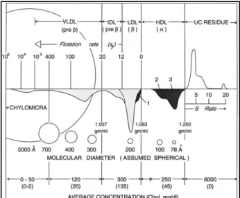
Two basic types of lipoproteins have been postulated. These are micellar lipoproteins and the pseudomolecular lipoproteins.60 The micellar type contains less than 30 % of proteins and includes chylomicrons, VLDL and LDL. These are thought to consist of a hydrophobic core of TG and CE surrounded by a hydrophilic coat of protein, phospholipids, and free cholesterol. The pseudomolecular type contains more than 30 % of protein and includes HDL.
Figure 1. Schematic presentation of the major lipoproteins in normal human plasma. From Olson 1998.61
1.2.2 Lipoprotein metabolism
10
and transport of lipids within the body. In this case, “forward” transport indicates the arrival of cholesterol in the blood from diet (exogenous pathway) and from liver (endogenous pathway) and the carriage back to the liver, whereas “reverse” transport (reverse cholesterol transport (RCT) pathway) is the movement of cholesterol in the opposite direction, the efflux from peripheral tissues back to the liver. They are interdependent pathways and disturbances in one will affect the function and products of the other.
Chylomicrons are the largest lipoproteins and are synthesized, assembled and secreted in the intestinal epithelial cells to transport dietary TG and cholesterol from the site of absorption to the various cells of the body (Figure 2).62 Therefore, chylomicrons are the molecules formed to mobilize dietary lipids (exogenous pathway). The main structural apolipoprotein of chylomicrons is ApoB-48 while others may be found in small amounts.
Figure 2. Overview of lipoprotein metabolism: (1) transport of diet lipids (exogenous pathway). Diet lipids
11 The catabolism of chylomicrons occurs at the endothelial surfaces of capillaries in adipose tissue and muscle: the TG in the core of the chylomicrons is hydrolyzed into free fatty acids and glycerol by the action of lipoprotein lipase (LPL).64This free fatty acids may be then absorbed by the tissues, used as an energy source by various cells or taken up by adipocytes and stored as TG. After the action of LPL, chylomicrons shrink in size becoming chylomicron remnants with lipid cores, having a relatively high concentration of CE.
Chylomicron remnants are taken up by liver cells via receptor-mediated endocytosis by a process equivalent to the mechanism of uptake of LDL and are further metabolized by hepatic lipases (HL) releasing the cholesterol to the endoplasmic reticulum where it becomes part of the cellular cholesterol pool.65,66 This cholesterol can be used for bile acid synthesis or resterified by acyl coenzyme A : cholesterol transferase (ACAT) and packaged along with TG within VLDL particles.
The endogenous pathway of lipoprotein metabolism refers to hepatic secretion and metabolism of VLD, IDL and LDL (Figure 2).
VLDL are TG rich particles with small amounts of CE, phospholipids, ApoB-100 and others apolipoproteins synthesized by the liver.67 ApoB-100 also synthesized by the liver is essential for the assembly of VLDL particles and their secretion into the circulation68,69Like chylomicrons, VLDL acquires in the bloodstream ApoCs and ApoE from circulating HDLs. Within the plasma compartment, the TG of VLDL are hydrolyzed by LPL to free fatty acids, generating a series of smaller, cholesterol-enriched lipoproteins. The free fatty acids may be delivered to cardiac or skeletal muscle cells for β-oxidation or to adipose tissue for TG resynthesis and storage. The circulating VLDL particles become progressively smaller as their core is removed by lipolysis whereas surface materials, including phospholipids, free cholesterol, ApoC’s and some ApoE are transferred to HDL. The smaller VLDL remnant are released from the endothelial cells surface and become IDL.
IDL are essentially composed by CE and a small percentage of TG, having ApoB-100 and ApoE as their main apolipoproteins. IDL may become further enriched by CE derived from HDL by mediation of the cholesterol ester transfer protein (CETP). IDL have two metabolic fates following interaction with HL: (1) to be uptake by hepatocytes after binding to the LDL receptor in a both ApoB-100 and ApoE mediated process, or (2) are subject to further lipase activity continuing to lose TGs and are released into the circulation as LDL.70 In humans this is
12
LDL are cholesterol-enriched lipoproteins, essentially CE, phospholipids, small amounts of TG, and ApoB-100 (the only protein component of LDL) make up the remainder of the particle. In comparison with the originally secreted VLDL, which range from 35 to 80 nm, LDL particles are much smaller having an average diameter of 22 nm, which allows them to cross the vascular endothelium and enter the tissue fluid delivering the cholesterol to the tissues. LDL delivers CE to peripheral (extrahepatic) cells (about 1/3 of LDL that is produced daily) or to hepatocytes within the liver (about 2/3 of LDL that is produced daily).
LDL particles are transferred from circulation to the liver and peripheral tissues via two pathways: 1) LDL receptor pathway which is regulated according to the cholesterol requirement of each individual cell; 2) non-receptor mediated pathways that depends almost entirely on the extracellular concentrations of LDL.
The LDL receptor is a single-chain transmembrane glycoprotein that is mainly expressed by hepatocytes (75 %) but is also present on adrenal and adipose tissue. Although capable of binding ApoEcontaining lipoproteins, LDL receptor usually binds to the ApoB100 containing lipoproteins, in particular LDL. After binding the LDL receptor-ligand complex is internalised within the cell by endocytosis, where it undergoes lysosomal degradation of LDL particles. ApoB is hydrolysed to its constituent aa and the CE is hydrolysed to free cholesterol by acid lipase. Free cholesterol is released to the cytoplasm where is re-esterifed by ACAT and stored as CE, reused for lipoprotein synthesis or converted into bile acid and vitamin D.20 Following that the receptor is recycled back to the cell surface and is again able to bind lipoproteins. Expression levels of LDL receptors are regulated by a sensitive feedback control through the intracellular cholesterol content of hepatocytes and the need for cholesterol. Transcription levels of LDL receptor gene is controlled by promoters with sterol regulatory elements, stimulation of this mechanism causes the release of a sterol regulatory element binding protein (SREBP) which binds to DNA and switches on the transcription.71
13 Via the non-receptor-mediated pathway, LDL binds to cell membranes at sites other than those at which LDL receptors are located and some of it passes through the membrane by pinocytosis. HDL is able to compete with LDL for this type of cell-membrane association.
In addition, LDL may also be removed from the circulation by a number of receptors other than the classical LDL receptor, the scavenger receptors (CD36 and SR-A) which are responsible for the clearance of only relatively minor amounts of LDL. Since they bind modified LDL and are mainly present on the macrophages scavenger receptor have gained considerable interest because they may have a central role in atherogenesis.
Further to the exogenous and endogenous pathways above explained lipid transport also has an important third pathway, the RCT. HDL particles have the capacity to mediate cellular cholesterol efflux by acting as primary acceptors, thereby facilitating RCT, a process in which cholesterol is transferred from peripheral tissues, including from macrophages on the arterial wall, to the liver for the excretion into the bile.73,74
As HDL particles are the main interest of this dissertation these will be described below in more detail, including the RCT pathway since it is considered one of the most important mechanisms by which HDL protects against atherosclerosis.
1.2.3 Lipoproteins and cardiovascular disease (CVD)
The first significant study that associated lipid abnormalities with CVD risk was the Framingham Heart Study, carried out by the National Heart Institute in the small town of Framingham, Massachusetts. It begun in 1948 and it is still ongoing to this day.75,76 The original cohort of the Framingham Heart Study consisted of 5209 subjects of a random sample of 2/3 of the adult population of Framingham with 30 to 62 years of age. Measurements were made of most of the potentially relevant risk factors for CVD known at the time. These included data on cigarette smoking, blood pressure, total serum cholesterol, LDL-C, HDL-C and presence of type 2 diabetes. Since then, the subjects have continued to return to the study every two years for a detailed medical history, physical examination, and laboratory tests with the last examination being set between May of 2008 and February of 2010.
14
factors, such as high blood pressure, smoking, obesity, diabetes and physical inactivity, and that any combination of these risk factors were at least additive.77
15
1.3
High Density Lipoprotein (HDL)
1.3.1 Structure (heterogeneity) and composition
Structurally, HDL represents the smallest (Stoke’s diameter of 7–12 nm) and the densest (ranging from 1.063 to 1.21 /mL) plasma lipoprotein (Table 1), which is produced by the liver and small intestine.79 It is a heterogeneous fraction, comprising a wide range of circulating particles that differ in shape, size, density, composition and surface charge/electrophoretic mobility.80
The basic structure of HDL involves a lipid core of CE and TG surrounded by a surface containing a phospholipid bilayer, free cholesterol and a number of apolipoproteins. HDL proteins constitute more than half of its mass, unlike the others lipoproteins which are mainly composed by cholesterol and /or TG.
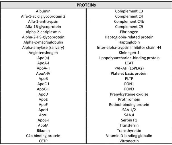
Table 2. Proteins detected in high density lipoproteins (HDL) by mass spectrometry.
PROTEINs
Albumin Complement C3
Alfa-1-acid glycoprotein 2 Complement C4
Alfa-1-antitrypsin Complement C4b
Alfa-1B-glycoprotein Complement C9
Alpha-2-antiplasmin Fibrinogen
Alpha-2-HS-glycoprotein Haptoglobin-related protein
Alpha-2-macroglobulin Haptoglobin
Alpha-amylase (salivary) Inter-alpha-trypsin inhibitor chain H4
Angiotensinogen Kininogen-1
Apo(a) Lipopolysaccharide-binding protein
ApoA-I LCAT
ApoA-II PAF-AH (LpPLA2)
ApoA-IV Platelet basic protein
ApoB PLTP
ApoC-I PON1
ApoC-II PON3
ApoD Prenylcysteine oxidise
ApoE Prothrombin
ApoF Retinol-binding protein
ApoH SAA 1/2
ApoJ SAA 4
ApoL-I Serpin F1
ApoM Transferrin
Bikunin Transthyreitin
C4b binding protein Vitamin D-binding globulin
CETP Vitronectin
16
Studies have allowed the identification of more than 50 proteins in human HDL (Table 2). In addition to the apolipoproteins and enzymes, numerous proteins involved in acute-phase response were found in HDL as well as proteins involved in complement regulation and protease inhibition,86,87 raising the possibility that HDL may play a previously unsuspected role in host defence mechanisms and inflammation. It is important to keep in mind, however, that the content of all these proteins in HDL is much lower than that of major HDL apolipoproteins, i. e., ApoA-I and ApoA-II (70% and 20%, respectively).
1.3.1.1 HDL subpopulations
On the basis of shape most of the HDL are spherical particles, although there is also a minor subpopulation of discoidal HDL (Figure 3 a). Discoid HDL are small ( 8 nm) particles and represent a nascent form of HDL, being lipid-poor and presenting two or three ApoA-I molecules arranged around a phospholipid bilayer. These relatively unstable and short-lived particles will be converted into mature spherical HDL. Spherical HDL particles are larger ( 8 nm) and contain a hydrophobic core of esterified cholesterol and TG surrounded by an outer part composed by two or more ApoA-I molecules, with or without ApoA-II, phospholipids and free cholesterol.80
On the basis of isopycnic ultracentrifugation density HDL may be classified into three subfractions: HDL1 (mean density (d) 1.05 g/mL), HDL2 (1.063 < d < 1.125 g/mL) and HDL3 (1.125 < d < 1.21 g/mL). HDL2 and HDL3 subfractions are spherical and mature particles. The composition of the HDL3 particle is a monolayer of phospholipids, mainly phosphatidylcholine, and a small amount of free cholesterol, ApoA-I and ApoA-II and a core containing esterified cholesterol. HDL3 collects cholesterol to form the larger HDL2 particles without ApoA-II, which can exchange both cholesterol and TG with LDL and VLDL particles.
17
Figure 3. High density lipoproteins (HDL) heterogeneity. The HDL in human plasma consist of several
subpopulations of particles which differ in shape (a), apolipoprotein composition (b), density and size (c) and surface charge in electrophoretic mobility (d). Modified from Rye KA et al 200989 and Tabet F & Rye KA 2009.90
On the basis of electrophoresis HDL can exhibit alpha (α), pre-beta () or gamma () mobility (Figure 3 d). The α–electrophoretic mobility (faster) particles are spherical lipoproteins that include both the HDL2 and HDL3 subfractions as well as the LpA-I and LpA-I/A-II subpopulations and account for the major proportion of HDL in human plasma. Pre- migrating (slower) HDL are either lipid-poor ApoA-I particles, with a single molecule of ApoA-I as a free molecule or in association with a few molecules of sphingomyelin and phosphatidylcholine, or nascent discoidal particles consisting of one or two molecules of ApoA-I complexed with phospholipids and possibly a small amount of unesterified cholesterol. In addition, migrating HDL are lipid-poor particles containing ApoE or ApoA-IV.91,92
18
Figure 4. Nomenclature of the high density lipoproteins (HDL) subclasses determined by different
methods. From Asztlos et al, 2011.93
Due to the increasing need to understand, validate, and quantify the diverse roles of HDL particles in the atherosclerotic process several investigators initiated an effort to uniform the nomenclature for HDL subpopulations. Recently, they proposed the development of a new classification system that defines five HDL subclasses on the basis of physical and chemical properties that includes very large HDL particles (VL-HDL), large HDL particles (L-HDL), medium HDL particles (M-HDL), small HDL particles (S-HDL), and very small HDL particles (VS-HDL). The very small HDL subclass includes pre-β-1, discoidal, or nascent HDL (Table 3).94
Table 3. Classification of high density lipoproteins (HDL) by physical properties. From Rosenson et al.
19 1.3.1.2 Major components
1.3.1.2.1 Apolipoprotein A-I (ApoA-I)
Human ApoA-I circulates in plasma primarily as a component of HDL (70 % of total HDL protein). It is also found in chylomicrons and VLDL.95 ApoA-I has two major sites of synthesis: the intestine and the liver. The intestinal derived ApoA-I enters the circulation associated with chylomicrons but is rapidly transferred to HDL particles during lipase hydrolysis of chylomicrons. Hepatic ApoA-I enters the circulation associated with nascent HDL particles and is the major contributor to the plasma ApoA-I pool.
ApoA-I is a 28 kDa single polypeptide synthesized as a prepropeptide (267 aa residues) and is cleaved to release 24 aa residues. Mature, circulating ApoA-I consisting of 243 aa residues is encoded by exon 3 (residues 1–43) and exon 4 (residues 44–243) of a gene located on the long arm of chromosome 11. Analysis of its aa sequence reveals that with the exception of the 44 aa that form the (N)-terminal region, the protein is organized into eight α-helical amphipathic domains of 22 aa with two repeats of 11 aa that are frequently separated by proline residues. These α-helices contain the lipid-binding carboxyl (C)-terminal domain that confers ApoA-I the capacity of avidly binding to lipids and also to move between lipoproteins.95-97 ApoA-I does not undergo post-translational modifications such as glycosylation, phosphorylation98 and has no disulfide linkages.
ApoA-I is polymorphic in plasma and is composed of a series of isoforms of similar molecular weights but different isoelectric points (pI). The protein is made up of one major isoform ApoA-I1 (pI 5.6) and two minor isoforms ApoA-I2 and ApoA-I3 (pI 5.53 and 5.46).99,100
The concentration of ApoA-I in plasma is about 100-150 mg/dL58 and has a plasma half-life of about 4 days.101
ApoA-I exists in plasma in three general forms: lipid poor ApoA-I, nascent discoidal HDL particles and mature spherical HDL forms. 95,102,103
In 1999 Segrest et al104proposed the “double-belt model” for discoidal HDL that consists
of two ring-shaped ApoA-I molecules wrapped around a leaflet of a disk-like patch of lipid bilayer in an anti-parallel orientation. Despite some debate on details of certain regions of ApoA-I in the discs, the majority of the most recent theoretical and experimental data supports the general features of the “double belt model” 105-107
In 1997, Borhani et al108 hypothesized a “Faberge Egg model” for spherical HDL,
20
“double-belt model” in discs) are also present in HDL spheres. However, one model recently presented, using cross-linking chemistry and mass spectrometry , indicates that the “ double-belt model” is a common organizational motif for ApoA-I in both discs and spheres.109 These authors also showed that particles with a diameter superior to 93 Å (which is comparable with human HDL3) present one extra molecule of ApoA-I, but the cross-linking patterns in reconstituted spheres were highly similar to those in the discs, regardless of whether they contained three molecules of ApoA-I in each particle or only two. The most promising model seems the “trefoil model” where the phospholipids in the sphere surface are broken into
three equal slices with angles of 120○.109
The understanding of the ApoA-I spatial arrangement in both discs and spheres is critical because it offers insights into how the ApoA-I structure modulates the metabolism and function of HDL. In fact, Apo A-I is essential for the correct assembly, overall stability of HDL and the regulation of HDL metabolism.
1.3.1.2.2 Apolipoprotein A-II (ApoA-II)
ApoA-II is the second most important HDL apolipoprotein and represents approximately 15-20 % of total HDL protein. About half of the HDL particles may contain ApoA-II. ApoA-II circulates as a 17 kDa protein and its synthesis takes place mainly in liver.110 Like ApoA-I it is also synthesized as a propeptide (100 aa residues) from a gene on chromosome 1 and is posterior cleaved to release 23 aa residues.111 ApoA-II transcription is upregulated by nuclear receptors such as peroxisomal proliferative-activated receptor-alfa (PPAR-), retinoid X receptor (RXR) and SREBP-2.112,113
ApoA-II circulates as a homodimer of two identical polypeptide chains, each containing 77 aa and linked by a single disulfide bond.111 The presence of a cysteine residue allows ApoA-II to form heterodimers with other cysteine containing apolipoproteins, such as ApoE and ApoD. Although it has amphipathic properties, ApoA-II is more hydrophobic than ApoA-I.