MUNIBE (Antropologia-Arkeologia) Nº 46 123-132 SAN SEBASTIAN 1994 ISSN1132-2217
Aceptado: 1994-01-28
Ungueal Morphology and Pathology of the human mummy
found in the Val Senales (Eastern Alps, Tyrol, Bronze Age)
Morfología Ungueal y Patología de la momia humana hallada en Val Senales
(Alpes Orientales, Tirol, Edad del Bronce)
KEY WORDS: Fingernail, paleopathology, Beau's lines.
PALABRAS CLAVE: Uña, paleopatología, líneas de Beau-Reil.
Luigi CAPASSO *
* Laboratory of Anthropology National Archaeological Museum Chieti (Italy).
SUMMARY
The author describes a nail plate from a finger of the by now well known Val Senales mummy. The nail had become detached in an- cient times, and was recovered in the course of the archaeological excavation conducted in the area in August 1992.
This nail plate (find Nº 168/92) is extremely interesting from a paleopathological standpoint. Several destructive lesions that are con- centrated over part of the distal region of the dorsal surface of the nail appear to be attributable, both because of their typology (onycore- xis and microgrooves) and topography, to microtraumas that occurred in life, as a result of the use of the nail as a tool.
In addition, the presence of three sets of Beau's lines have allowed us to determine the seriousness, time of onset, and duration of three periods of intense stress that the subject underwent during the last months of his life the nature of these stresses remains unk- nown, even though their repetition suggests they are somehow related to a disease with acute episodes. During the last five months of his life. The subject underwent three such episodes; they occurred four, three, and two months before death. The final episode was the most serious, and the systemic upheaval it produced lasted at least two weeks.
RESUMEN:
El autor describe una lámina de uña perteneciente a un dedo de la, actualmente bien conocida, momia de Val Senales. La uña había quedado separada del resto en tiempos antiguos y fue recuperada en el curso de la excavación arqueológica llevada a cabo en el área en agosto de 1992.
Esta lámina de uña (hallazgo nº 168/92), es de un enorme interés desde el punto de vista paleopatológico. Varias lesiones destructivas concentradas sobre parte de la región distal de la superficie dorsal de la uña parecen ser atribuibles, tanto por su tipologia (onicorrexis y microsurcos) como por su topografía, a microtraumas que tuvieron lugar en el transcurso de la vida del sujeto, como resultado de la utili- zación de la uña como instrumento.
Además, la presencia de tres grupos de líneas de Beau-Rei1 nos ha permitido determinar la gravedad, tiempo de ataques o arremeti- das y duración de tres periodos de intensa tensión que el sujeto experimentó durante los últimos meses de su vida. El sujeto sufrió tres de dichos episodios, que tuvieron lugar cuatro, tres y dos meses antes de su muerte. El último episodio fue el más serio, y el trastorno sistemático que produjo duró al menos dos semanas.
LABURPENA
Val Cenales-eko momia ezagunaren hatzazalaren ijelki bat aztertzen du artikuluaren egileak. Hatzazala gorputzetik banatua gertatu zen aintzinean eta 1992ko abuztuan bertan egindako indusketan berreskuratua izan zen.
Hatzazal ijelki hau (168/92 zkia.) paleopatologiaren ikuspegitik oso interesgarria suertatu da. Hatzazal honen gainaldean eta ertz-distale- an azaltzen diren lesio txikitzaileak, beren tipologiari (onicorrexia eta mikrohildoak) eta topografiari begiratuz, badirudi gizaki honen bizitzan zehar hatzazala tresna bezala erabitzeagatik sortutako mikrotrauma batzuen ondorio liratekeela.
Gainera, Beau-Reuil-en hiru lerro-multzo azaldu direnez, gizon honek bizitzako azken hilabetetan jasan izan zituen hiru estualdi edo ten- tsio uneen larritasuna, erasoaldien denbora eta iraupena azertu ahal izan dira. Gizakihonek horrelako hiru estualdi jasan zituen, hil baino lau- garren, hirugarren eta bigarren hilabeteetan. Azkeneko estualdi edo tentsio-unea izan zen gogorrena eta honek eragindako nahaste siste- matikoak gutxienez bi aste iraun zuen.
INTRODUCTION
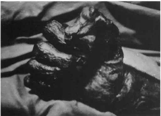
The detailed anthropologic examination of the human mummy found in the Val Senales in Septem- ber 1991 revealed that it had lost all the nails of its finger (Fig. 1) and toes. The loss of the nails follo-
wing death has also been noted in other ancient mummies.
Figure 1. Detail of the right hand of the Val Senales mummy (Tyrol). The total absence of the nails (lost post mortem) and the good preser- vation of the nail bed are evident.
and pathological observations on this type of tissue as well.
The study of this kind of human remain is extre- mely important and can furnish a surprising amount of information on the life style of the subject, the sort of work the subject performed, and, finally, the systemic diseases the subject suffered from, if any. As Brothwell (1992) notes, the study of human nail plates from archaeological sites "opens new hori- zons to bio-archaeological research, and raises new questions about this unusual line of research".
In the present paper we accepted this invitation, and were astounded by the quantity and quality of the paleobiological informations obtained from the study of a single specimen.
MATERIALS AND METHODS
The specimen examined in this study is find Nº 168 ("human fingernail"), found on August 15, 1992 at Hauslabjoch (Val Senales) in the course of the ar- chaeological excavation directed by Dr. DAL RI, Dr.
NOTHDURFTER, and Prof. LIPPERT.
The archaeological site has been dated to the Bronze Age, while the radiometric analysis of the mummy, carried out with the 14C-AMS method by the Radiocarbon Laboratory of Zurich, has yielded an absolute age of about 5,300 years, (BONANI et al.,
1992).
The fingernail was found a few meters down slo- pe from the point from which the mummy was ex- tracted the year before, during the filtration of the
melt water from the ice up slope. Consequently the stratigraphic location of the find within the site is un known.
To date, only the external morphology of the fin- gernail has been 'examined, using a binocular ste- reoscopic microscope capable of zooming from 6x to 50x.
During the observation and photography of the specimen, it was kept completely immersed in a ste- rile saline solution, at a temperature of about +0.5ºC.
It has not been possible to observe the nail with a scanning electron microscope, because the nail must be kept wet, lest it rapidly develop deep cracks and fissures. At present the feasibility of making re- sin casts of the nail for use with the SEM is being evaluated. A basic chemical analysis is being carried out, and the results will be published shortly.
We must note that the mechanisms that might have produced the detachment of the nail plate from its bed are unknown. It is clear, however, that the mechanism operated equally on both the fingers and toes, all of which lost their nails.
There is only one other example mentioned in the literature that can be compared with this, the so- called "Lindow Man", a mummy from the bogs of Schleswing (Germany) that has been dated to about 300 B.C. (BROTHWELL, 1992). An initial phase of de-
UNGUEAL MORPHOLOGY AND PATHOLOGY OF THE HUMAN MUMMY FOUND IN THE VAL SENALES (EASTERN ALPS, TYROL, BRONZE AGE) 125
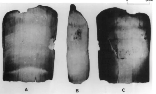
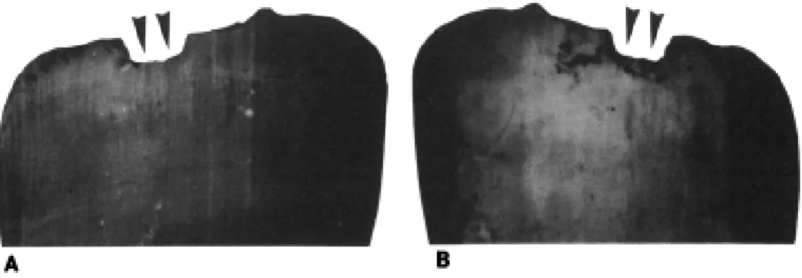
Figure 2. Human fingernail, (find Nº 168/1992) from the archaeolo- gical excavations carried out at Val Senales (Tyrol) by the Ar- chaeological Superintendency of Bolzano A: ventral side of the nail B: profile. C: dorsal side of the nail. The distal edge is up. The photographs (and the rest as well) were taken through a stereo microscope while the specimen was completely immersed in a 0.5ºC bath of saline solution.
It is not difficult to imagine that a similar mecha- nism may have operated in the case of the "Ice Man". Indeed, in this case the absence of burial un- doubtedly favored the initial phases of decomposi- tion, including the loosening of the nail plates from their beds. During thaws, superficial water currents may have carried the nails considerable distances from the body.
DESCRIPTION
Find Nº 168/1992 is a human nail plate; it is tra- pezoidal in shape, and elongate longitudinally (Fig. 2). Its dimensions are:
Maximum lenght: 15 mm Maximum width (close to the tip at the
distal end) 12 mm Minimum width (at the base of the nail bed) 10 mm The nail plate reaches its maximum thickness (about 0.8 mm) at the distal end; at the proximal end its thickness is reduced to a tenth of a millimeter. The major axis of the nail is oriented proximodistally, and the general shape of the nail is subrectangular. This shape is typical of the nails of the fingers, while those of the toes are generally much squarer.
The nail plate is convex, like the crystal of a watch, and its convexity is developed both longitudi- nally and transversally. However, the longitudinal convexity is minimal (Fig. 2b), because the curvature of the matrix is slight. The transversal curvature of the nail is slightly greater. Over all, therefore, the nail is only slightly convex: this is typical of the nails of the fingers, where the low convexity is a response to the more elongate, flatter morphology of the phalan- ges of the hands (GONZÀLEZ-SERVA, 1990).
The general shape of the nail and its convexity suggest that it came from a finger. The identification of the finger or the side of the body from which it ca- me is impossible. We can, however, exclude the lit- tle finger (because its nail tends to be small).
The dimensions of our sample fall within the fields of variability for the nails of the thumb, forefin- ger, index finger, and ring finger of a modern Euro- pean male whose height is between 155 and 165 cm (HAMILTON et al., 1955).
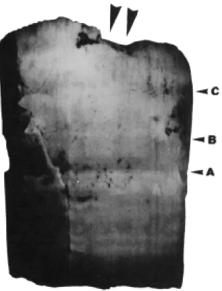
The dorsal face of the nail is an almost uniform light brown in color. Two lighter colored striations, both slimmer than 1 mm, cross the entire nail about 2 mm from its proximal edge. These lines (marked with arrows in Fig. 4A) may be related to the attach- ment of the cuticle to the dorsal face of the nail. The dorsal face of the nail is also crossed by three trans- verse furrows. The most proximal furrow ("furrow A" in Fig. 3) is also the deepest; it is about 1 mm wi- de, and is located about 6.9 mm from the proximal edge of the nail.
A second, more distal furrow, which is much shallower than the first, is located 8.6 mm from the proximal edge of the nail ("furrow B" in Fig. 3).
The third furrow, whose width and depth are in- termediate between those of the first two, is located about 10.8 mm from the proximal edge of the nail ("furrow C" in Fig. 3).
Figure 3. Locations of the three Beau’s lines on the dorsal side of the nail (the letters refer to the description given in the text). The arrows point to the edge with a large loss of onycokeration.
Figure 4. Detail of the proximal part of the finger nail. A: dorsal si- de, B: ventral side. The arrows point to two colored lines which may be where the ungueal cuticle was attached. On the ventral si- de, the letter M indicates the extent of the nail matrix, and L the extent of the lunula.
nish deposits that do not display crystalline structure under high magnification, but rather appear to be amorphous encrustations. These encrustations are more abundant in the centre of the nail and towards the tip, where they occur both in the center and to- wards the lateral edges. The same encrustations are also present on the ventral surface of the distal ed- ge, and, to a greater degree, on a 2 mm wide band along the ventral surface of the tip.
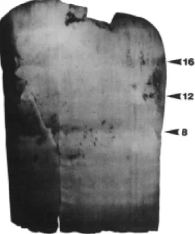
The ventral face of the nail is evenly concave, and is evenly crossed by the furrows of the nail bed, which are more closely spaced and more pronoun- ced towards the sides of the nail (Fig. 5). These fu- rrows tend to deepen and become more closely spa- ced with increasing age (HAMILTON et al., 1955). The
transverse furrows present on the dorsal side of the nail are not matched by any sort of mark on the ven- tral side. The nail does not present any features, either chromatic or morphological, that can be rela- ted to the presence of the onycodermic band. The nail matrix is clearly evident only on the ventral face of the nail. Said matrix is light yellow at the center of the nail, and tends towards brown along the sides (Fig. 4B). The distal margin of the matrix is gently rounded and convex distally, and has almost straight edges towards the sides (lunula). The hypocromia of the nail, including the lunula, is not at all evident on the dorsal side of the nail (Fig. 4A). This is because
the keratogenous zone (which is lighter) is in direct contact with the nail bed, and in other words is loca- ted on the ventral side of the nail, while on the dorsal side the width of the layer (wich is darker) is extre- mely reduced throughout the whole area of the nail matrix (GONZÀLEZ-SERVA, 1990).
With regards to the edges of the nail, the proxi- mal edge is almost straight, but slightly saw-toothed (Fig. 6), probably as a result of the post mortem loss of small fragments. The lateral edges are sub-para- Ilel. and diverge slightly in the distal direction. They are almost knife-like in the region of the matrix, and are rounded and thick towards the tip.
The tip of the nail is gently rounded and convex distally, and parallels the lunula. At the intersections between the distal edge of the nail and lateral edges, the nail is smoothly rounded (Fig. 5).
UNGUEAL MORPHOLOGY AND PATHOLOGY OF THE HUMAN MUMMY FOUND IN THE VAL SENALES (EASTERN ALPS, TYROL, BRONZE AGE) 127
Figure 5. Latero-anterior detail of the ventral face of the nail, sho- wing the well developed furrows of the nail bed and the well roun- ded edges of tip. The arrows point to the zones with heavier depo- sits of brown amorphous matter ("dirt").
Figure 6. Highly enlarged view of the proximal edge of the nail, which is slightly saw-toothed, as a result of the post mortem loss of small fragments.
As we have already noted, these encrustations form a border that appears to be uniform along the ventral face of the tip of the nail.
The distal edge is interrupted by a large square notch located in its center. The detachment surface is inclined with respect to the surface of the nail, ra- ther than perpendicular, and thus the outline of the notch is considerably larger on the dorsal side of the nail (indicated by arrows in Fig. 3) than it is on the ventral side. It is important to note that the detach- ment surfaces are encrusted with brownish amorp- hous particles that appear to be identical to those present along the rest of the tip.
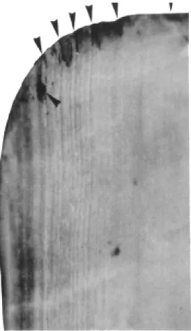
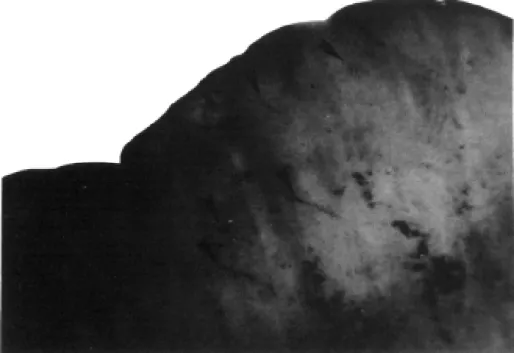
Along a band about 5 mm from the tip, on the dorsal side, there are minute grooves that can only be observed under high magnification and with tan- gential light (Figs. 8 and 9). These grooves are sha- llow, from 1 to 2.5 mm long, are not parallel to each other, and are always oriented at a slight angle with respect to the tip. No grooves oriented parallel to the tip were observed. Around the detachment zone of the onychokeratin in the center of the tip (indicated with arrows in Fig. 3), these grooves appear to be concentrated and arranged radially. The beds of many of these grooves are covered by brown amorp- hous encrustations.
OBSERVATIONS
Several important conclusions can be drawn from the nail plate described above, both with res- pect to the furrows present on the dorsal surface, and with respect to the features found on the tip.
Figure 7. Tip of the nail. A: ventral side. B: dorsal side. Dark amorphous encrustations are clearly evident on the ventral side, where they are tightly bonded to the onycokeratin, especially on the beds of the furrows of the nail bed. On the dorsal side. the chip in the onycokeratin that presumably formed during the life of the individual is clearly evident (see Fig. 8). The arrows point to the edge with a large loss of onycokera- tin (see text).
Figure 8. Detail of the tip of the nail at the point where it is chip- ped. Note the presence of amorphous brown encrustations on the detachment surface, and the presence of grooves ("professional scratches"), which also contain encrustations, in the vicinity of the chip (arrows). Asterisk point to the edge with a large loss of onyco- keratin (see text).
ness is also reduced during the period of stress. These transversal furrows provide a precious record of the occurrence of a disease (BEAU, 1846).
The factors that can cause a reduction in the growth rate and thickness of the nails are quite va- ried, and can be either local or systemic. Among the local factors, there are ungueal psoriasis (BASUK et al., 1990), and traumas or injuries to the region of the proximal nail fold (DANIEL III et al., 1990).
However, these localized pathologies rarely pro- duce transversal furrows that extend across the enti- re nail, since the stress they cause is almost always limited to a single sector of the nail matrix. Therefo- re, these localized factors generally produce disconti- nuous Beau's lines, with depressions that are limited to a single sector of the nail. In addition, traumatic in- juries never result in multiple Beau's lines.
Systemic causes can on the other hand result in the formation of more extensive and regular Beau's lines. They are produced by diseases that result in a temporary reduction or halt in the development or growth of the nail. However, only fairly serious sys- temic diseases are capable of producing the degree of suffering of the nail matrix necessary to produce a reduction in the growth rate of the nail that can be observed macroscopically. We are therefore dealing with some sort of serious systemic disease that las- ted at the very least for several days (DE NICOLA et al., 1974).
UNGUEAL MORPHOLOGY AND PATHOLOGY OF THE HUMAN MUMMY FOUND IN THE VAL SENALES (EASTERN ALPS, TYROL, BRONZE AGE) 129
Figure 9. Detail showing the mor- phology and orientation of the grooves present on the distal part of the dorsal side of the nail. These grooves are related to the use of the nail during work ("pro- fessional scratches").
eth of the adult as a permanent record of a period of stress in infancy (GOODMAN & CAPASSO, 1992).
As is the case with dental hypoplasiae, not all of the serious systemic diseases which can result in ungueal hypoplasiae (Beau's lines) are known. It would seem reasonable that Beau's lines, like dental hypoplasiae, could be the result of generic forms of stress whose major characteristics are seriousness, systemicity, and duration.
Diseases or other stressful conditions, which can also be of extremely different natures, are capable of producing the same type of hypoplastic lesion.
For example, Beau's lines can appear on new- born infants around the first month, and persist until the age of four months (TURANO, 1968). This tempo-
rary halt in growth is rarely observed, is analogous to the dental birth-line (from ameloblastic stress related to birth; SEOW, 1992), and is probably similar to the
telogen effluvium that strikes many babies during the same period. These neonatal Beau's lines are certainly related to the stress resulting from the adaptation to extrauterine life (SILVEMAN, 1990). No
correlations have been found, however, between Seau's lines and either intrauterine malnutrition, as- phyxia at birth, or feeding with artificial milk. Howe- ver, in a case discussed by WOLF et al. (1982), neona-
tal Beau's lines were shown to have formed follo- wing intrauterine asphyxia.
In adults, Beau's lines have been correlated with generalized suffering due to a variety of diseases. LONG (1981) observed them in cases of parathyroidal
disease, WEISMANN (1977) has correlated them with
zinc deficiencies, and COLVER & DAWBER (1984) have
found them in association with dysmenorrhea. The appearance of Beau's lines in subjects suffering from serious chronic diseases of the digestive system, with or without complications affecting the liver, is more common (DE NICOLA et al., 1974).
BEAVEN & BROOKS (1991) have shown that deep Beau's lines can form a few weeks after a serious episode in which the patient nearly dies. Even though there is no confirmed proof, many workers have observed that a serious febrile disease lasting several days or a less serious metabolic upset that lasts a week is necessary to produce these furrows. Diseases of short duration that are sudden and extre- mely serious, such as a life threatening hemorrhage with hypotension, are capable of producing transver- se lines that can be distinguished on all the nails (BEAVEN & BROOKS, 1991).
No matter the nature of the stress, there is pro- bably a direct correlation between the intensity of the stress suffered and the probability that a subject will develop Beau's lines (DANIEL III et al., 1990).
Likewise, the width of the hypoplastic line and its depth could be directly related to the duration and seriousness of the stress (LEWIN, 1965).
On our nail it has been possible to measure the distance between the proximal margin of the nail matrix and the Beau's lines; this measurement is ex- tremely interesting, because the growth rate of fin- ger nails, which appears to be constant (BEAN, 1974),
is rather well known.
Figure 10. Graph showing the possible chronological relationships between the Beau's lines and the approximate number of weeks before death.
to a maximum of 4.5 mm per month, but tends to re- main constant in a given individual, and to be similar among members of a given family (HAMILTON et al.,
1955). The generally accepted average is 3 mm per month, in other words about 0.1 mm per day for the nails of the fingers (SAMMAN & FENTON, 1986); in a ge-
nerally cooler climate, the average rate may have be- en lower (GEOGHEGAN et al., 1958; Le Gros CLARK &
BUXTON, 1938).
Applying these parameters to our case, we find that the stress which produced the most distal Beau Line ("furrow C" in Fig. 3) began about four months before the death of the individual, the one that pro- duced the second line began about three months be- fore death, and, finally, the episode that caused the formation of the most proximal line began about two months before the death of the individual (Fig. 10).
The final episode was undoubtedly the most se- rious, since the Beau's line it produced is the widest and deepest of the three. The furrow is about a milli- meter wide, which would suggest a period of stress that lasted at least ten days. However, experiments have shown that the growth rate of nails decreases in the course of infectious or circulatory diseases (BEAN, 1974); It is therefore possible that the stress
in this case lasted longer, perhaps as long as two weeks.
It is evident that the interpretation proposed here is limited by a series of factors:
Indeed, systemic stresses cause the simultaneous appearance of similar Beau's lines on all the nails. Some workers have highlighted the fact that for Beau's lines to be correlable with a serious illness, they must be observable on all the nails of one hand, or, preferably, of both hands (BEAVEN & BROOKS,
1991).
b) The data available only reveal the stesses that affected the individual during the final five months of his life (about 150 days, considering that the total length of the nail is 15 mm); we do not know if he suffered prior episodes of stress.
c) It is impossible to establish correlations bet- ween the episodes of stress, even though the possi- bility that the three lines were caused by three episo- des of the same kind of stress (for example acute at- tacks of a chronic disease) would seem reasonable. Neither is it possible to establish a correlation, at this level of observations, between the preceding periods of stress and the cause of death.
The examination of the tip of the nail has produ- ced other interesting revelations. In particular, the significance of the three following items must be in- terpreted: a) the brown amorphous encrustations, b) the detachment of onycokeratin from the center of the distal edge of the nail, and c) the microstriae pre- sent on the distal portion of the dorsal side of the nail (so-called "professional scratches"; RAMRAKHIANI, 1978).
If the brown amorphous encrustations were a post-mortem deposit, one would expect them to be randomly distributed over the entire surface of the nail, especially if it became detached shortly after death. The dorsal surface, however, is completely free from these encrustations. They are instead con- centrated along the distal end of the ventral surface of the nail, so heavily as to create a dark rim that pa- rallels the distal edge (Figs. 5 and 7A). It would appe- ar that this amorphous material is the remains of dust that accumulated under the fingernail, the way it still does today, when a person's nails are dirty.
The loss of material along the distal end of the nail takes the form of a chip (onycorexis); in addition, the detachment surfaces are encrusted with the sa- me brown amorphous substance.
UNGUEAL MORPHOLOGY AND PATHOLOGY OF THE HUMAN MUMMY FOUND IN THE VAL SENALES (EASTERN ALPS, TYROL, BRONZE AGE) 131
the nail. These microstriae are, instead, present only on the dorsal surface of the nail, and are concentra- ted towards the distal end, in the vicinity of the chip- ped region. They should therefore be considered "professional scratches" (RAMRAKHIANI, 1978).
If we observe the general topographic distribu- tion of the chip and the microstriae (Figs. 8 and 9), al- so taking the orientation of the striae into account, it becomes clear that one part of the tip of the nail was more subject to traumas, perhaps as a result of work performed by the individual.
The only comparisons possible are with another archaeological specimen, which was described by BROTHWELL (1992). The finger nails of the so-called "Lindow Man", already mentioned above, have mi- crostriae on the distal most parts of their dorsal sides. In this case, however, the microstriae are all oriented the same way, parallel to the distal edge of the nails. BROTHWELL therefore interpreted these grooves to be
the traces left by a nail file. Thus, the "Lindow Man" has been portrayed as a dignitary deserving of mani- cures!
The striations of the nail of the "Ice Man" sug- gest the reverse: the lesions are compatible with much more traumatizing activities, for example he- avy manual labor (KERN, 1990). In fact, a comparison
between the typology and topography of the lesions present on the Ice Man's fingernail and the ample documentation on professional lesions published, for example, by BEAVEN & BROOKS (1991), shows that
they bear the closest resemblances with the lesions found on the nails of farmers, stone masons, mecha- nics, etc. Therefore, our nail appears to have belon- ged to a coarse laborer, who used the extremities of his fingers as tools (one must note that the heavy wear of the anterior teeth shows that they too were used for extramasticatory activities; CAPASSO et al.,
1992).
CONCLUSIONS
Find Nº 168/1992 is a fingernail that fell from one of the fingers of the Ice Man, probably as a result of the initial post-mortem decomposition of the nail bed. The nail displays several morphological and pat- hological features that shed light on some aspects of the paleobiology of the individual.
The deep grooves present on the nail are a form of ungueal alteration that correlates with age (HAMIL- TON et al., 1955), a fact that should be taken into con-
sideration by the anthropologists studying the mum- my, who have proposed an extremely uncertain age estimate for the individual, between 25 and 40 years (SJVOLD, 1992).
The presence of three Beau's lines suggests that the individual suffered three serious bouts of physi-
cal stress, respectively four, three, and two months before death. The final episode, which occurred two months before the time of death, was the most se- rious, and the causative agent (or its pathological ef- fect on the individual) lasted for at least two weeks. All these suggests that a chronic infectious disease was possible involved.
Finally, the examination of the distal end of the nail has revealed the presence of brown particles at- tached to the distal end of the ventral side of the nail ("dirt", accumulated under the finger nail), and, mo- reover, traces of a series of traumatic lesions con- centrated on part of the dorsal side of the tip of the nail (chips and "professional scratches"), which, if they occurred in life, show that the individual used this nail when he worked.
ACKNOWLEDGEMENTS
The author is indebted to Dr. H. STAMPFER, supe-
rintendent of the District of Bolzano, and Dr. L. DAL
RI, director of the Archaeological Offices of the Dis-
trict of Bolzano, for allowing him to carry out the an- thropological examination of the human remains re- covered from the Val Senales in the course of the ex- cavations carried out in 1992.
In addition, the author thanks Mrs. NADIA RABO- TTINI for her patient editing. Finally, the author wishes
to express his thank to Dr. KYLE PHILLIPS, for transla-
ting the text into English.
This work is partially supported by Consiglio Nazionale delle Ricerche.
REFERENCES
BASUK, P.J.; SCHER, R.K.; RICCI, A.R.
1990 Dermatological Diseases of the nail-unit. In: Scher, R.K. & Daniel III, C.R. (Eds.): Nails: Therapy, Diagnosis, Surgery. W.B. Saunders Company, Philadelphia.
BEAN, W.B.
1974 Nail growth: 30 years of observation. Arch Intern. Med. 134, 497.
BEAU, J.H.S.
1846 Certain caracteres de semeiologie retrospective, presen- tés par les ongles. Archives General Medicine 9, 447. BEAVEN, D.W. & BROOKS, S.E.
1991 L'unghia nella diagnosi clinica. Wolfe Medican Publication Ltd., Harofarma, U.K.
BONANI, G.; IVY, S.D.; NIKLAUS, T.R. & SUTER, M.
1992 Altersbestimmung von Milligrammproben der Otztaler Gletscherleiche mit der Bescheleunigermassenspektro- metrie-Methode (AMS). Veroffentlichungen der Universi- tat Innsbruck 187, 108-116.
BROTHWELL, D.R.
COLVER, G.B. & DAWBER, R.P.R.
1984 Multiple Beau's lines due to dysmenorrea? Br. J. Derma- tol. 111, 111.
DANIEL III, C.R.; SAMS, W.M. & SCHER, R.K.
1990 Nails in systemic diseases. In: Scher, R.K. & Daniel III, C.R. (Eds.): Nails: Terapy, Diagnosis, Surgery. W.B. Saunders Company, Philadelphia.
DAWEBER, R. & BARAN, R. 1987 Nail Growth. Cutis 39, 99.
DE NICOLA, P.; MORSIANI, M. & ZAVAGLI. G.
1974 Nail Diseases in Internal Medicine. C.C. Thomas, Spring- field.
GEOGHEGAN, B.; ROBERTS, D.F. & SANFORD, M.R.
1958 A possible climatic effect on nail growth. J. Appl. Physiol. 13, 135.
GONZALES-SERVA, A.
1990 Structure and Function. In: Scher, R.K. & Daniel III, C.R. (Eds): Nails: Therapy, Diagnosis, Surgery. W.B. Saunders Company, Philadelphia.
GOODMAN, A.H. & CAPASSO, L.
1992 Recent Contribution to the Study of Enamel Develop- mental Defects. Journal of Paleopathology (Monographic Publication) 2, 1.
HAMILTON, J.B.; TERADA, H. & MESTLER, G.E.
1955 Studies of growth throughout the lifespan in Japanese: growth and size of nails and their relationship to age, sex, hereditary, and other factors. J. Gerontol. 10, 401. KERN, D.G.
1990 Professional Diseases. In: Scher, R.K. & Daniel III, C.R. (Eds.): Nails: Terapy, Diagnosis, Surgery. W.B. Saunders Company, Philadelphia.
LE GROS CLARK, W.E. & BUXTON. L.H.
1938 Studies in nail growth. Br. J. Dermatol. 50, 221.
1981 The clinical spectrum of parathyroid disease. J. Am. Acad. Dermatol. 5. 733.
RAMRAKHIANI, M.
1978 Identification and hardness studies of human nails. Indian Journal of Biochemistry and Biophysics 15(4), 341. RONCHESE, F.
1969 Occupational nails. Cutis 5, 164. SAMMAN, P.D. & FENTON, D.A. (Ed.)
1986 The Nails in Disease, ed 4. Yearbook Medical Publishers, Chicago.
SEOW, W.K.
1992 Dental enamel defects in low birth weight children. Jour- nal of Paleopathology (Monographic Publication) 2, 321. SILVEMAN, R.A.
1990 Pediatric Pathology. In: Scher, R.K. and Daniel III, C.R. (Eds.): Nails: Terapy, Diagnosis, Surgery. W.B. Saunders Company, Philadelphia.
SJVOLD, T.
1992 The Stonne Age Man from the Austrian-Italian Alps. Dis- covery, Description and Current Research. Collegium An- tropologicum 16(1), 1.
TURANO, A.F.
1968 Transverse nail ridging in early infancy. Pediatricis 41, 996.
WEISMANN, K.
1977 Lines of Beau: possible markers of zinc deficiency. Acta Dermato-Venereologica (Stockh) 57 (1), 88.