Smart Soup, a Traditional Chinese Medicine Formula,
Ameliorates Amyloid Pathology and Related Cognitive
Deficits
Yujun Hou1., Ying Wang2., Jian Zhao1
*., Xiaohang Li1
, Jin Cui1, Jianqing Ding2, Ying Wang3, Xianglu Zeng1, Yun Ling4, Xiaoheng Shen5, Shengdi Chen2, Chenggang Huang4, Gang Pei1,6*
1State Key Laboratory of Cell Biology, Institute of Biochemistry and Cell Biology, Shanghai Institutes for Biological Sciences, Graduate School of the Chinese Academy of Sciences, Chinese Academy of Sciences, Shanghai, China,2Institute of Neurology, Ruijin Hospital, Shanghai Jiaotong University School of Medicine, Shanghai, China,
3Department of Neurology, Ruijin Hospital, Shanghai Jiaotong University School of Medicine, Shanghai, China,4Shanghai Institute of Materia Medica, Chinese Academy of Sciences, Shanghai, China,5Department of Traditional Chinese Medicine, Ruijin Hospital, Shanghai Jiaotong University School of Medicine, Shanghai, China,6Shanghai Key Laboratory of Signaling and Disease Research, School of Life Science and Technology, Tongji University, Shanghai, China
Abstract
Alzheimer’s disease (AD) is a progressive neurodegenerative disease that causes substantial public health care burdens. Intensive efforts have been made to find effective and safe disease-modifying treatment and symptomatic intervention alternatives against AD. Smart Soup (SS), a Chinese medicine formula composed of Rhizoma Acori Tatarinowii (AT), Poria cum Radix Pini (PRP) and Radix Polygalae (RP), is a typical prescription against memory deficits. Here, we assessed the efficacy of SS against AD. Oral administration of SS ameliorated the cognitive impairment of AD transgenic mice, with reduced Ablevels, retarded Abamyloidosis and reduced Ab-induced gliosis and neuronal loss in the brains of AD mice. Consistently, SS treatment reduced amyloid-related locomotor dysfunctions and premature death of AD transgenic Drosophila. Mechanistic studies showed that RP reduced Ab generation, whereas AT and PRP exerted neuroprotective effects against Ab. Taken together, our study indicates that SS could be effective against AD, providing a practical therapeutic strategy against the disease.
Citation:Hou Y, Wang Y, Zhao J, Li X, Cui J, et al. (2014) Smart Soup, a Traditional Chinese Medicine Formula, Ameliorates Amyloid Pathology and Related Cognitive Deficits. PLoS ONE 9(11): e111215. doi:10.1371/journal.pone.0111215
Editor:Hua Zhou, Macau University of Science and Technology, Macao
ReceivedMarch 23, 2014;AcceptedSeptember 28, 2014;PublishedNovember 11, 2014
Copyright:ß2014 Hou et al. This is an open-access article distributed under the terms of the Creative Commons Attribution License, which permits unrestricted use, distribution, and reproduction in any medium, provided the original author and source are credited.
Data Availability:The authors confirm that all data underlying the findings are fully available without restriction. All relevant data are within the paper and its Supporting Information files.
Funding:This work was supported by National Natural Science Foundation of China (31371419), Chinese Academy of Sciences (KSCX2-EW-Q-1-01, KSCX2-YW-R-252), National Science and Technology Support Program (2012BAI10B03), the Ministry of Science and Technology (2011CB910202) and Shanghai Municipal Commission for Science and Technology (13401900600). The funders had no role in study design, data collection and analysis, decision to publish, or preparation of the manuscript.
Competing Interests:The authors have declared that no competing interests exist. * Email: gpei@sibs.ac.cn (GP); jzhao@sibs.ac.cn (JZ)
.These authors contributed equally to this work.
Introduction
Alzheimer’s disease (AD) is a fatal and progressive neurodegen-erative disease which is characterized by the persistent worsening of cognitive function and daily living performance [1,2], and it has been considered one of the most significant health, social and economic crises of this century. AD is a complex disease, and its pathological hallmarks include senile plaques formed with amyloid peptides, tangles containing twisted tau proteins, destroyed synapses, abnormal brain inflammation, eventual neuronal death and severe brain shrinkage [3]. Current FDA-approved Alzhei-mer’s drugs are only symptomatic interventions, such as acetyl-cholinesterase inhibitors and N-methyl-D-aspartate (NMDA) receptor antagonists, which are effective only for about half of the patients for approximately 6–12 months [4]. Up till now, no disease-modifying medications have been approved in the last 10 years. Considering that, as with many other complex diseases, the pathogenesis of AD has a multifactorial basis that includes both genetic and environmental risk factors, a successful therapeutic
strategy against this disease might be a set of concerted pharmacological interventions that interact with multiple targets including Ab/tau pathology, abnormal inflammation and neuro-nal loss.
composed of Rhizoma Acori Tatarinowii (AT), Poria cum Radix Pini (PRP) and Radix Polygalae (RP). Each component is frequently used in different TCM formulae for their pharmaco-logical efficacies against dysfunctions of the central nervous system (CNS, Table S1). AT has been shown to exhibit a neuroprotective action and attenuates learning and memory deficits [7]. PRP has been reported to possess sedative activity [8]. RP shows repairing effects on the memory and behavioral deficits in rats [9], exhibits neuroprotective effects [10,11], enhances cognition and memory in elderly adults [12,13]. This study was to assess the efficacy of SS against AD.
Materials and Methods
Ethics Statement
All animal experiments were performed according to the National Institutes of Health Guide for the Care and Use of Laboratory Animals. The animal protocols were approved by the Biological Research Ethics Committee, Shanghai Institutes for biological Sciences, Chinese Academy of Sciences. Animal pain and discomfort were minimized with efforts.
Preparation and the quality analysis of SS
The drug materials were purchased and identified according to the rigid specifications set by Chinese Pharmacopeia (2010 Edition). The CFDA-approved single-herb granules of Rhizoma Acori Tatarinowii (AT), Poria cum Radix Pini (PRP) and Radix Polygalae (RP) were obtained from Tianjiang Pharmaceutical, Jiangyin, China. The granule-mixed Smart Soup (SS-G) were prepared by mixing 10 g of AT, 10 g of PRP and 10 g of RP granules to a concentration of 1 g/ml in water.
The chemical constituent identification of each batch of SS was performed using HPLC-TOF/MS. In detail, an aliquot of 1 ml of SS-G was centrifuged at 12,000 rpm. The supernatant was filtered and used for analysis. HPLC-TOF/MS was performed on a 1200 Series HPLC instrument (Agilent, Waldbronn, Germany) coupled with an Agilent 6224 Accurate-Mass TOF LC/MS. The chromatographic separations were performed at 25uC on an Apollo-C18 reversed-phase column (4.66250 mm i.d., 5mm, Grace) connected to an EasyGuard Kit C18 guard column (462 mm, Grace). The separation was conducted with an acetonitrile/water gradient with 0.5% formic acid. The injection volume was 20ml for MS analyses.
Quality analysis of SS using HPLC fingerprints
To assure the quality and thereby warrant the safety and effectiveness of the SS, the chromatographic fingerprints of SS were established and characterized using HPLC. Detection was performed at a wavelength of 320 nm at room temperature. Similarity analysis was performed using similarity evaluation system for TCM chromatographic fingerprints (Version 2004A, Chinese Pharmacopeia Commission) as recommended by CFDA.
APP/PS1 transgenic mice and drug treatment
The APPswe/PS1dE9 (APP/PS1) double-transgenic mice (The Jackson Laboratory, stock number 004462) were used in our investigation [14–16]. The mice were maintained and genotyped according to the guidance of Jackson Laboratory. The transgene-negative wild type (WT) littermates were used as age-matched controls.
APP/PS1 and WT mice were chronically administered 200ml of SS (1 g/ml) or vehicle only (water) per 20 g mouse body weight by gavage once per day from 7 to 9 months old (n = 8–12 mice per group).
Morris water maze test
The Morris water maze (MWM) was performed as described [17–19]. The apparatus was a circular pool of 120 cm diameter filled with water with small white plastic balls maintained at 23.060.5uC. A transparent platform of 11 cm diameter 1 cm below the water surface was placed at a fixed point of one quadrant. Animals were brought to the behavior room, acclima-tized and trained.
The training consisted of 10 consecutive days, with four trials per day. On day 4 and 7, a probe trial was performed, followed by four training trials. On the 11th day, a single probe trial was conducted. Swim paths were monitored using an automated tracking system (Ethovision XT software).
Objective recognition test
Tests were performed as previously described [20,21] with modifications. The detailed procedures are schematically repre-sented in Fig. S1. The apparatus consisted of an evenly illuminated soundproof box with a Plexiglas box (25 cm625 cm625 cm) inside. The procedure included four phases: pre-habituation, habituation, training and testing. The animals were familiarized with the environment for at least one day. On the 1st day of the experiment, the mice were randomly ordered and habituated to the empty box for 5 min. On the 2nd and 3rd day, each mouse was allowed to freely explore two identical objects, which were located at points with same distance from the nearest corner. On the 4th day, during the training phase, each mouse was allowed to explore the identical objects for 10 min first. After a one-hour interval, during the 10-min testing phase, the mouse was returned to the same box with one familiar object switched to a novel one. To preclude the existence of olfactory cues, each mouse had its own packing paper in the box that wiped thoroughly before each testing.
Object exploration time was the length of time when a mouse was sniffing, directing its nose to and pawing the object. The exploration time was recorded in a double-blinded manner. The location preference in the training phase and recognition index in the testing phase were calculated as following: Location preference means the time exploring one object relative to the time exploring two objects, and Recognition index means the time exploring the novel object relative to the time exploring two objects.
Immunohistochemistry and image analysis
The mice were anesthetized and transcardiac perfused with phosphate-buffered saline (PBS) buffer and then with 4% paraformaldehyde (PFA) in PBS. 30-mm thick brain sections were prepared and immunostained using a mouse antibody against Ab (6E10, Covance) for amyloid plaques, a polyclonal rabbit antibody against GFAP (DAKO) for astrocytes, a mouse antibody against CD11b (BD) to assess microglia or anti-NeuN antibodies (Millipore) to assess neurons. Images were captured using a microscope (Carl Zeiss). Quantification was performed using Image-Pro Plus 5.1 software (Media Cybernetic), and the percentage of antibody-immunoreactive area was calculated. The plaques were identified as area.1,500mm2, and the number of plaques were also calculated as three groups: 1,500mm2, plaque size ,3,000mm2; 3,000mm2,plaque size ,6,000mm2; plaque size .6,000mm2. Four to five coronal sections were analyzed per mouse.
ELISA for Ab
these extracts was quantified using ELISA kits (ExCell Bio). Fly head extracts were prepared and the Ab levels were assessed as reported [23]. Ab40 and Ab42 in SK-N-SH-APPsw cell or HEK293-APPsw cell culture medium were also measured with ELISA.
Drosophila culture and drug treatments
We used the pan-neuronal elav-GAL4 to express transgenes as described [24]. The upstream activating sequence (UAS) trans-genic lines of Ab42 and APP/BACE were provided by Dr. FD Huang and have been described in detail [25]. Canton S (CS) flies were used as WT controls. These flies were kept in a 25uC
incubator with 65% humidity and 12 h light/12 h dark circle. The drugs were mixed with liquid food, 0.5 g of instant food (Q/ SCQC0005S, Nestle) and 0.05% methyl p-hydroxybenzoate (Sinopharm) in a total of 1.4 ml of sterile distilled water. Every 3 days, the fly food was changed.
Survival assay
Sixty flies of each group were cultured at 25uC. The number of dead flies was recorded every day. Survival rates were calculated using the Kaplan–Meier estimation.
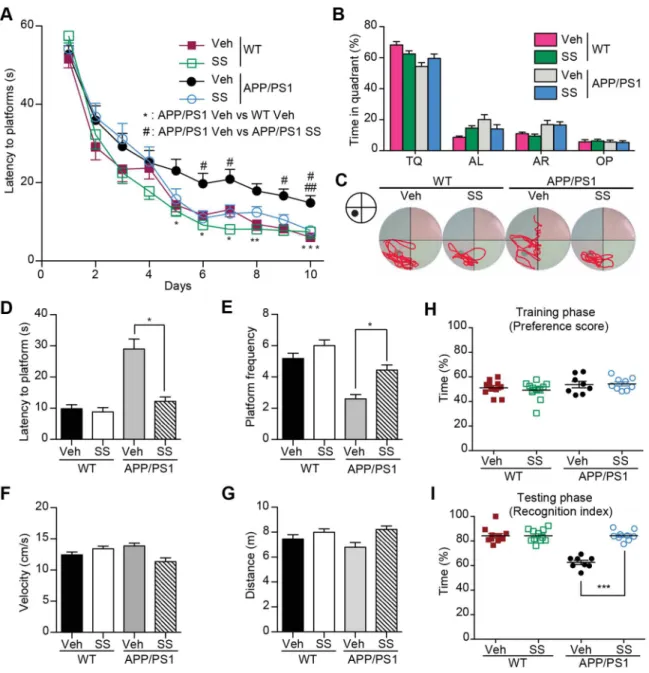
Figure 1. SS treatment ameliorates learning and memory impairment in Morris Water Maze and Object recognition test.(A) MWM test for SS and vehicle-treated APP/PS1 and WT mice. The mean escape latency was given for different test days. (B) The mean percent time in probe trial of MWM on day 7. TQ: Target quadrant; AL: Adjacent left; AR: Adjacent right; OP: Opposite. (C) Representative mice search paths from different groups. (D and E) The latency to target quadrant (D) and the frequency to pass the target position (E) in probe trial are shown. (F and G) The swimming velocity (F) and distance (G) in probe trial are shown. (H and I) Novel object recognition analysis. Preference scores of training phase (H) and Recognition Index of testing phase (I) during a 10-min testing phase are shown, respectively. n = 9–12 for each group. *P,0.05, **P,0.01, ***P, 0.001,#P,0.05,##P,0.01,###P,0.001.
doi:10.1371/journal.pone.0111215.g001
Locomotor assay
The locomotor assay of flies was performed as described previously with minor modifications [26]. Briefly, treated ten male flies (n = 30 for each group) were placed in a plastic 25-ml tube. After a 30-min recovering phase, flies were gently tapped to the bottom of the tube. The fly behavior was recorded with a video
camera. After 10 s of climbing, the number of flies between the 0, 5, 10, 15, 20 and 25 ml scale marks were recorded (Fig. S2). The results for each group of flies are calculated by the formula below: Climbing Index = (flies above 20 ml scale mark)61+ (flies between 15 and 20 ml scale marks)60.8+(flies between 10 and
Figure 2. SS treatment alleviates Ablevels and amyloid plaque burden, reduces gliosis and neuron loss in APP/PS1 mice.(A–C) Representative half brain sections of WT mice, vehicle or SS-treated APP/PS1 mice stained with antibody against Ab(6E10) and double staining of GFAP and 6E10 are shown. Scale bar, 1 mm. (BandC) Quantitative analysis of the number of 6E10-positive amyloid plaques (B) and Abcovered area (C). n = 5 animals per group. (DandE) ELISA of soluble and insoluble Ab40and Ab42levels in cortical and hippocampal tissues of APP/PS1 mice. n = 6 for each group. (F, I and J) Representative images of WT mice, vehicle- and SS- treated APP/PS1 mice hippocampus and cortex double immunostaining of GFAP and 6E10 (F), CD11b (I) and NeuN (J). Arrows indicate astrocytes surrounding the amyloid plaques. Scale bar, 200mm. (H) Coincidence of GFAP and Abburden in the brains of SS-treated APP/PS1 mice (red; n = 17) and vehicle-treated APP/PS1 mice (black; n = 17;P, 0.0001). (G,KandL) The histograms depict the mean GFAP (G), CD11b (K), and NeuN (L) positive area6S.E.M. in three groups. *P,0.05, **P,0.01, ***P,0.001.
15 ml scale marks)60.6+(flies between 5 and 10 ml scale marks) 60.4+(flies below 5 ml scale mark)60.2.
Western blot analyses
Fly head lysate preparation and western blot analyses were performed as described previously [23,27]. Briefly, thirty fly heads from each group were collected and lysed in RIPA buffer containing protease inhibitors. HEK293-APPsw cell lysates were also prepared. Proteins were separated on 16% tricine gel and probed with antibodies against APP C-terminal and b-actin (Sigma-Aldrich).
Preparation of Abpeptides
The peptides were prepared according to the protocols described by Stine [28]. Briefly, hexafluoroisopropanol (HFIP)-treated Ab42 peptides (Anaspec) were resuspended in dimethyl sulfoxide (DMSO). For the oligomeric conditions, the peptide was then diluted to a concentration of 100mM with Ham’s F12 and incubated at 4uC for 24 hours. After centrifugation 10 min at 14,000 g, the supernatant with soluble Ab42oligomers was added to cultures. The soluble oligomeric Abpeptides were confirmed using atomic force microscopy (AFM) and western blot analysis.
Thioflavin-T fluorescence assay
The Thioflavin-T (Th-T, Sigma-Aldrich) fluorescence assay was performed to measure amyloid fibril formation [29]. Th-T was prepared as a stock at 2 mM (avoid exposure to light) and filtered through a 0.22mm filter. The Th-T stock solution was diluted into PBS on the day of analysis. HFIP-treated Ab peptides were dissolved in DMSO at 5 mM, and then diluted in Th-T/PBS with a final concentration of 15mM (Ab) and 20mM (Th-T). The Ab/ Th-T solutions were incubated at 37uC with or without TCM for six days in a black 96-well-plate (PerkinElmer). Fluorescence was measured using an EnVision multilabel plate reader (PerkinEl-mer), at excitation and emission wavelengths of 440 nm and 490 nm, respectively. Fluorescence of 20mM Th-T/PBS solution was measured and used as blank. The fluorescence intensity reflects the degree of Abaggregation.
Primary culture of neuron
Primary neurons were cultured as described previously [30,31]. The cortices and hippocampi obtained from newborn C57BL/ 6 mice were used. 56104 cells per well in DMEM/Ham’s F12 containing 10% FBS (Invitrogen) were plated onto poly-D-lysine (Sigma-Aldrich) coated 96-well plates and maintained at 37uC for four hours, and the culture medium was then switched to B27/ Neurobasal medium (Invitrogen). On day 6, the cells pre-treated
Figure 3. SS treatment improves locomotor functions and prolongs lifespan of AD transgenicDrosophila.(A, C and E) CS, APP/BACE and Abtransgenic flies were cultured on food containing different concentrations of SS (the triangle symbol stands for concentrations from low to high: 0.2, 0.6 and 2 mg/ml) or Memantine (120mM). Survival curves for flies treated with either SS or Memantine. The data are presented as mean6 S.E.M. The right panel shows the mean survival days calculated according to the survival curves. (B, D and F) The climbing ability of CS, APP/BACE and Abtransgenic flies treated with SS or Memantine at day 30 (for CS and APP/BACE flies) and day 20 (for Abflies). Values are mean6S.E.M. Each value represents the mean of three experiments. *P,0.05, **P,0.01, ***P,0.001vs.Ctrl group. Mem = Memantine.
doi:10.1371/journal.pone.0111215.g003
with TCMs for two hours were incubated with 5mM Ab42 oligomers and TCMs for another 48 hours. Staining of primary neurons with a Tuj1 antibody (Covance) was imaged and analyzed using Operetta (PerkinElmer).
CellTiter-Glo assay
The primary neurons, SK-N-SH-APPsw cells and HEK293-APPsw cells were assessed for viability using the CellTiter-Glo luminescent cell viability assay (Promega) following the manufac-turer’s instructions.
TUNEL assay
The assay was performed using the Kit from Roche following the manufacturer’s protocol. Primary neurons were pre-treated with TCMs for two hours followed by incubation with 5mM Ab42 oligomers and TCMs for another 24 hours. The total numbers of DAPI-stained or TUNEL-positive cells were counted.
Atomic Force Microscope (AFM)
Amyloid peptides for AFM analysis were prepared according to the protocols described by Stine [28]. First, HFIP-treated Ab42 peptides (Anaspec) were dissolved in DMSO. 5 mM Ab42 in DMSO were diluted in H2O to 100mM and used immediately as Ab42 monomers. 5 mM Ab42in DMSO were diluted in Ham’s F12 to 100mM and incubated at 4uC for 24 hours and used as Ab42oligomers. Ab42fibrils were prepared by diluting 5 mM Ab42 in DMSO with 10 mM HCl to 100mM and incubated at 37uC for 24 hours. For AFM analysis, Ab42monomers, oligomers and fibrils samples were further diluted to a final concentration of 10mM with distilled H2O and spotted on freshly cleaved mica disks for 3 minutes. After being rinsed three times with deionized water, the mica disks were dried overnight at room temperature. AFM was performed in tapping mode using a Molecular Force Prove 3D (MFP-3D, Asylum Research) with NSC11 cantilevers (Mikro-Masch, 48 Newton/meter spring constant). The data were analyzed with Igor-Pro (Wavemetrix). The resonance frequency was applied in the 330 kHz range, the scan rate was 1 Hz, and the resolution was 5126512 pixels.
Statistical analyses
GraphPad Prism 5.0 was used. The data are shown as the mean 6S.E.M. The Kaplan-Meier test was employed to compare the differences between the survival curves using SPSS 16.0. Group differences were analyzed with one-way ordinary or repeated-measures analysis of variance (ANOVA) followed by Tukey multiple comparisons test. Student’s independentttest was applied for comparisons of two groups. Differences were significant when
P,0.05.
Results
SS treatment ameliorates learning and memory deficits in APP/PS1 mice
Traditionally, TCM formulae are prepared as decoctions, i.e., the herbs are boiled for a fixed time, and the liquid is used. Recently, CFDA-approved single-herb granules have been widely accepted as alternatives for decoctions. These CFDA-approved single-herb granules are clearly documented for their chemical fingerprints and pharmacokinetic parameters along with standard manufacturing protocols, which are not only suitable for large-scale industry production but are also more appropriate for evaluation of their efficacies and exploration of the underlying molecular mechanisms. Thus, as described in the Methods section, we prepared SS using the CFDA-approved single-herb granules according to the recipe (granule-mixed SS, SS-G). We first analyzed the SS-G by HPLC-TOF/MS. The total ion current chromatograms corresponding to positive and negative signals of SS were obtained, as shown in Fig. S3 and Table S2. We found that the major chemical constituents identified in the SS-G were similar to the traditionally prepared SS decoctions (SS-D) [32]. Therefore, we used the SS-G for the following assays. Then, we compared the HPLC fingerprints of four different batches of SS-G. The similarity indices of these four batches of SS were between 0.943 and 0.982 (Fig. S4), demonstrating that SS was produced consistently with good quality.
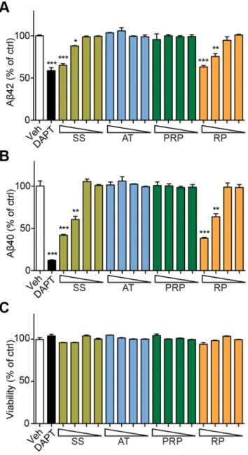
Figure 4. RP reduces the Abgeneration in SK-N-SH-APPsw cells.
Ab42(A) and Ab40(B) in SK-N-SH-APPsw cell culture medium and cell viability (C) after treatment with SS, AT, PRP, RP for 24 hours, respectively (the triangle symbol stands for concentrations from high to low: 3000, 1000, 300 and 100mg/ml for SS; 1000, 300, 100 and 30mg/ ml for AT, PRP and RP). *P,0.05, **P,0.01, ***P,0.001; DAPT: ac -secretase inhibitor.
The age-dependent Abaccumulation was found in the brains of APP/PS1 mice, and memory deficits were began to show since the age of 6 months [33]. To explore the potential therapeutic effects of SS, we began the oral administration of SS to the 7-month-old APP/PS1 mice for two months. There were no obvious adverse effects or body weight loss (Table S3). We compared the mice behavioral phenotypes to assess the cognitive function of these transgenic AD mice after drug administration. We assessed the spatial memory of these mice in the MWM. The MWM is a common behavioral task used to determine spatial learning and reference memory deficits. SS treatment did not influence the swimming velocity and distance (Fig. 1, F and G). However, the APP/PS1 mice showed defects in learning compared with the WT
mice (Fig. 1A). On day 5, vehicle-treated APP/PS1 mice spent more time than WT littermates to locate the hidden platform (P, 0.05, APP/PS1 mice vs. WT mice, Fig. 1A). This difference constantly appeared on day 6, 7, 8 and 10, reflecting deficits in spatial memory of APP/PS1 mice, as reported previously [34]. Interestingly, since day 6, SS-treated APP/PS1 mice showed an improved performance compared with vehicle-treated APP/PS1 mice (P,0.01, Fig. 1A), indicating that the SS treatment alleviated the impairment of spatial learning in APP/PS1 mice. To assess the memory strength of spatial learning, we administered the probe trials on day 4, 7 and 11. The mice in all group showed a random swimming pattern on day 4 (Fig. S5). However, on the day 7 probe trial, compared with the vehicle-treated APP/PS1
Figure 5. RP treatment improves locomotor functions, prolongs lifespan and reduces Ablevels of AD transgenicDrosophila.CS, Ab and APP/BACE transgenic flies were cultured on food containing different concentrations of RP (the triangle symbol indicates concentrations from low to high: 0.2, 0.6 and 2 mg/ml) or Memantine (120mM). (A) Survival curves of flies treated with either RP or Memantine. The data are presented as the mean6S.E.M. (B) The climbing ability of flies (right panels) was assessed at day 30 for CS and APP/BACE flies and at day 20 for Abflies. The values are the mean6S.E.M. Each value represents the mean of three experiments. (CandD) Aband APP/BACE transgenic flies were cultured on SS, AT, PRP or RP (2 mg/ml). Ab40and Ab42levels in 500 fly heads were measured by ELISA assay. Mem = Memantine. *P,0.05, **P,0.01, ***P,0.001vs.the control group.
doi:10.1371/journal.pone.0111215.g005
mice, the SS-treated group spent more time searching for the platform in the target quadrant (Fig. 1, B and C), took less time to reach the position of the missing platform (P,0.05, Fig. 1D), and increased frequency of crossing within the position of the platform (P,0.05, Fig. 1E). These results suggest that administration of SS alleviates the deficient spatial reference memory of APP/PS1 mice.
To further assess the learning and recognition memory processes of these AD mice, we applied the object recognition test (Fig. S1). There were no obvious differences among mice in all groups regarding which object they preferred or the location of the objects as the preference scores were all approximately 50% (Fig. 1H). In the testing phase, the WT mice significantly increased the time exploring the novel object (P,0.001, Fig. 1I). In contrast, the time exploring the novel object of vehicle-treated APP/PS1 mice did not increased obviously. Meanwhile, the SS-treated
APP/PS1 mice explored the novel object longer than vehicle-treated APP/PS1 mice (P,0.001) indicating improved memory retention by SS treatment.
SS treatment reduced Ablevels, Ab amyloidosis, gliosis and neuron loss in the brains of APP/PS1 mice
APP/PS1 mice begin to develop cerebral amyloidosis at 2 months of age, and the Ablevels or Abdeposits in the brain can be detected since 6 months of age. Histology was performed on fixed brain tissues. APP/PS1 mice showed 6E10-positive amyloid plaques (Fig. 2A), which were significantly reduced in the brains of SS-treated APP/PS1 mice. Detailed plaque size distribution analysis revealed a reduction of different size plaques in SS-treated APP/PS1 mice compared with that of control APP/PS1 mice (Fig. 2B). The overall amyloid plaques of SS-treated APP/PS1 mice was reduced by 18.1% compared with that of control APP/
Figure 6. AT and PRP improves locomotor function and prolongs lifespan of AD transgenicDrosophila.CS and Abtransgenic flies were cultured on food containing different concentrations of AT (0.2, 0.6 and 2 mg/ml), PRP (0.2, 0.6 and 2 mg/ml) or Memantine (120mM). (A, C) Survival curves for flies treated with either AT, PRP or Memantine. (B, D) The climbing ability of flies was assessed. The values are the mean6S.E.M. Each value represents the mean of three experiments. Mem = Memantine. *P,0.05, **P,0.01, ***P,0.001vs.the control group.
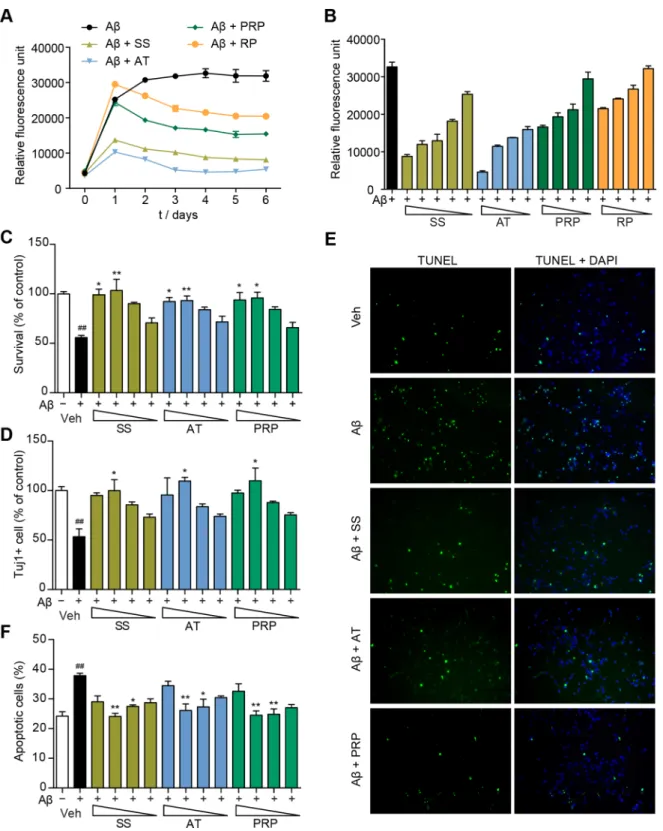
Figure 7. AT and PRP inhibit Ab42aggregation and exert neuroprotective effects against Ab42in primary neurons.(A) The effects of SS (3000mg/ml), AT (1000mg/ml), PRP (1000mg/ml) or RP (1000mg/ml) on Ab42aggregation, as measured by Th-T fluorescence assay. (B) SS, AT, PRP and RP (3000, 1000, 300, 100 and 30mg/ml for SS; 1000, 300, 100 and 30mg/ml for AT, PRP and RP) inhibited Ab42aggregation in dose-dependent manner. (CandD) Cell viability of primary cultured neurons pre-treated with SS or AT, PRP for two hours followed by incubation with Ab42oligomers (5mM) for another 48 hours (300, 100, 30 and 10mg/ml for SS; 100, 30, 10 and 3mg/ml for AT and PRP). Viable cells were quantified using a CellTiter-Glo assay (A). Tuj1-positive cells were counted and presented (D). *P,0.05 compared with the Ab42-treated group. (EandF) TUNEL analysis of the primary neurons pre-treated with SS (100mg/ml), AT (30mg/ml), or PRP (30mg/ml) for two hours followed by incubation with Ab42oligomers (5mM) for another 24 hours. The green, TUNEL-positive cells are merged with blue DAPI-positive cells (E). The TUNEL-positive cells and DAPI-positive cells pre-treated with SS or AT, PRP (300, 100, 30 and 10mg/ml for SS; 100, 30, 10 and 3mg/ml for AT and PRP) were counted in three independent experiments (F). *P,0.05, **P,0.01, ***P,0.001vs.the Ab42-treated group,##P,0.01vs.the control group.
doi:10.1371/journal.pone.0111215.g007
PS1 mice (P,0.01, Fig. 2C), indicating that SS treatment prevented the formation of plaques and/or enhanced the clearance of amyloid plaques.
Soluble Ab oligomers are deleterious, and related to cognitive deficits in AD [35–40]. To quantify Ablevels, we used an ELISA assay. ELISA analysis of SDS-soluble fractions and SDS-insoluble fractions demonstrated that there were high Ab levels in both fractions from the hippocampi and cortices of APP/PS1 transgenic mice. SS treatment reduced cortical SDS-soluble Ab40and Ab42 levels (30.4% reduction for Ab42,P,0.05, and 33.7% reduction for Ab40,P,0.05, Fig. 2D). We also observed a similar reduction of hippocampal SDS-soluble Ab42level after SS treatment (17.0% reduction,P,0.01, Fig. 2E). There were no significant differences between the SDS-insoluble formic acid-extractable fractions from vehicle-treated and SS-treated mice (Fig. 2, D and E) suggesting that SS reduces the level of toxic soluble Ab. These data indicate that SS treatment reduces Ablevels and the correlated amyloidosis in APP/PS1 mice.
Abnormal neuroinflammation, including accumulation of acti-vated microglia and astrocytes, is a pathological characteristic of the neurodegenerative disease [41]. In the brains of AD patients and APP/PS1 transgenic mice, amyloid plaques are surrounded by activated microglia and reactive astrocytes [41]. Consistent with previous reports [42], we noted that GFAP-positive astrocytes surrounded the amyloid plaques in APP/PS1 mice (Fig. 2, A and F, arrows), and the GFAP-positive staining intensity in APP/PS1 mice was higher than that of WT mice (7.8060.25vs.1.8960.22 percent area,P,0.001, Fig. 2, F and G). Furthermore, coincident GFAP and Abstaining was obvious in the brains of vehicle-treated APP/PS1 mice (R2= 0.72, Fig. 2H). CD11b-positive microglia were also observed (Fig. 2I). We found significant reductions of astrocytes (staining intensity: 6.30% vs.7.80%, P,0.01; coinci-dence: 0.68vs. 0.72,P,0.001, Fig. 2, G and H) and microglia (staining intensity: 1.37%vs.2.34%, Fig. 2, I and K) in the brains of SS-treated mice, demonstrating that the AD-like pathological gliosis has been significantly moderated by SS treatment in APP/ PS1 mice.
As one of the majorin vivoneurotoxic properties of Ab, severe neuronal loss is also observed in APP/PS1 transgenic mice since the age of 10 months [43]. Consistently, in addition to the accumulation of microglia and astrocytes, we also observed significant less neurons (NeuN-positive cells) in the brains of APP/PS1 mice compared with WT mice (Fig. 2, J and L). Interestingly, we found an increase in neurons (NeuN-positive cells) in SS-treated APP/PS1 mice compared with control APP/ PS1 mice (P,0.05, Fig. 2L). Together, these results suggest that in APP/PS1 mice, SS treatment retards the Ab-related pathological gliosis and neuronal loss.
SS treatment improves locomotor functions and prolongs lifespan of AD transgenicDrosophila
TransgenicDrosophilamodels expressing human Ab, APP or secretases have been investigated to gain insight into disease mechanisms as well as to elucidate potential therapeutic approaches [44]. AD transgenic Drosophila expressing human Ab42show memory deficiencies and premature death [45]. Very recent reports have also shown that the generation of Aboligomers through the APP processing by secretases in APP/BACE flies display a very similar pathology as that in AD patients [46]. Because the AD-like pathologies are evident within a few days in these Drosophilamodels, we performed a rapidin vivo compar-ison of SSvs.Memantine, the clinically approved medication for treatment of Alzheimer’s disease, using transgenic flies carrying Ab42or APP/BACE.
We applied the climbing assay to assess the CNS dysfunction of these AD transgenic flies. The climbing assay is a behavioral test based on the negative geotaxis response of Drosophila. Thus, assessing the climbing ability of flies in a fixed time period can reflex their locomotor function.
Consistent with previous reports, flies expressing human APP and BACE or flies expressing Ab42 showed premature death compared with CS flies (Fig. 3, A, C and E). We also observed significant decreases in the climbing ability of these flies (Fig. 3, B, D and F). Interestingly, when the Ab42and APP/BACE transgenic flies were cultured on either SS or Memantine, Ab-induced premature death was significantly reduced (Fig. 3, C and E). Furthermore, SS or Memantine treatment considerably rescued the climbing ability of these transgenic flies (Fig. 3, D and F). These results indicate that SS treatment improves locomotor functions and reduces premature death of AD transgenic flies, similar to that of Memantine.
RP reduces Abgeneration
To explore which granule in SS might reduce Ab levels, we treated SK-N-SH-APPsw cells with different concentrations of each granule in SS. SK-N-SH-APPsw cells stably express APP protein carrying a Swedish mutant (K595N/M596L) and show elevated Ab40 and Ab42secretion. As shown in Fig. 4, Ab40and Ab42 levels in the culture medium of cells treated with SS were reduced in a dose-dependent manner. Interestingly, the same dose of RP but not that of AT or PRP exhibited similar Ab40and Ab42 reduction effects as SS (Fig. 4, A and B). The viability of cells was not altered by any of these treatments (Fig. 4C). Similar results were observed using HEK293-APPsw cells (Fig. S6).
Then, we assessed whether RP ameliorates Ab-related pathol-ogy by reducing Abgenerationin vivo. We found that RP showed no obvious beneficial effects in the Abtransgenic flies expressing Ab42directly (Fig. 5A). Interestingly, APP/BACE transgenic flies that were cultured on food containing RP showed considerably reduced Ab-induced premature death (Fig. 5B). Consistently, the climbing ability of these RP-treated APP/BACE transgenic flies was enhanced than that of vehicle-treated APP/BACE transgenic flies. Ab levels in APP/BACE and Ab transgenic flies were monitored by ELISA. As shown in Fig. 5C, APP/BACE flies cultured on food containing SS or RP produced significantly lower levels of Ab40and Ab42than control. Moreover, Ablevels in Ab transgenic flies cultured on food containing SS or RP were not different from that of control Ab transgenic flies (Fig. 5D). This result suggests that RP may affect human APP proteolysis. To further assess the proteolysis process of APP in APP/BACE flies, full-length APP and APP C-terminal fragments (APP-CTF) in fly heads were monitored using western blots. There was no significant difference in full-length APP and APP-CTF levels in APP/BACE flies (Fig. S7). These results suggest that RP can reduce Ab generation and thus moderate Ab-related pathology
in vivo.
AT and PRP moderate Ab-induced neurotoxicityin vivo
andin vitro
different concentrations of AT or PRP. As shown in Fig. 6, both AT and PRP prolonged lifespan and improved the climbing activity of Ab42 flies (Fig. 6, A–D). Both AT and PRP also prolonged lifespan and improved climbing activity of APP/BACE flies but to a less significant extent (Fig. S8).
To investigate whether SS inhibits Ab aggregation, we performed a Th-T fluorescence assay. Th-T fluorescence signals were gradual increased over time. Both SS and AT inhibited the fluorescence intensity almost completely (Fig. 7A), while PRP showed a less significant effect. SS, AT and PRP inhibited Ab42 aggregation in a dose-dependent manner (Fig. 7B). To directly assess the neuroprotective effects of AT and PRP, we further monitored Ab-induced cell death of primary mouse cortical neurons. Ab42oligomer preparations consistently provided small oligomers and expected structure as monitored by western blot and AFM (Fig. S9), respectively. Ab42 oligomers effectively induced the death of cortical neurons within 48 hours as measured by the CellTiter-Glo assay (Fig. 7C) and Tuj1 staining (Fig. 7D). SS significantly prevented neurons from Ab42oligomers-induced cell death. Consistent with thein vivoresults above, both AT and PRP improved primary neuron survival in the presence of toxic Ab42 oligomers (Fig. 7, C and D). Ab42 oligomers-induced cell apoptosis was further monitored using a TUNEL assay. As shown in Fig. 7E, approximately 40% of cells showed severe DNA fragmentation upon Ab42treatment. In the presence of SS, AT or PRP, the number of apoptotic cells was significantly reduced (Fig. 7, E and F). Together, these data indicate that both AT and PRP protect neurons from Ab42-induced toxicity.
Discussion
Our findings in this study demonstrate that SS, the three-herb TCM formula, can ameliorate AD-related pathological symptoms. More interestingly, our results indicate that SS treatment not only ameliorated AD-related symptoms but also exhibited disease-modifying effects such as reduction of Ab levels and Ab amyloidosis, retardation of neuronal loss and decrease in astrocytes and microglia. Collectively, our study suggests that it is possible to alleviate symptoms and modify the disease simultaneously, and this ancient formula provides an example of such effects. Further extensive investigation is required to identify the active functional constituents as well as to elucidate the correlate molecular mechanisms, which should finally lead us to a systematic therapeutic strategy against this complicated disease.
It is characteristic of TCM that each formula comprises several herbs with different or distinct functions that work synergistically at multiple targets of a complicated disease. Our studies indicate that three herbs of SS have different therapeutic targets of AD: RT reduced Ab generation, and AT and PRP protected neuron against Ab-induced toxicity. RP reduced Ab40 and Ab42 generation in SK-N-SH-APPsw cells, showed beneficial effects in flies expressing APP/BACE but did not significantly affect locomotor function or survival in flies expressing Ab directly. Consistently, RP treatment reduced Ab40and Ab42levels in APP/ BACE transgenic fly heads, but had no significant effects in Ab transgenic fly heads, indicating that RP reduced Ab generation and affected the proteolysis of human APP. Furthermore, AT and PRP showed inhibitory effects on Ab42aggregation and protected primary neurons against Ab42oligomers-induced cell toxicity and apoptosis. In AD transgenic flies, AT and PRP treatment alleviated Ab-induced premature death and motor neuron dysfunction in both Ab transgenic flies and APP/BACE flies, suggesting that AT and PRP protect neurons through other mechanisms but may not affect APP cleavage or processing.
Unfortunately, the functional active constituents of each herb are unclear in the present study. Obviously, further elucidation of the detailed molecular mechanisms should provide more efficient screening models to identify those functional constituents in each herb. Furthermore, it is of note that SS treatment improved locomotor activity and prolonged lifespan in AD Drosophila
models, which makes it possible to quickly identify/verify functional constituents of SS in the near future. On the other hand, SS is one of many TCM formulae documented against memory loss and cognitive impairment. We must evaluate other formulae using current molecular, cellular and animal models as performed in this study, which may eventually lead to a more effective treatment against AD.
TCMs have been orally administered as decoctions, such as this formula for the so-called Smart Soup. Recently, CFDA-approved single-herb granules have been widely used due to their ease of large-scale industry production with stringent manufacturing protocols, better quality control with well-documented chemical fingerprints, and reproducible pharmacokinetic parameters. In our study, we found that the granule preparation (a mixture of three single-herb granules) was as effective as the traditional decoctions (the ‘‘soup’’ preparation), which simplifies future studies of individual herbs or their combination.
Although SS has been prescribed by Chinese medical physicians to patients with aging-related cognitive impairment, a multicenter, double-blind, randomized, placebo-controlled study should be carried out to evaluate the overall efficacies of SS against AD. Furthermore, whether SS should be prescribed alone or combined with other FDA-approved drugs should be assessed in future clinical trials. Moreover, it remains to be determined whether SS or other TCM formulae can be applied for MCI or for preventing neurodegenerative diseases.
Supporting Information
Figure S1 A schematic diagram of novel object recogni-tion.
(TIF)
Figure S2 A sketch of the locomotor assay.
(TIF)
Figure S3 MS total ion current chromatograms of SS (SS-G).Negative-ion (A) and positive-ion (B) modes were selected for TOF/MS analysis. Forty-five compound peaks were tenta-tively identified on the basis of mass measurements and retention times. Of the 45 peaks of SS, AT accounts for 5 of 45 peaks, PRP accounts for 8 of 45 peaks, and RP accounts for 32 of 45 peaks. (TIF)
Figure S4 HPLC fingerprints of four batches of SS (from bottom to top: 201102, 201205, 201301 and 201311).The peaks are the characteristic and representative chemical constit-uents detected in SS. The similarity indices of four batches of samples were between 0.943 and 0.982.
(TIF)
Figure S5 Representative raw data of the MWM search paths of SS- or vehicle-treated APP/PS1 mice or WT littermates.
(TIF)
for SS; 1000, 300, 100 and 30mg/ml for AT, PRP and RP). *P, 0.05, **P,0.01, ***P,0.001; DAPT, ac-secretase inhibitor. (TIF)
Figure S7 SS and RP do not alter APP expression in APP/BACE transgenicDrosophila.Western blot of human APP in APP/BACE transgenic flies cultured on SS, AT, PRP or RP (2 mg/ml). Lane 1: HEK293-APPsw cell lysates; Lane 2–6: head lysates of APP/BACE flies. Full-length APP (,110 kD), APP-CTFs (,10–12 kD) and Appl-CTFs (,15 kD) were detected. (TIF)
Figure S8 AT and PRP improves locomotor functions and prolongs lifespan of APP/BACE transgenic Dro-sophila. APP/BACE transgenic flies were cultured on food containing different concentrations of AT, PRP (0.2, 0.6, or 2 mg/ ml) or Memantine (120mM). (A and C) Survival curves for flies treated with either AT, PRP or Memantine. (B and D) The climbing ability of flies was assayed. The values are the mean6 S.E.M. Each value represents the mean of three experiments. Mem = Memantine. *P,0.05, **P,0.01, ***P,0.001 vs. the control group.
(TIF)
Figure S9 Representative Western blots and Atomic Force Microscope (AFM) for Ab42 oligomers. (A)
Repre-sentative western blots of Ab42monomers and oligomers separated
by SDS-PAGE using 16% tricine gel and probed with the antibody 6E10 are shown. (B) AFM images of Ab42 monomers, oligomers and fibrils are shown. The sample was scanned with AFM analysis at 10mM. Scale bar, 0.5mm.
(TIF)
Table S1 Herbal Ingredients in the SS.
(PDF)
Table S2 Compounds identified in SS by HPLC-TOF/ MS.
(PDF)
Table S3 The detailed information of each mouse in each group.
(PDF)
Acknowledgments
We thank all members of the laboratory for sharing reagents and ideas.
Author Contributions
Conceived and designed the experiments: YH JZ GP. Performed the experiments: YH YW(Institute of Neurology) XL JC XZ YL. Analyzed the data: YH YW(Institute of Neurology) JZ. Contributed reagents/materials/ analysis tools: GP JD YW(Department of Neurology) XS SC CH. Contributed to the writing of the manuscript: YH JZ GP.
References
1. Hardy J, Selkoe DJ (2002) The amyloid hypothesis of Alzheimer’s disease: progress and problems on the road to therapeutics. Science 297: 353–356. 2. Selkoe DJ (2002) Deciphering the genesis and fate of amyloid beta-protein yields
novel therapies for Alzheimer disease. The Journal of clinical investigation 110: 1375–1381.
3. Selkoe DJ (1997) Alzheimer’s disease: genotypes, phenotypes, and treatments. Science 275: 630–631.
4. Winslow BT, Onysko MK, Stob CM, Hazlewood KA (2011) Treatment of Alzheimer disease. American family physician 83: 1403–1412.
5. Howes MJ, Houghton PJ (2003) Plants used in Chinese and Indian traditional medicine for improvement of memory and cognitive function. Pharmacology, biochemistry, and behavior 75: 513–527.
6. Tian XY, Liu L (2012) Drug discovery enters a new era with multi-target intervention strategy. Chinese journal of integrative medicine 18: 539–542. 7. Lee B, Choi Y, Kim H, Kim SY, Hahm DH, et al. (2003) Protective effects of
methanol extract of Acori graminei rhizoma and Uncariae Ramulus et Uncus on ischemia-induced neuronal death and cognitive impairments in the rat. Life sciences 74: 435–450.
8. Tong XG, Liu JL, Cheng YX (2010) A new pregnane steroid from Poria cum radix pini. Journal of Asian natural products research 12: 419–423. 9. Chen YL, Hsieh CL, Wu PH, Lin JG (2004) Effect of Polygala tenuifolia root on
behavioral disorders by lesioning nucleus basalis magnocellularis in rat. Journal of ethnopharmacology 95: 47–55.
10. Park CH, Choi SH, Koo JW, Seo JH, Kim HS, et al. (2002) Novel cognitive improving and neuroprotective activities of Polygala tenuifolia Willdenow extract, BT-11. Journal of neuroscience research 70: 484–492.
11. Lin Z, Gu J, Xiu J, Mi T, Dong J, et al. (2012) Traditional chinese medicine for senile dementia. Evidence-based complementary and alternative medicine: eCAM 2012: 692621.
12. Lee JY, Kim KY, Shin KY, Won BY, Jung HY, et al. (2009) Effects of BT-11 on memory in healthy humans. Neuroscience letters 454: 111–114.
13. Shin KY, Lee JY, Won BY, Jung HY, Chang KA, et al. (2009) BT-11 is effective for enhancing cognitive functions in the elderly humans. Neuroscience letters 465: 157–159.
14. Jankowsky JL, Slunt HH, Ratovitski T, Jenkins NA, Copeland NG, et al. (2001) Co-expression of multiple transgenes in mouse CNS: a comparison of strategies. Biomolecular engineering 17: 157–165.
15. Reiserer RS, Harrison FE, Syverud DC, McDonald MP (2007) Impaired spatial learning in the APPSwe+PSEN1DeltaE9 bigenic mouse model of Alzheimer’s disease. Genes, brain, and behavior 6: 54–65.
16. Bero AW, Yan P, Roh JH, Cirrito JR, Stewart FR, et al. (2011) Neuronal activity regulates the regional vulnerability to amyloid-beta deposition. Nature neuroscience 14: 750–756.
17. Morris R (1984) Developments of a water-maze procedure for studying spatial learning in the rat. Journal of neuroscience methods 11: 47–60.
18. Alamed J, Wilcock DM, Diamond DM, Gordon MN, Morgan D (2006) Two-day radial-arm water maze learning and memory task; robust resolution of
amyloid-related memory deficits in transgenic mice. Nature protocols 1: 1671– 1679.
19. Teng L, Zhao J, Wang F, Ma L, Pei G (2010) A GPCR/secretase complex regulates beta- and gamma-secretase specificity for Abeta production and contributes to AD pathogenesis. Cell research 20: 138–153.
20. Bevins RA, Besheer J (2006) Object recognition in rats and mice: a one-trial non-matching-to-sample learning task to study ‘recognition memory’. Nature protocols 1: 1306–1311.
21. Liu X, Zhao X, Zeng X, Bossers K, Swaab DF, et al. (2013) beta-arrestin1 regulates gamma-secretase complex assembly and modulates amyloid-beta pathology. Cell research 23: 351–365.
22. Lazarov O, Robinson J, Tang YP, Hairston IS, Korade-Mirnics Z, et al. (2005) Environmental enrichment reduces Abeta levels and amyloid deposition in transgenic mice. Cell 120: 701–713.
23. Chakraborty R, Vepuri V, Mhatre SD, Paddock BE, Miller S, et al. (2011) Characterization of a Drosophila Alzheimer’s disease model: pharmacological rescue of cognitive defects. PloS one 6: e20799.
24. Brand AH, Perrimon N (1993) Targeted gene expression as a means of altering cell fates and generating dominant phenotypes. Development 118: 401–415. 25. Zhao XL, Wang WA, Tan JX, Huang JK, Zhang X, et al. (2010) Expression of
beta-amyloid induced age-dependent presynaptic and axonal changes in Drosophila. The Journal of neuroscience: the official journal of the Society for Neuroscience 30: 1512–1522.
26. White KE, Humphrey DM, Hirth F (2010) The dopaminergic system in the aging brain of Drosophila. Frontiers in neuroscience 4: 205.
27. Mhatre SD, Satyasi V, Killen M, Paddock BE, Moir RD, et al. (2014) Synaptic abnormalities in a Drosophila model of Alzheimer’s disease. Disease models & mechanisms 7: 373–385.
28. Stine WB Jr, Dahlgren KN, Krafft GA, LaDu MJ (2003) In vitro character-ization of conditions for amyloid-beta peptide oligomercharacter-ization and fibrillogen-esis. The Journal of biological chemistry 278: 11612–11622.
29. Lu JH, Ardah MT, Durairajan SS, Liu LF, Xie LX, et al. (2011) Baicalein inhibits formation of alpha-synuclein oligomers within living cells and prevents Abeta peptide fibrillation and oligomerisation. Chembiochem: a European journal of chemical biology 12: 615–624.
30. Cartier AE, Djakovic SN, Salehi A, Wilson SM, Masliah E, et al. (2009) Regulation of synaptic structure by ubiquitin C-terminal hydrolase L1. The Journal of neuroscience: the official journal of the Society for Neuroscience 29: 7857–7868.
31. Cisse M, Halabisky B, Harris J, Devidze N, Dubal DB, et al. (2011) Reversing EphB2 depletion rescues cognitive functions in Alzheimer model. Nature 469: 47–52.
33. Jankowsky JL, Fadale DJ, Anderson J, Xu GM, Gonzales V, et al. (2004) Mutant presenilins specifically elevate the levels of the 42 residue beta-amyloid peptide in vivo: evidence for augmentation of a 42-specific gamma secretase. Human molecular genetics 13: 159–170.
34. Cao D, Lu H, Lewis TL, Li L (2007) Intake of sucrose-sweetened water induces insulin resistance and exacerbates memory deficits and amyloidosis in a transgenic mouse model of Alzheimer disease. The Journal of biological chemistry 282: 36275–36282.
35. Lesne S, Koh MT, Kotilinek L, Kayed R, Glabe CG, et al. (2006) A specific amyloid-beta protein assembly in the brain impairs memory. Nature 440: 352– 357.
36. Walsh DM, Selkoe DJ (2007) A beta oligomers - a decade of discovery. Journal of neurochemistry 101: 1172–1184.
37. Walsh DM, Klyubin I, Fadeeva JV, Cullen WK, Anwyl R, et al. (2002) Naturally secreted oligomers of amyloid beta protein potently inhibit hippocampal long-term potentiation in vivo. Nature 416: 535–539.
38. Shankar GM, Li S, Mehta TH, Garcia-Munoz A, Shepardson NE, et al. (2008) Amyloid-beta protein dimers isolated directly from Alzheimer’s brains impair synaptic plasticity and memory. Nature medicine 14: 837–842.
39. Cheng IH, Scearce-Levie K, Legleiter J, Palop JJ, Gerstein H, et al. (2007) Accelerating amyloid-beta fibrillization reduces oligomer levels and functional deficits in Alzheimer disease mouse models. The Journal of biological chemistry 282: 23818–23828.
40. Lesne S, Kotilinek L, Ashe KH (2008) Plaque-bearing mice with reduced levels of oligomeric amyloid-beta assemblies have intact memory function. Neurosci-ence 151: 745–749.
41. McGeer EG, McGeer PL (2003) Inflammatory processes in Alzheimer’s disease. Progress in neuro-psychopharmacology & biological psychiatry 27: 741–749. 42. Kraft AW, Hu X, Yoon H, Yan P, Xiao Q, et al. (2012) Attenuating astrocyte
activation accelerates plaque pathogenesis in APP/PS1 mice. FASEB journal: official publication of the Federation of American Societies for Experimental Biology.
43. Hamilton A, Holscher C (2012) The effect of ageing on neurogenesis and oxidative stress in the APP(swe)/PS1(deltaE9) mouse model of Alzheimer’s disease. Brain research 1449: 83–93.
44. Muqit MM, Feany MB (2002) Modelling neurodegenerative diseases in Drosophila: a fruitful approach? Nature reviews Neuroscience 3: 237–243. 45. Bilen J, Bonini NM (2005) Drosophila as a model for human neurodegenerative
disease. Annual review of genetics 39: 153–171.
46. Iijima K, Iijima-Ando K (2008) Drosophila models of Alzheimer’s amyloidosis: the challenge of dissecting the complex mechanisms of toxicity of amyloid-beta 42. Journal of Alzheimer’s disease: JAD 15: 523–540.
47. Dahlgren KN, Manelli AM, Stine WB Jr, Baker LK, Krafft GA, et al. (2002) Oligomeric and fibrillar species of amyloid-beta peptides differentially affect neuronal viability. The Journal of biological chemistry 277: 32046–32053.