chroococcum
: A Spectroscopic Characterization
Aulie Banerjee., Subhrangshu Supakar., Raja Banerjee* Department of Bioinformatics, West Bengal University of Technology, Salt Lake, Kolkata, W.B., India
Abstract
Melanins, the ubiquitous hetero-polymer pigments found widely dispersed among various life forms, are usually dark brown/black in colour. Although melanins have variety of biological functions, including protection against ultraviolet radiation of sunlight and are used in medicine, cosmetics, extraction of melanin from the animal and plant kingdoms is not an easy task. Using complementary physicochemical techniques (i.e.MALDI-TOF, FTIR absorption and cross-polarization magic angle spinning solid-state13C NMR), we report here the characterization of melanins extracted from the nitrogen-fixing non-virulent bacteriumAzotobacter chroococcum, a safe viable source. Moreover, considering dihydroxyindole moiety as the main constituent, an effort is made to propose the putative molecular structure of the melanin hetero-polymer extracted from the bacterium. Characterization of the melanin obtained fromAzotobacter chroococcumwould provide an inspiration in extending research activities on these hetero-polymers and their use as protective agent against UV radiation.
Citation:Banerjee A, Supakar S, Banerjee R (2014) Melanin from the Nitrogen-Fixing BacteriumAzotobacter chroococcum: A Spectroscopic Characterization. PLoS ONE 9(1): e84574. doi:10.1371/journal.pone.0084574
Editor:Vishal Shah, Dowling College, United States of America
ReceivedSeptember 5, 2013;AcceptedNovember 24, 2013;PublishedJanuary 9, 2014
Copyright:ß2014 Banerjee et al. This is an open-access article distributed under the terms of the Creative Commons Attribution License, which permits unrestricted use, distribution, and reproduction in any medium, provided the original author and source are credited.
Funding:This study was supported by the Department of Biotechnology, Govt. of India, sanction number BT/BI/25/001/2006-Vol. II. The funders had no role in study design, data collection and analysis, decision to publish, or preparation of the manuscript.
Competing Interests:The authors have declared that no competing interests exist.
* E-mail: [email protected]
.These authors contributed equally to this work.
Introduction
Melanins are found widely dispersed in the animal and plant kingdoms. They have a variety of biological functions, including protection against the UV radiation of the sunlight and energy transduction [1]. Melanins influence human skin and hair colour and are found in the medulla and zona reticularis of the adrenal gland, the inner ear, and in pigment-bearing neurons within areas of the brain stem, such as thesubstantia nigra. Melanins can also protect microorganisms, such as bacteria and fungi, against thermal as well as chemical (e.g. heavy metals and oxidizing agents) and biochemical (e.g. host defenses against invading microbes) stresses [2] that involve cell damage by the solar UV radiation through generation of reactive oxygen species. A potentially novel role of melanins as photosynthetic pigments in some fungi, enabling them to capturec-rays [3] and harness their
energy for growth, has recently been described [4]. Organisms of the genus Azotobacter are free-living, non-virulent, nitrogen-fixing obligate aerobes [5]. Among various species of this genus,
Azotobacter chroococcumhas been most commonly isolated from the soils worldwide. The production of melanin by this bacterium has been reported [6–8]. Although the intensity of melanogenesis does not appear to be directly correlated with the nitrogenase activity, it is possible that Azotobacter employs melanogenesis to enhance oxygen utilization and is able to maintain the reducing conditions necessary to bind atmospheric nitrogen. The presence of iron and copper ions in the medium significantly increases the Azotobacter
melanization process [9].
Melanins, classified as eumelanins, allomelanin, pheomelanin, pyomelanin, neuromelanin, are biosynthesized from different
sources through different biochemical pathways (e.g. eumelanins from tyrosine in the presence of tyrosinase enzymes, while allomelanin from cathecol in the presence of polyphenol oxidase) [9]. These widely dispersed pigments are amorphous, heteroge-neous, insoluble and resistant to crystallization. In spite of being responsible for a wide range of biological functions, this pigment has not been amenable to easy chemical and structural analyses. The poor solubility of these pigments severely limits the range of techniques useful for their investigation. However, some structural information of melanins has been derived largely from the extensive chemical degradation studies [10–12].
As extraction of pure melanins from the animal and the plant kingdoms is not an easy task, the focus of the present study is to characterize melanins extracted from a safe and easy source: non-virulent, nitrogen-fixing bacteriumAzotobacter chroococcum. Towards establishing the extracted compound as melanin, results from complementary physicochemical techniques have been employed to infer about the constituent functional groups of the pigment and have been compared with the available results used for charac-terization of different melanins. Further, on the basis of the results obtained, we have extended our effort to propose putative model structure of the constituent protomolecules for the melanins extracted from bacteriumAzotobacter chroococcum.
Results
The dark-brown compound(s) obtained from the bacterium
Azotobacter chroococcumhave been investigated by elemental analysis, UV-VIS, MALDI-TOF mass spectrometry, FTIR spectroscopy and solid-state13C NMR spectrometry.
I. Elemental Analysis
Presence of the nitrogen in the dark-brown compound(s) obtained has been confirmed from the Lassaigne’s test (dark blue coloration) [23]. The result is further supported by the C:H:N analysis [carbon (47.7218%), hydrogen (2.9707%) and nitrogen (6.9024%)]. However, appearance of no coloration/precipitate in the respective Lassaigne’s test may indicate the absence of sulfur and halogen. Further, as the sample is insoluble in water and does not contain sulfur and halogen, chances of interferences from other elements and ionized radicals can be ignored [24].
II. UV-VIS spectrum
The compound(s) is soluble in 1N NaOH and shows a broad spectrum in the range of 650–200 nm (Figure S1 in File S1).
III. Matrix-assisted laser desorption/ionization-time of flight (MALDI-TOF) analysis
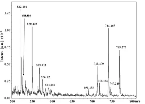
MALDI-TOF analysis of the dark-brown compound(s) demon-strates the presence of several molecular ions (from the observed m/z values) in the spectrum (e.g.m/z values: 522.101, 528.014, 550.139, 569.913, 576.12, 591.958, 691.193, 713.170, 719.183, 741.165, 747.210, 769.275) (Figure 1), which indicate the presence of mixture of compounds. No m/z peak has been observed beyond the value of 800. Out of these several peaks observed, m/z values corresponding to 528.014, 569.913, 691.193, 719.183 and 747.210 may be designated as [M+H]+molecular ions, while the
m/z values 550.139, 591.958, 713.170, 741.165 and 769.275 represent the corresponding sodiated [M+Na]+species.
IV. FTIR absorption analysis
The FTIR absorption spectrum in KBr pellet (in complete dry conditions using a nitrogen atmosphere) shows intense peaks at 3435, 2926, 2361, 1716, 1622, 1406 and 1194 and 1120 cm21 (Figure 2) which indicate the presence of several functional groups (e.g.C = O of -COOH, C-O of -COOH, carbonyl C = O, C = N, aromatic C = C, -OH and –NH) [25].
V.13C-NMR chemical shift analysis
Cross-polarization magic-angle spinning (CPMAS) solid-state 13
C NMR technique has been employed for further characteriza-tion, as the material is almost insoluble in water as well as in organic solvents. The overall spectrum (Figure 3) can be deconvoluted broadly into three parts: a) 160–200 ppm; b) 100–
150 ppm; c) 10–90 ppm which would be attributed to carbonyl, aromatic and aliphatic carbon containing functionalities, respec-tively [11,26–28].
Discussion
I. Characterization of the extracted compound as melanin Towards characterization of the dark brown compound(s) obtained from the bacterium Azotobacter chroococcum, all the attempts for purification using the HPLC technique failed. This negative result strongly indicates the presence of a polydisperse, complex heterogeneous mixture (supported by the MS spectrum and the appearance of a broad spot in TLC). However, appearance of two sets of m/z values in the range of 500 and 700 with a difference of m/z 191 (Figure 1, Figure 4) supports the view that two sets of polymers (where the number of monomeric unit varies) exists in the compounds. The possibility of existence of strongly acidic (e.g.-COOH) and/or weakly acidic (e.g.phenolic -OH group) functionality may be concluded from the solubility of the compound in 1N NaOH. The strong band at,3435 cm21in
the FTIR spectrum can be assigned to the vibration of non-hydrogen bonded NH groups [16,29–30] and the broad band observed between 3200–2000 cm21 may be related to O-H stretching vibrations associated to intra/intermolecular hydrogen bonds [25–26]. Appearance of peaks at 2926 cm21 and 1622 cm21in the FTIR spectrum can be assigned to the aromatic C-H and C = C stretching modes, respectively [25] pointing towards the presence of an aromatic system in the compound(s). This conclusion is further supported by appearance of the 13C peaks in the range of 110–150 ppm, corresponding to aromatic moieties. An intense peak at ,140.3 ppm emphasizes the
existence of deshielded aromatic carbon atoms, while the peak at,110 ppm (111.3 ppm) may be considered as the characteristic
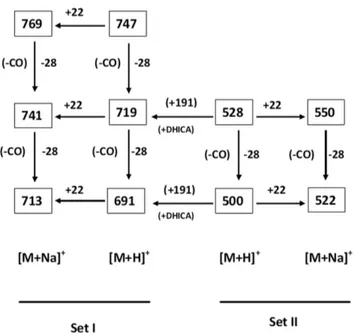
signature of indole/pyrrole carbons [31] (presence of nitrogen is confirmed through C:H:N analysis and elemental analysis using Lassaigne’s test). M/z values having a difference of 28 in both the sets [e.g.for [M+H]+: 747, 719, 691 (for Set I) and 528, 500 (Set II)
while for [M+Na]+: 769, 741, 713 (for Set I) and 550, 522 (for Set
[17]. The 13C chemical shift value at ,167 ppm indicates the
presence of carbonyl groups of carboxylic acids similar to that observed for an indole carboxylic acid moiety [27]. Further, appreciable transmittance,1700–1650 cm21in the IR spectrum
suggests the presence of C = O (carbonyl) group or C = N group or both, that may be associated in intra/intermolecular hydrogen bonds. Presence of strongly H-bonded secondary or tertiary amide C = O (may be due to presence of proteinaceous species) cannot be ruled out from the appearance of strong band,1650–1600 cm21.
However, appearance of the13C peak at,159 ppm may suggest
the presence of C = N group, which is probably arising from the indole/pyrrole system. Thus, the obtained IR spectra of the compound(s) under study matches very well with the solid-state
FTIR absorption spectrum of the indole-2-carboxylic acid reported in literature [34] and with those of a few melanins extracted from other natural sources [35–38]. A closely related FTIR absorption spectrum is also obtained by us (observed peaks at: 3385, 3205, 2910, 2362, 1714, 1622, 1396, 1295 cm21) for the synthetic melanin, purchased from Sigma-Aldrich (CAS No. 8049-97-6) (Figure S2 in File S1).
The 13C NMR spectrum obtained for the compound(s) extracted from the bacteriumAzotobacter chroococcumresembles that of 5,6-dihydroxyindole, an important constituent of the pigment melanin [13,16,27,39]. 13C-chemical shift value at ,195 ppm
may be associated to the carbonyl group from the quinone tautomer of the dihydroxyindole compound. This hypothesis is inferred from the theoretically calculated chemical shift values of different tautomers of 5,6-dihydroxyindole and 5,6-dihydroxyin-dolecarboxylic acid using the ChemBioDraw Ultra 12.0 (Figure S3 in File S1). The appearance of peaks at,10–80 ppm (in particular
intense peak between 50 and 35 ppm) establishes the presence of aliphatic carbon atoms and matches well with the chemical shift values of aliphatic carbons of several model compounds (L-dopa, dopamine, 2-methoxycarbonyl-3-ethoxycarbonyl-4-methylpyrro-le,ethyl5,5-dimethoxyindole-2-arboxylate etc.) used for the eluci-dation of melanin structure [27–28]. The overall 13C spectrum obtained from the compound(s) under investigation is very similar to that of the melanins obtained fromSepiamelanin, human hair melanin, dopa melanin and melanoma melanin (Table 1) [27–28]. Moreover, the 13C NMR spectrum of Sepia melanin, in the region from 80 to 20 ppm, shows a broad resonance due to many overlapping peaks. Similar spectrum is also observed for the compound(s) under study. Absence of peak at ,90–105 ppm
found in the spectrum of the compound under study (reported for the model compound 5,5-dimethoxyindole-2-carboxylate [26]) indicates substitutions at the aromatic carbons, emphasizing co/ hetero polymerization at the indole moiety. Such substitutions found in the aromatic carbons ofSepiamelanin and human hair melanin is responsible for the formation of the polymeric structures of the respective melanins. The broad features of the Figure 1. Partial MALDI-TOF MS [M+H]+ and [M
+Na]+spectrum of the dark brown compound(s) obtained from the bacterium Azotobacter chroococcum.
doi:10.1371/journal.pone.0084574.g001
Figure 2. FTIR absorption spectrum (in KBr pellet under complete dry conditions in a nitrogen atmosphere) of the dark brown compound(s) obtained fromAzotobacter chroococ-cum.
observed spectrum, similar to those of Sepiamelanin and human hair melanin, may indicate heterogeneity in the polymer, a well-known aspect of melanin structure, as well as the presence of free radicals. Moreover, the poor signal-to-noise ratio in the spectrum reported in Figure 3 may be the result of dipolar line broadening due to the presence of unpaired electrons, as found in Sepia
melanin and noted in an EPR study [40].
As the overall FTIR absorption and 13C NMR spectra emphasize the occurrence of a hydroxy-indolecarboxylic acid moiety in the extracted compound(s), one can conclude that 5,6-dihydroxyindole-2-carboxylic acid along with its tautomeric form, reported as the main constituents of melanins [10,12], represents the basic units of the dark brown compound(s) extracted from the bacterium Azotobacter chroococcum. In addition, as the extracted compound(s) is chemically similar to the melanins obtained from several different sources; using the complementary techniques one can unambiguously establish that the dark brown compound(s) extracted from Azotobacter chroococcum would be none other than melanins, constituted by the hydroxyindole moiety, as reported for eumelanins.
Finally, presence of the proteinaceous material in the compound seems to be a logical conclusion from the appearance of peaks at
,165–200 ppm in the 13C NMR spectrum along with the
Figure 3. Natural abundance CPMAS solid-state13C-NMR 1D-spectrum of the dark brown compound(s) obtained from bacterium
Azotobacter chroococcum.
doi:10.1371/journal.pone.0084574.g003
Figure 4. m/z values of the compound(s) obtained from the bacteriumAzotobacter chroococcumin MALDI-TOF MS spectrum representing molecular ions corresponding to two sets of [M+H]+and [M
+Na]+having difference of 191amu (one unit
5,6-dihydroxyindole-2-carboxylic acid).
doi:10.1371/journal.pone.0084574.g004
Table 1.Comparison of the13C resonances of the
compound(s) (melanin) extracted from bacteriumAzotobacter chroococcumwith other type of melanins obtained from different sources [aMagn. Reson Chem. (2008)46, 471;b
Magn. Reson Chem. (2003)41, 466].
Compounds 13C resonances
Carbonyl Aromatic Aliphatic
Dopa melanina 172 143-118 35
Melanoma melaninb 173 125 53,33
Sepia melaninb 200-160 150-110 90-30
Sepia Melanin Free Acidb 200-160 150-110 90-30
Human hair melaninb 200-170 135-110 90-30
Compound(s) fromA. chroococcum 200-160 150-115 90-25
doi:10.1371/journal.pone.0084574.t001
appearance of strong band ,1650–1600 cm21 in the FTIR
absorption spectrum (due to amide carbonyls). However, from the almost comparable peak intensity ratio of the aliphatic to aromatic signals obtained in the 13C spectrum, one can justify that the presence of the proteinaceous material, if any, is of minor significance. This conclusion can be validated from the literature survey of the13C CPMAS studies of theSepiamelanin and human hair melanin, which shows that the intensity ratio of aliphatic to aromatic signals for Sepia melanin is comparable (amino acids account for 6.17%) while that for human hair melanin is substantially larger (amino acids account for 66.8%) [27].
II. Towards model structure(s)
Melanins are considered to be heteropolymers constituted of the indole moieties and linked via carbocycles or heterocycles, predominantly polymerized through C-C linkages [12,41]. Nev-ertheless, so far no molecular structure of melanin has been proposed, as the molecular weight of this heteropolymer was not obtained with a reliable accuracy. The dark brown compound(s) melanins, extracted from the bacterium Azotobacter chroococcum, contain nitrogen but neither sulfur nor halogen. Difference of m/z of 191amu between set-I and set-II (Figure 1, 4) of the extracted melanin in MALDI-TOF experiment, a very good tool for
ascertaining the molecular weight of the compound(s), can be attributed to single unit of 5,6-dihydroxyindole-2-carboxylic acid (DHICA), recognized as an important constituent of melanins (eumelanin). These results would clearly corroborate and justify that DHICA would act as the basic constituent of the heteropoly-mer (melanins) under study. Further, the observed m/z values are quite similar to the m/z values (e.g. 524, 552, 576, 598, 698, 767, 787) obtained from the chemical and enzymatic oxidations of 5,6-dihydroxyindole-2-carboxylic acid (DHICA) using the MALDI-TOF technique [42].
From the complementary physicochemical studies, it can be reasonably concluded that, like other melanins reported in literature, 5,6-dihydroxyindole and/or 5,6-dihydroxyindole-2-car-boxylic acid/ester would be the constituent monomeric units for the melanins extracted from the bacteriumAzotobacter chroococcum
and the compound is under the category of eumelanins although obtained from a bacterium, which may be a strain related phenomenon. However, recent studies on production of melanin revealed that in some cases even bacteria can produce eumelanins [43–44].
At this point, on the basis of the information obtained from various complementary spectroscopic techniques described above, we are extending an effort to propose a putative model structure Figure 5. Proposed putative structure(s) of the protomolecules of melanin hetero-polymers, obtained from the nitrogen-fixing soil bacteriumAzotobacter chroococcum.
for melanins protomolecules obtained from Azotobacter chroococcum
having m/z values of [M+H]+: 528.014, 569.913, 576.12, 747.210
respectively [as the others are generated from loss of CO from their immediate precursor (Figure 4)] (Figure 5). These proposed model structures (Figure 5) would be well justified and validated by their respective calculated m/z values [M+H]+as well as by the
theoretically predicted/calculated chemical shift values from13C NMR spectroscopy using the Chem Ultra software (Figure S4 in File S1). However, it should be stated that the putative structures of melanins proposed here may not exactly match with those of the naturally occurring hetero-polymers, as there may be partial degradation/oxidation of these polydisperse compound(s) during the extraction process. Characterization of melanins obtained from a nitrogen-fixing, non-virulent bacterium Azotobacter chroo-coccum, thus leads to a safe and easy source for this photo-protective pigment. These results would allow expansion of experimental studies on melanins as protective agent against UV radiation and development of novel ways to administer this pigment in hypo- as well as hyper-pigmentation.
Methods
The soil sample was collected from a farmland at Howrah, West Bengal, India (22u359240N and 88u189360E), dried and pulverized aseptically. The farmland is the family/ancestral property of author Ms Aulie Banerjee and as a family member she, one of the owners of the land, would not require any permission for sample collection from her own land. The species used here is not a protected or endangered one. 10 gm were shaken in 90 ml sterile distilled water for 15 min. 1 ml of the suspension was diluted in 9 ml of 1% mannitol and 1 ml of it was plated onto the Burk nitrogen free agar medium [45–47] and was allowed to grow for 7 days at 30uC. A few dark black/brown spots were observed measuring around 1.5 cm in diameter. The isolates were purified by streaking on Petri plates and the purified isolates were grown in liquid media [48]. The media (pH 7) was kept at 30uC. Based on the observations of the colony morphology and coloration, cell nature, mean dimensions of the cells along with the flagella pattern and the pigment produced, the strain was identified and characterized [5,49].
Extraction of melanins
Isolation of melanin from the cells of Azotobacter chroococcum
through the protocol described here has been reported earlier [7]. The culture was centrifuged at 1000 g for 5 min to pallet the cells. The cell pallet was extracted three times with 5% trichloroacetic acid, washed twice with ether-ethanol (1:1 volume/volume) then washed once with absolute ether to remove impurities. The residual material was then dissolved in 0.05M sodium carbonate by treatment in a 100uC water bath for 10 min. Further centrifuge the solution to remove insoluble material. After that the mixture was stored and suspended at room temperature for 15 minutes. The brown-black material was washed three times with deionised water and freeze dried to obtain a brown-black powder which is used for further experiments.
MALDI-TOF
MALDI-TOF experiment was performed as positive mode in a Bruker Daltonics Autoflex TOF/TOF instrument. a
-cyano-4-hydroxycinnamic acid was used as the matrix and the Flex Analysis software was used for analyzing the [M+H]+results.
FTIR absorption spectroscopy
Melanin powder (obtained from the bacterium) and KBr (purchased from Sigma) were mixed in a 1:100 w/w. The mixture was ground using a mortar pestle till it achieved a uniform color indicating its homogeneity. FTIR absorption spectrum was obtained at 25uC using a model Bx Perkin-Elmer FTIR spectrophotometer Spectrum 1000, using 4 cm21resolution with 8 number of scans.
CPMAS13C NMR spectrometry
The NMR data were recorded on a Bruker DSX 300, 7.04 Tesla, solid-state NMR spectrometer using 5-mm probes. For the 13
C CPMAS experiments, .100 mg of sample were tightly packed using 4-mm rotors with teflon spacers and spun at a typical speed of 1.2 kHz. 13C CPMAS experiments were conducted with a1H decoupling strength of 50 kHz, a delay time of 1 s between successive acquisitions, a line broadening of 50– 100 Hz, and contact times of 2 ms to establish the ratios of rigid carbon moieties [50]. Chemical shift referencing of 13C NMR studies was performed by setting the glycine -CO- at 176 ppm in a separate experiment using a pure glycine sample.
Supporting Information
File S1 Supporting figures.Figure S1, UV-VIS absorption spectrum of the dark brown compound(s) obtained fromAzotobacter chroococcumin NaOH. Figure S2, FTIR spectrum (in KBr pellet under complete dry condition in nitrogen atmosphere) obtained from synthetic melanin purchased from SIGMA ALDRICH. Figure S3, a) Calculated13C-NMR chemical shift values using the Chem Ultra software for 5,6-dihydroxyindole and its tautomer b) Calculated13C-NMR chemical shift values using the Chem Ultra software for 5,6-dihydroxyindole-2-carboxylic acid and its tauto-mer, Figure S4, Calculated13C-NMR chemical shift values using the Chem Ultra software for the proposed putative structure(s) of the protomolecules of melanin hetero-polymers obtained from the nitrogen-fixing soil bacteriumAzotobacter chroococcum.
(DOC)
Acknowledgments
Authors gratefully acknowledge Prof. Rahul Banerjee, SINP, Kolkata for allowing his laboratory facilities to be used during extraction of melanins from Azotobacter chroococcum. Authors extend their sincere thanks to the MALDI-TOF facility of Bose Institute, Kolkata and MCBL of IISc Bangalore. The Solid-state Unit, IISc Bangalore is acknowledged for recording the FTIR data and the NMR Research Center, IISc Bangalore for the solid-state13C-NMR spectrum. We are indebted to BIF-DBT at Department of Bioinformatics, WBUT for pursuing the calculations and the modeling part to determine the molecular structure of the bacterial melanins.
Author Contributions
Conceived and designed the experiments: RB. Performed the experiments: AB SS RB. Analyzed the data: RB SS AB. Wrote the paper: RB.
References
1. Hill HZ (1992) The function of melanin or six blind people examine an elephant. BioEssays 14: 49–56.
2. Hamilton AJ, Gomez BL (2002) Melanin in fungal pathogens. J Med Microbiol 53: 189–191.
3. Castelvecchi D (2007) Dark Powder: Pigment seems to put radiation to good use. Science News 171: 325.
4. Dadachova E, Bryan RA, Huang X, Moadel T, Schweitzer AD, et al. (2007) Ionizing Radiation Changes the Electronic Properties of Melanin and Enhances the Growth of Melanized Fungi. PLoS ONE 2(5): e457. doi:10.1371/journal.-pone.0000457.
5. Becking JH (1981) The familyAzotobacteraccae. In: Starr MP, Stolp H, Truper HG, Balows A, Schlegel HG. The Prokaryotes. Berlin: Springer-Verlag 1: pp. 795–817.
6. Jensen HL (1954) The Azotobacteriaceae. Bacteriol Rev 18: 195–214. 7. Shivaprasad S, Page W (1989) Catechol Formation and Melanization by Na+
-DependentAzotobacter chroococcum: a Protective Mechanism for Aeroadaptation? J Appl and Env Microbiol 55(7): 1811–1817.
8. Thompson JP, Skerman VBD (1979) Azotobacteraceae: the Taxonomy and Ecology of the Aerobic Nitrogen-Fixing Bacteria. London: Academic Press. 277 p.
9. Plonka PM, Grabacka M (2006) Melanin synthesis in microorganisms – biotechnological and medical aspects. Acta Biochim Pol 53: 429–443. 10. Nicolaus RA, Piattelli M, Fattorusso E (1964) The structure of melanins and
melanogenesis. IV. On some natural melanins. Tetrahedron 20: 1163–1172. 11. Chedekel MR (1982) Photochemisrty and photobiology of melanins. Photochem
photobiol 35: 881–885.
12. Pezzella A, d’Ischia M, Napolitano A, Palumbo A, Prota G. (1997) An integrated approach to the structure of Sepia melanin. Evidence for high proportion of degraded 5,6-dihydroxyindole-2-carboxylic acid units in the pigment backbone. Tetrahedron 53: 8281–8286.
13. Duff GA, Roberts JE, Foster N (1988) Analysis of the structure of synthetic and natural melanins by solid-phase NMR. Biochemistry 27: 7112–7116. 14. Swan GA (1974) Structure, chemistry and biosynthesis of the melanin. Fortschr
Chem Org Natrust 31: 521–582.
15. Schnitzer M, Chan YK (1983) Structural Characteristics of a fungal melanin and a soil humic acid. Soil Sci Soc Am J 50: 67–71.
16. Reinheimer P, Hirschinger J, Granger P, Breton P, Lagrange A, Gilard P, Lefebvre M A, Goetz N (1999) Cross-polarization/magic-angle-spinning nuclear magnetic resonance in selectively 13C-labeled synthetic melanin. Biochim Biophys Acta - General Subjects 1472(1–2): 240–249.
17. Tire A, Guillaume P, Massat A, Aaron JJ (1998) Infrared study of indolecarboxylic acids associations with lanthanide acetates. Spectrochim Acta A 54: 1451–1459.
18. Moores OT (1995) The science of Melanin. Maryland USA: Beckham Pubs. Silver Spring. 158 p.
19. Baraboi˘ VA (1999) Melanin: structure, biosynthesis, biological functions. Ukrainian Biochem J 71: 5–14.
20. Lindgren J, Uvdal V, Sjo¨vall P, Nilsson DE, Engdahl A, Schultz BP, Thiel V (2012) Molecular preservation of the pigment melanin in fossil melanosomes. Nat Commun 3: 824.
21. Katritzky AR, Akhmedov NG, Denisenko SN, Denisko OV (2002) 1H NMR spectroscopic characterization of solutions of Sepia melanin, Sepia melanin free acid and human hair melanin. Pigment Cell Res 15(2): 93–97.
22. Chedekel MR, Ahene AB, Zeise L (1992) Melanin standard method: empirical formula 2. Pigment Cell Res 5(5): 240–246.
23. Gower RP, Rhodes IP (1969) A review of the Lassaigne sodium-fusion. J Chem Educ 46: 606.
24. Clarke HT (1975) A handbook of organic analysis 5thEd. Hodder & Stoughton Educational. 1975, H.T. Clarke, revised by B. Haynes.
25. Kemp W (1987) Organic spectroscopy. 2ndEd. Hong Kong: ELBS/Macmillan Education Ltd. 19 p.
26. Herve´ M, Hirschinger J, Granger P, Gilard P, Deflandre A, Gotez N (1994) A 13C solid-state NMR study of the structure and auto-oxidation process of natural and synthetic melanins. Biochim Biophys Acta 1204: 19–27. 27. Adhyauru BB, Akhmedov NG, Katritzky AR, Bowers CR (2003) Solid-state
cross-polarization magic angle spinning13
C and15
N NMR characterization of
Sepiamelanin,Sepiamelanin free acid andHuman hairmelanin in comparison with several model compounds. Magn Reson Chem 41: 466–474.
28. Ghiani S, Baroni S, Burgio D, Digilio G, Fukuhara M, Martino P, Monda K, Nervi C, Kiyomine A, Aime S (2008) Characterization of human hair melanin and its degradation products by means of magnetic resonance techniques. Magn Reson Chem 46: 471–479.
29. Pysh ES, Toniolo C (1997) Conformational analysis of protected norvaline oligopeptides by high resolution proton magnetic resonance. J Am Chem Soc 99: 6211–6219.
30. Toniolo C, Bonora GM, Bavoso A, Benedetti E, Di Blasio B, Pavone V, Pedone C, Barone V, Lelj F, Leplawy MT, Kaczmarek K, Redlinski A (1988) Structural versatility of peptides from Ca,a
-dialkylated glycines. II. An IR absorption and
1
H-nmr study of homo-oligopeptides from Ca,a
-diethylglycine. Biopolymers 27(3): 373–379.
31. Peter MG, Fo¨rster H (1989) On the structure of eumelanins: identification of constitutional patterns by solid-state NMR spectroscopy. Angew Chem Int Ed Engl 28741–743.
32. Finar IL (1986) Organic Chemistry Volume 1: The Fundamental Principles. UK : Longman Group. 700 p
33. Dobrzynska D, Turoska-Tyrk I (1997) 9,1-Dihydro-9-oxo-10acridineacetic Acid. Acta Crystallogr C 53: 238–239.
34. Morzyk-Ociepaa B, Michalskab D, Pietraszko A (2004) Structures and vibrational spectra of indole carboxylic acids. Part I. Indole-2-carboxylic acid. J Mol Struc 688: 79–86.
35. Zecca L, Mecacci C, Seraglia R, Parati E (1992) The chemical characterization of melanin contained in substantia niagra of human brain. Biochim and Biophys Acta 1138: 6–10.
36. Double KL, Zecca L, Costi P, Mauer M, Griesinger C, Ito S, Ben-Shachar D, Bringmann G, Fariello RG, Riederer P, Gerlach M (2000) Structural characteristics of human substantia nigra neuromelanin and synthetic dopamine melanins. J Neurochem 75: 2583–2589.
37. Hewedy MA, Ashour SM (2009) Production of a Melanin like Pigment by
Kluyveromyces marxianusandStreptomyces chibaensis. Aust J Basic Appl Sci 3(2): 920– 927.
38. Magarelli M, Passamonti P, Renieri C (2010) Purification, characterization and analysis of sepia melanin from commercial sepia ink (Sepia Officinalis). Rev CES Med Vet Zootec 5: 18–28.
39. Schneider HJ (1989) Additives of Electrostatic and Hydrophobic interactions in Host-Guest Complexes. Theis I Angew Chem 101: 757.
40. Enochs WS, Nilges MJ, Swartz HM (1993) A standardized test for the identification and characterization of melanins using electron paramagneti-c(EPR) spectroscopy. Pigment Cell Res 6: 91–99.
41. Pezzella A, Napolitano A, d’Ischia M, Prota G (1996) Oxidative polymerization of 5,6-dihydroxyindole 2-carboxylic acid units to melanin: a new insight. Tetrahedron 52: 7913–7920.
42. Napolitano A, Pezzella A, Prota G, Seraglia R, Traldi P (1996) Structural analysis of synthetic melanins from 5,6-dihydroxyindole by MALDI mass spectrometry. Rap Comm in Mass Spec 10: 204–208.
43. Solano F, Garcı´a E, de Egea EP, Sanchez-Amat A (1997) Isolation and characterization of strain MB-1 (CECT 4803), a novel melanogenic marine bacterium. Appl Environ Microbiol 63: 3499–3506.
44. Geng J, Yuan P, Shao C, Yu SB, Zhou B, Zhou P, Chen XD (2010) Bacterial melanin interacts with double-stranded DNA with high affinity and may inhibit cell metabolism in vivo. Arch Microbiol 192(5): 321–329.
45. Bark D (1930) The influence of oxygen gas upon the organic catalysis of nitrogen fixation byAzotobacter. Jr Phy Chem 34: 1195–1209.
46. Bark D, Lineweaver H (1930) The influence of fixed nitrogen onAzotobacter. J Bacteriol 19: 389–414.
47. Wilson PW, Knight SG (1952) Experiments in bacterial physiology. Minneap-olis, USA: Burgess Publishing Co.
48. Dalton H, Postage JR (1968) Effect of oxygen onAzotobacter chroococcumin bath and continuous cultures. J Gen Microbiology 34: 463–469.
49. Becking JH (2006) Chapter 3.3.26. In: Dworkin M, Falkow S, Rosenberg E, Schleifer KH, Stackebrandt E. Prokaryotes. New York: Springer pp 759–783. 50. Schaefer J, Stekskal EO (1979) High-resolution13