IN VITRO AND IN VIVO EVALUATION
OF PIROXICAM LOADED CERAMIC
NANOPARTICLES
PAVANI VENGALA1, CVS SUBRAHMANYAM1, M GANGARAJU2
1
Department of Pharmaceutics, 2Department of Pharmacology Gokaraju Rangaraju College of Pharmacy, Hyderabad, Telangana-500090
Email: [email protected]
Abstract: The use of nanotechnology in drug delivery is spreading rapidly. The nanocarriers have been used for the enhanced delivery of a range of drugs. The present study was aimed at investigating the application of ceramic nanoparticles called as aquasomes for the delivery of drug, piroxicam. Piroxicam belongs to oxicam group of NSAID’s, commonly used for the treatment of arthritis. It is a BCS class II drug, with low solubility. There is a need to improve the dissolution property of piroxicam in order to enhance its therapeutic efficacy. Ceramic Nanoparticles were prepared by colloidal precipitation method. The ceramic core was coated with polysaccharide, cellobiose, followed by adsorption of drug. The drug loaded nanoparticles were evaluated for size, entrapment efficiency and drug release profile. The SEM studies indicated that the formed particles were with nanometric dimensions (185 nm). 21% drug loading was observed and more than 95% drug release was observed within 135 min in 0.1N HCl compared with pure drug which released 89% in 90 mins. In vitro dissolution studies indicated that the piroxicam ceramic nanoparticles released the drug in a controlled manner. Anti-nociceptive and anti-inflammatory studies were performed with piroxicam cellobiose aquasomes. Paw edema method was employed for assessing anti-inflammatory effect. The anti-inflammatory activity of aquasome formulation showed quicker effect up to 3 h compared to pure piroxicam.
Key words: Piroxicam, cellobiose, ceramic nanoparticles, analgesic, arthritis. Introduction:
Advances in drug discovery have led to an increasing number of new drugs great therapeutic potential. However they are with poor water solubility and thus poor and variable bioavailability, esp via oral administration. As most of the human body is made up of water, a drug must have certain water solubility and thus an acceptable bioavailability level. Poorly water soluble drugs tend to be eliminated from the gastrointestinal tract before they get the opportunity to fully dissolve and be absorbed into the blood circulation, which results in low bioavailability and poor dose proportionality. Many approaches have been developed to enhance the dissolution rate as well as bioavailability of poorly water soluble drugs, including modifications to the drug substance itself and the creation of specific formulations [1].
The most commonly used approach is size reduction to micron level which results in a modest increase in surface area that may not change the dissolution rate or saturation solubility to significantly impact bioavailability [2].
Nanoparticle formulation technologies have provided the pharmaceutical industry with options for addressing solubility and bioavailability issues associated with poorly soluble compounds [3]. One among them is the aquasome technology, a ceramic based biodegradable nanoparticulate system. Aquasomes are spherical in shape with 60–300 nm particles size. These are nanoparticulate carrier systems but instead of being simple nanoparticles these are three layered self assembled structures, comprised of a solid phase nanocrystalline core coated with oligomeric film to which biochemically active molecules are adsorbed with or without modification. These structures are self assembled by non covalent and ionic bonds. The solid core provides the structural stability, while the carbohydrate coating protects against dehydration and stabilizes the biochemically active molecules. The delivery system has been successfully utilized for the delivery of insulin, hemoglobin, and enzymes like serratiopeptidase etc [4].
Materials:
Piroxicam was received as a gift sample from Strides Arcolabs Ltd., Bangalore. Calcium Chloride dihydrate, disodium hydrogen orthophosphate and cellobiose were purchased from SD Fine Chemicals Ltd., Mumbai, India. All the other chemicals and reagents were of analytical grade.
Methods: Preparation of aquasomes:
Ceramic nanoparticles or otherwise called aquasomes are prepared by a three-step method that consists of production of nano cores, adsorption of cellobiose on core, and adsorption of active ingredient on sugar-coated core.
For the preparation of ceramic core, previously used procedure [7] was followed. Disodium hydrogen phosphate and calcium chloride were dissolved in water each separately and mixed. These were then sonicated (2 h at 4°C) using bath sonicator to yield the colloidal precipitate [8, 9]. After sonication, the mixture was centrifuged (15000 rpm) for 1 h. The supernatant was poured out and the precipitate was thoroughly washed three times using double distilled water. The precipitate was re-suspended in distilled water (50 ml), and then filtered through 0.2 µ membrane filter. The core was dried (100 ºC, 2 days). The percentage yield was calculated [9-11].
Adsorption of polyhydroxy-oligomer, cellobiose on the ceramic core
The core particles (prepared as above) were coated with polyhydroxy-oligomer by adsorption method [12] using sonication. Cellobiose was dissolved in double distilled water to which specific quantity of ceramic core was added. The solution was sonicated using probe sonicator (30% pulse and 18 W) for 30 min. This suspension was incubated for 3 hr time period (100 rpm, 25 °C). Non solvent (acetone, 1 ml) was added to the suspension and allowed the sugar to get adsorbed on to core by keeping the solution aside for approximately 20 min. The solution was then centrifuged (2000 rpm, 25 °C and 15 min). The supernatant was decanted; the sugar coated core was washed two times with double distilled water and dried at 70 °C in a hot air oven. The sugar coated core was quantified by tagging with the anthrone reagent and absorbance was estimated at max = 625 nm.
Quantification of cellobiose coating on core: Fifty mg of cellobiose coated core was precisely measured and dissolved in 5 ml of distilled water. From this stock, 2 ml of the solution was taken and 5.5 ml anthrone reagent was added and boiled (10 min, 100 °C). The solution was cooled rapidly and absorbance was estimated at max = 625 nm [13, 14].
Drug loading
Piroxicam solution (1.5% w/v in acetone) was taken in a volumetric flask containing an accurately weighed amount of sugar coated ceramic core (25 mg). The flask was stoppered and shaken vigorously at 130 rpm for 24 h to obtain three layered drug adsorbed aquasomes. The suspension was centrifuged at 15000 rpm for 5 minutes. Ceramic nanoparticles were separated and air dried.
Evaluation of ceramic nanoparticles
The three layered aquasomes were evaluated for particle size, shape, percent yield, drug loading efficiency,
in vitro drug release and in vivo pharmacological efficiency. Particle size and size distribution analysis
The average size and size distribution of aquasomes were measured using scanning electron microscope (Hitachi S-3000N)[15]. SEM pictures of piroxicam aquasomes was presented in Figure 1.
FTIR analysis
FTIR spectroscopy was used for the confirmation of all the three layers; core, sugar and drug. The method was reported earlier [16]. FTIR of final formulation (aquasomes of piroxicam) was compared with that of individual components and given in Table 1 and Figure 2.
Determination of drug payload
Weighed amount of the formulated aquasome was dissolved in acetone and diluted with suitable solution (0.1 N hydrochloric acid solutions, phosphate buffer, pH 6.8). Absorbance of the solution was measured spectrophotometrically for piroxicam at λ max = 334 nm and 354 nm respectively. Percent payload was
Estimation of percentage yield
After drying of the formulated drug-loaded ceramic nanoparticles, free-flowing powdered nanoparticles were obtained. The ceramic nanoparticles were collected carefully and accurately weighed. Percentage yield of the nanoparticles was calculated by the following equation.
In vitro release of drugs from ceramic nanoparticles
In vitro release studies were performed by accurately weighing dried drug loaded ceramic nanoparticles equivalent to 10 mg of pure drug, and transferring into empty capsules. Dissolution was performed using capsules as reported in USP/NF, by the use of USP type I (Basket) dissolution apparatus [17]. After preset time intervals, samples were collected and analyzed for cumulative percent drug dissolved/released.
To know the release kinetics, data acquired from in vitro drug release studies were plotted in different kinetics models to comprehend the linear relationship, i.e., kinetic principles. To study the release mechanisms, the data of in vitro drug release was checked using Higuchi, Hixson Crowell Cube root law and Korsemeyer Peppas models [18].
In vivo studies
Pharmacological method was used to confirm the activity of drug in the aquasomes. The Institutional Animal Ethics Committee (IAEC) has approved this study. Male Wistar rats were selected for in vivo efficacy studies according to the instructions set by CPCSEA. They were accommodated in a set of controlled conditions. The animals were placed separately in PP cages having sterile husk of paddy (obtained locally) as base all through the experiment. All animals were fed with sterile commercial pelleted rat chow (Sri sai Thirumala enterprises, Hyderabad, India) and had free access to water ad libitum. Animals were kept for fasting all night and weighed prior to the experiment. The study was undertaken after getting approval from Institutional Animal Ethics Committee. Doses were calculated according to conversion factor [19].
Carrageenan induced paw edema model
The anti inflammatory activity was estimated in Wistar rats employing the method of Winter et al [20]. Animals were kept for overnight fasting and were separated into control, standard and formulation test groups, each containing six rats. Animals in the standard group received piroxicam at the dose of 0.2 mg/ml by oral route. Aquasome formulation was administered to the test group, at equimolar doses of piroxicam. All test and standard compounds were administered as 0.5% CMC suspension. Rats in the control group were given the vehicle solution (without drug). One hour after test drug administration, rats in all the groups were challenged with 0.1 ml of 0.1% carrageenan in sub plantar region of right hand paw. A zero hour reading of rat paw volume was measured using plethysmometer immediately after the introduction of carrageenan for all groups. Paw volumes were measured at 1, 3 and 6 h and again measured at 24 h after the challenge of carrageenan. The increased paw volumes were presented as mean ± SEM. The percent inhibition of paw volume for each rat in treated group was calculated by comparing with mean paw volume of control group and expressed as mean ± SEM. ANOVA was carried out to establish the significance of the exhibited activity and percentage decline in paw volume was measured.
vt and vo represents the average volume in the hind paw of the rats before and after anti-inflammatory agent
treatment, respectively.
Results and Discussion:
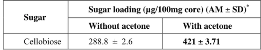
Colloidal precipitation technique was used for preparation of core and 50% yield was obtained. The ceramic core was further coated with sugar. Non solvent technique was employed to coat the sugar, cellobiose. The loading was increased with the addition of non-solvent (acetone). Acetone being more soluble in water, the water rejects the sugar (which was earlier soluble). Hence the loading of sugar can be enhanced.
Table 1: Effect of non-solvent addition on sugar loading onto the ceramic core
Sugar Sugar loading (µg/100mg core) (AM ± SD)
*
Without acetone With acetone
Cellobiose 288.8 ± 2.6 421 ± 3.71
Adsorption of piroxicam on the sugar coated core
Piroxicam was loaded onto cellobiose coated core using adsorption technique. The drug loading was found to be 21%. After drying, the drug-loaded ceramic nanoparticles were free-flowing. The dried ceramic nanoparticles were collected and weighed. Percentage yield of the nanoparticles was found to have a good yield (66.7%).
Evaluation of Piroxicam Aquasomes Particle size analysis and morphology
The SEM images of piroxicam loaded aquasomes showed spherical nanoparticles. The particle size was uniform and particles were mostly single (discrete) (Figure 1). The size was within nano range whereas that of pure drug was in micron range (Table 2).
Table 2: Average particle size of drug loaded aquasomes
* Each value represents the mean of 3 determinations
Figure 1: Scanning electron microscopy image of piroxicam cellobiose aquasomes
FTIR spectroscopic analysis
The FTIR spectra is shown in Figure 2, and the characteristic bands were reported in the Table 3 and all were found to be within the limits.
Figure 2: FTIR spectra of piroxicam cellobiose aquasomes
Particles Particle size, (nm)(AM ± SD)*
Piroxicam cellobiose aquasomes 178.40 ± 47.24
Table 3: Comparison of characteristic FTIR bands of piroxicam aquasomes
Characteristic bands Observed in this study, cm-1 Literature values21
, cm
-1
Core
Phosphate (P-O) 871.82 845-725
Phosphate (P=O) 1318.86 1300-1240
Sugars
CH stretching, symmetrical 2879.72 3200-3000
Piroxicam
NH, OH stretching of amide 3336.85 3330
Bending of amide carbonyl 1685.79 1635 or 1625
Bending of second amide gp 1527.62 1525
SO2-N 1060.85 1050-1070
In vitro piroxicam release in 0.1 N hydrochloric acid medium
The cumulative percent piroxicam release from pure piroxicam, ceramic nanoparticles at 37 ± 0.5 °C was carried out in 0.1 N HCl solution and was reported in Table 4. Pure piroxicam showed incomplete dissolution of 89% in 75 mins. In vitro dissolution studies indicated that the piroxicam ceramic nanoparticles released the drug in a controlled manner. Piroxicam loaded aquasomes gave 95% release 130 mins (Figure 3). The gradual release of piroxicam also indicated that there would be some type of interaction between piroxicam and lactose/cellobiose. There was no instantaneous dissolution. It indicated the absence of free piroxicam precipitation during incubation of core particles with piroxicam. The dissolution of piroxicam from pure piroxicam followed first order release (Table 4) and justified.
Table Error! No text of specified style in document.: Cumulative percentage release of piroxicam from the pure drug, ceramic
nanoparticles and plain nanoparticles and in 0.1 N hydrochloric acid solution
Time (min)
Cumulative drug release (%), (AM ± SD)*
Pure piroxicam Piroxicam cellobiose nanoparticles
0 0 0
5 5.64 ± 2.6 5.12 ± 1.6
15 40.21 ± 2.3 15.36 ± 2.8
30 62.82 ± 6.5 29.41 ± 4.6
45 75.34 ± 4.5 42.65 ± 3.2
60 84.32 ± 4.9 52.82 ± 3.1
75 89.64 ± 3.9 61.52 ± 1.2
90 - 72.16 ± 1.8
105 - 79.64 ± 2.2
120 - 89.82 ± 2.6
135 - 95.88 ± 1.5
Figure 3: In vitro piroxicam release profile from pure drug, aquasome formulations and plain nanoparticles in 0.1 N hydrochloric acid solution
Pharmacological Studies - Piroxicam aquasomes Anti inflammatory studies
Carrageenan generated paw edema model was used to test the anti-inflammatory effect of piroxicam and its aquasome formulation. Both drug and formulation showed statistically significant (P<0.0001) inhibitory effect, on mean increase in paw volume at all time points (1, 3, and 6 h) as shown in Table 5 and Figure 4. The aquasome formulation showed rapid anti inflammatory activity up to 6 h compared to control. The anti-inflammatory activity of formulation was slightly less than that of piroxicam alone. This may be due to slow release of piroxicam from the aquasomes. This phenomenon was also observed during dissolution in media.
Table 5: Anti inflammatory effect of piroxicam and its formulation on paw edema
Treatment Time, h Control
AM ± SEM*
Pure piroxicam AM ± SEM*
Piroxicam formulation AM ± SEM*
Increase in mean paw volume (mm)
0 0.1 ± 00 0.1 ± 00 0.1 ± 00
1 0.405±0.03 0.251±0.02**** 0.324±0.01****
3 0.628±0.03 0.19±0.01**** 0.25±0.01****
6 0.415±0.02 0.111±0.01**** 0.138±0.01****
24 0.1± 0.0 0.1± 0.0 0.1± 0.0
Figure Error! No text of specified style in document.: Anti-inflammatory activity of piroxicam and formulation on paw edema
Conclusion:
Aquasomes of piroxicam were prepared by colloidal precipitation method using cellobiose as the sugar. SEM studies indicated nanorange spherical particles. Release studies in both the media showed slow and complete release of the drug whereas pure drug showed incomplete release. The release was gradual without initial peak level which was further supported by anti-inflammatory studies. Aquasome technology thus can be a promising tool for the oral delivery of piroxicam and other poorly soluble drugs also.
Acknowledgement:
The authors would like to thank Dr Sneha JA and Mrs Suvarchala, GRCP for their timely help in conducting the
in vivo studies.
Conflicts of interest: the authors declare that they have no conflicts of interest. References:
[1] Mitali Kakran, Professor Lin Li, and Professor Dr. Rainer H. Müller, Overcoming the Challenge of Poor Drug Solubility,
Pharmaceutical Engineering, 2012, 32 (4), pp. 82-89.
[2] Vijaykumar N, Venkateswarlu V and Raviraj P, Research Journal of Pharmaceutical, Biological and Chemical Sciences. Development
of oral tablet dosage form incorporating drug nanoparticles. October – December 2010 RJPBCS 1(4) Page No. 952
[3] Yellela S.R. Pharmaceutical technologies for enhancing oral bioavailability of poorly soluble drugs. J Bioeq & Bioav.
2010;2(2):28-36.
[4] Jain NK, Jain SK. Advances in controlled and novel drug delivery. 1st ed. New Delhi (Inida): CBS Publishers; 2008. P. 317-31.
[5] Maryadele J. The Merck Index-An encyclopedia of chemicals, drugs and biologicals. 13th ed. Whitehouse Station (New Jersey): Merck
and Co. Inc; 2006. p.1709, 7435.
[6] Martindale. The Complete Drug Reference. London: Pharmaceutical Press; 2005: p. 761.
[7] Lactose coated ceramic nanoparticles for oral drug delivery. Pavani Vengala, Swetha Dintakurthi, Chavali Venkata Satya
Subrahmanyam journal of pharmacy research 7 (2013) 540-545
[8] Jain SK, Cherian AK, Rana AC. Self-assembled carbohydrate-stabilized ceramic nanoparticles for the parenteral delivery of insulin.
Drug Dev Ind Pharm. 2000;26(4):459-63.
[9] Kossovsky N, Gelman A, Sponsler EE, Hnatyszyn HJ, Rajguru S, Chow K, Chung A, Torres M, Zemanovich J, Anderson S, Yao G,
Wei K, Goodwin C. Control of molecular polymorphisms by a structured carbohydrate/ceramic delivery vehicle-aquasomes. J Con Rel. 1996;39:383-88.
[10] Rawat M, Singh D. Development and in vitro evaluation of alginate gel- encapsulated, chitosan coated ceramic nanocores for oral
delivery of enzyme. Drug Dev Ind Pharm. 2008;34:181-88.
[11] Rojas OI, Salazar LR, Gasga AR, Barreda CTQ. Elaboration and structural analysis of aquasomes loaded with indomethacin. Eur J
Pharm Sci. 2007;32:223-30.
[12] Hedge JE, Hofreiter BT. Determination of reducing sugars and carbohydrates. Analysis and preparation of sugars. Carbohydrate
chemistry. New York: Academic Press; 1962. p.48–59.
[13] Devor AW, Baker WC, Devor KA. Effect of toluene and similar compounds on the carbohydrate-anthrone reaction. Clin Chem.
1964;10:597-99.
[14] Raymond C, Paul J, Marian E. Handbook of pharmaceutical excipients. Washington: Pharmaceutical press and Americian pharmacist
assosiation; 2009. p.159-160, 373-74.
[15] Vernon-Parry KD. Scanning electron microscopy: an introduction. III-Vs Review. 2000 Aug;13(4):40-41.
[16] Sergei G, Kazarian, Chan A. ATR-FTIR spectroscopic imaging: recent advances and applications to biological systems. Analyst.
2013;7.
[17] USP/NF-The official compendia of standards. Asian ed. Rockville (MD): United States Pharmacopoeial Convention, Inc.; 2003. p.
1486-87, 2303.
[18] Dash S, Padala NM, Nath L, Chowdhury P. Kinetic modeling on drug release from controlled drug delivery systems. Acta Pol Pharm
Drug Res. 2010;67(3):217-23.
[19] Turner RA. Screening methods in pharmacology, New York and London: Academic press; 1965.
[20] Chakraborty A, Devi RKB, Rita S. Preliminary studies on anti-inflammatory and analgesic activities of spilanthes acmella in
experimental animal models. Ind. J. Pharmacol. 2004;36:148‐50.