*Correspondence: Panikumar Anumolu Durga. Gokaraju Rangaraju College of Pharmacy, Department of Pharmaceutical Analysis, Hyderabad, 500090 - Andhra Pradesh, India. E-mail: [email protected]
A
vol. 50, n. 2, apr./jun., 2014 http://dx.doi.org/10.1590/S1984-82502014000200012
Development of dissolution test method for a telmisartan/
amlodipine besylate combination using synchronous derivative
spectrofluorimetry
Panikumar Durga Anumolu
1,*, Sirisha Neeli
2, Haripriya Anuganti
2, Sathesh Babu Puvvadi
Ranganatham
2, Subrahmanyam Chavali Venkata Satya
21R & D Center, Jawaharlal Nehru Technological University, Department of Pharmaceutical Sciences, Hyderabad, Andhra
Pradesh, India 2Gokaraju Rangaraju College of Pharmacy, Osmania University, Hyderabad, Andhra Pradesh, India
The dissolution process is considered an important in vitro tool to evaluate product quality and drug release behavior. Single dissolution methods for the analysis of combined dosage forms are preferred to simplify quality control testing. The objective of the present work was to develop and validate a single dissolution test for a telmisartan (TEL) and amlodipine besylate (AML) combined tablet dosage form. The sink conditions, stability and speciicity of both drugs in different dissolution media were tested to choose a discriminatory dissolution method, which uses an USP type-II apparatus with a paddle rotating at 75 rpm, with 900 mL of simulated gastric luid (SGF without enzymes) as the dissolution medium. This dissolution methodology provided good dissolution proiles for both TEL and AML and was able to discriminate changes in the composition and manufacturing process. To quantify both drugs simultaneously, a synchronous irst derivative spectroluorimetric method was developed and validated. Drug release was analyzed by a luorimetric method at 458 nm and 675 nm for AML and TEL, respectively. The dissolution method was validated as per ICH guidance.
Uniterms: Combined dosage forms/quality control. Dissolution test/combined dosage forms. Telmisartan. Amlodipine besylate. Spectroluorimetry/quantiication analysis.
O processo de dissolução é considerado como uma importante ferramenta in vitro para avaliar a qualidade do produto e o comportamento de liberação do fármaco. Prefere-se um ensaio único de dissolução para formas farmacêuticas contendo associação de fármacos pela simpliicação dos testes de controle de qualidade. O objetivo do presente trabalho foi desenvolver e validar um teste de dissolução único para forma farmacêutica comprimidos contendo telmisartana (TEL) e besilato de anlodipino (AML) associados. Condições “sink”, estabilidade e especiicidade de ambos os fármacos nos diferentes meios de dissolução foram avaliadas para selecionar um método de dissolução discriminatório, que utiliza um aparato do tipo II da USP, com pás girando a 75 rpm e 900 mL de luido gástrico simulado (SGF sem enzima) como o meio de dissolução. Estas condições proporcionaram bons peris de dissolução para ambos, TEL e AML, sendo capaz de discriminar as mudanças na composição e processo de fabricação. Para quantiicar os dois fármacos simultaneamente, um método de luorescência derivada sincronizado foi desenvolvido e validado. A quantidade de fármaco liberado foi analisada pelo método luorimétrico em 458 e 675 nm para a AML e TEL, respectivamente. O método de dissolução foi validado de acordo com a orientação da ICH.
Unitermos: Forma farmacêutica com associação/controle de qualidade. Teste de dissolução/forma
INTRODUCTION
Drug dissolution testing an important analytical technique to evaluate product quality, to assess drug release behavior and to discriminate changes in the formulation and manufacturing process (Kulkarni et al., 2012). The strategy to determine the solubility and permeability
properties of drugs uses a biopharmaceutical classiication
system to classify drugs into four basic groups (Amidon
et al., 1995). The development of a dissolution method for a drug product with limited water solubility and combinations of drugs has been a challenge for both the pharmaceutical industry and regulatory agencies (Soni et al., 2008; Dressman et al., 1998; Oliveira et al., 2009). Currently, there is increased demand for biorelevant dissolution media, which have the ability to discriminate changes in the formulation and manufacturing process (He et al., 2004; Panikumar et al., 2012; Menegola et
al., 2007). In vitro dissolution media is formulated to be biorelevant, as it should be able to serve as a surrogate of the in vivo environment. In vitro dissolution media are made biorelevant by adding various levels of bio-salts, lecithin and fatty acids (Galia et al., 1998). A single
dissolution method for the analysis of multiple API active components in combinations present in a dosage form is preferred to simplify quality control testing procedures
(Vignaduzzo et al., 2010; Huang et al., 2011; Panikumar et al., 2013; Zongyun et al., 2011).
Telmisartan (TEL) (Figure 1A), chemically known as 4′-[(1,4′-dimethyl-l-2′-propyl[2,6′-bi-1H
-benzimidazol]-1′yl)methyl]-[1,1′-biphenyl]-2–carboxylic
acid, is a angiotensin–II (AT1) receptor antagonist used in the treatment of hypertension and myocardial infarction.
Amlodipine besylate (AML) (Figure 1B), chemically known as 3-ethyl-5-methyl 2-(2-amino ethoxy methyl)-4-(2-chloro phenyl)-1,4-dihydro-6-methyl pyridine-3,5-dicarboxylate benzene sulfonate, is a calcium channel
blocker used in the treatment of hypertension and angina
pectoris (Anthony , David, Brian, 2004). TEL and AML have been formulated in a ixed-dose combination used in
the treatment of hypertension. To the best of our knowledge, no single dissolution test has been reported for TEL and AML in a combined tablet dosage form. Therefore, the objective of the present investigation was to develop and validate a single discriminating dissolution test method for TEL and AML in a combined tablet dosage form. A literature survey revealed that a few analytical methods
are available for the simultaneous quantiication of TEL
and AML by spectrophotometry (Pratap et al., 2012) and high performance liquid chromatography (Mhaske et al.,
2012; Kottai et al., 2010). Chromatographic methods
are complex, as they require expensive instrument setup
and skilled operators (Basavaiah, Raghu, Vinay, 2012). Spectrophotometry methods are unsuitable for the evaluation of drugs in multi-component analysis because
of the lack of speciicity (Mark, Workman Jr., 2003).
Spectroluorimetry has assumed a major role in drug
analysis because of its greater sensitivity and selectivity
than absorption spectrophotometry (Gomez-Hens, 1991).
The synchronous first derivative spectrofluorimetry technique is superior in terms of sensitivity, spectral
discrimination, and provides more reliable identiication
of chemical species in multi-component analysis without
interference from formulation excipients and components
of the dissolution media (Andrade et al., 2010; Belal
et al., 2011; Ei-wassef et al., 2009). Therefore, we developed and validated a simple synchronous first
derivative spectroluorimetric method for the simultaneous quantiication of TEL and AML in dissolution samples.
MATERIAL AND METHODS
Material
All chemicals and reagents were of analytical grade. Telmisartan (TEL) and amlodipine besylate were gift samples from Dr. Reddy’s Laboratories Ltd, Hyderabad.
Telsartan-AM and Sartel-AM formulations (TEL 40 mg
and AML 5 mg) were purchased from local pharmacies. Hydrochloric acid, ortho-phosphoric acid, potassium
dihydrogen orthophosphate, sodium hydroxide and sodium chloride were purchased from SD Fine Chemicals Ltd, Mumbai, India; sodium lauryl sulfate (SLS), Tween 80, cetrimide, lecithin and sodium taurocholate were
purchased from Himedia Ltd, Mumbai, India.
Instrumentation
The fluorescence spectra and measurements
were recorded using a Shimadzu (Japan) RF-5301 PC
FIGURE 1- Chemical structure of telmisartan (A) and
spectroluorophotometer, equipped with a 150 W Xenon arc lamp. A 1 cm quartz cell was used, connected to RFPC software. The instrument was operated both at low and high sensitivity with the excitation and emission
slit width set at 5 nm. A dissolution apparatus (Electro
lab TDT-08L), analytical balance (Shimadzu AUX 220, Japan), pH meter (Elico), tablet compression machine
(Lab Press, CIP Machineries, Ahmedabad, India) and hardness tester (Secor, Hyderabad, India) were used for the study.
Analytical method
Standard solutions of TEL and AML were diluted appropriately with 1 molar (M) hydrochloric acid
(HCl) to obtain solutions containing TEL (4 µg/mL) and AML (4 µg/mL). The luorescence spectra of these
diluted solutions were scanned in the spectral range of
350 to 800 nm. The normal spectra of TEL and AML
were transformed to corresponding synchronous first
derivative spectra in the range of 350 to 800 nm and overlapped. The irst derivative spectrum of TEL had zero intensity at 458 nm, whereas AML gave a signiicant
derivative response. The derivative spectrum of AML had
zero intensity at 675 nm, whereas TEL gave a signiicant derivative response. Therefore, 458.0 nm was selected for the estimation of AML and 675 nm was selected for
the estimation of TEL in the co-formulation and in vitro
dissolution studies.
Dissolution test conditions
Dissolution testing of TEL (40 mg) and AML (5 mg) bulk drug-illed capsules (12 units) was performed
using a paddle-type USP tablet dissolution apparatus, in 900 mL of various buffers, such as 0.1 M HCl, acetate
buffer (pH 2.7 and 4.7), phosphate buffer (pH 3.6/5.6/6.8 and 7.4) and like simulated gastric luid without enzymes (SGF); modified fasted and fed state intestinal fluids (MFaSSIF and MFeSSIF) and blank fasted and fed state intestinal luids (FaSSIF and FeSSIF) at 50 rpm and at 37±0.5 °C for 60 min. Aliquots of 5.0 mL were withdrawn at 5 min interval up to 60 min, and replaced
with an equal volume of fresh medium to maintain sink conditions. At the end of the test, the withdrawn samples
were iltered, diluted with 1 M HCl and quantiied by the developed and validated spectroluorimetric method. The
dissolution studies were conducted three times using four capsules in each of the media (12 units). The amount of dissolved drugs was computed from the respective calibration curves and then plotted against time. The
media in which highest drug release occurred for the TEL and AML bulk drugs was the medium chosen for the in vitro dissolution studies of the tablet dosage form (Telsartan-AM and Sartal-AM).
Validation of the dissolution method
The method was validated by the analysis of
speciicity, linearity, accuracy and precision as per ICH
guidelines (2005).
The specificity of the proposed method was evaluated through the analysis of a placebo solution, which
was prepared with the common excipients (lactose, starch,
microcrystalline cellulose, magnesium stearate, titanium
dioxide and talc) of the pharmaceutical formulation. Thus, the mixture of inert components was prepared in their usual
concentrations employed in tablets (concentrations were determined based on the Handbook of Pharmaceutical
Excipients and calculated for the medium weight of the
contents) (Raymond, Paul, Sian, 2007). The developed method was applied in order to check if any component of the formulation could generate a response or generate an emission band similar to the drugs.
Linearity was determined by constructing the plot between analyte intensity vs. concentration to calculate
the regression line for standard dilutions of 4-14 (TEL) and 1-6 µg/mL (AML) using the linear least squares
methodology.
The precision of the method was determined by intra-day precision and inter-day precision variations as per ICH guidelines. The intra-day precision and inter-day
precision were assessed after subjecting six tablets to the
dissolution test conditions, on the same day and on three different days respectively. The % RSD was calculated.
The accuracy was carried out by adding known
amount of standard drug at 80, 100 and 120% of the
nominal assay of TEL and AML to the placebo sample in the dissolution media and then subjected to the proposed
dissolution method. The experiment was conducted in
triplicate. The percentage recovery and percentage relative standard deviation (%RSD) were calculated for each concentration.
Stability determination
Sample solutions were prepared in the optimized
dissolution media and at the same dissolution test conditions. Aliquot samples were collected initially and
at 24 h intervals for 2 days and analyzed by the proposed
analytical method. The drug concentrations were
RESULTS AND DISCUSSION
Development of dissolution method
The selection of a dissolution test method was based on screening studies using a USP type II apparatus at a paddle speed of 50/75 rpm. The selection of a dissolution medium to provide adequate solubility and stability of both TEL and AML was critical for the selected dissolution
method. The log P values of TEL and AML were 6.66 and
3.0, respectively, indicating the poor water solubility and lipophilic character of both drugs. TEL is soluble in strong acid and basic media, but strong basic media are not suitable as dissolution media because of the gastrointestinal pH
range of 1.0-7.4. AML solubility was similar in all buffered
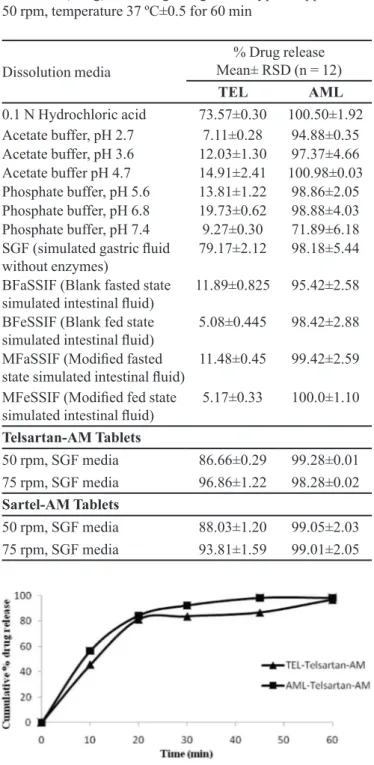
media (pH 1.2-7.5). The screening results showed that the dissolution rates of both bulk drugs were higher in simulated
gastric luid (SGF) media (pH 1.2) than other media (Table
I), due to the interaction between lone pairs of electrons on
nitrogen atoms and the ionizable groups present in TEL and AML with NaCl, HCl molecules present in SGF media (prepared as per USP-2007, without enzymes, consisting of
2 gm sodium chloride, 0.2M hydrochloric acid to adjust pH to 1.2 and volume make upto 1000 mL). Thus, this media was useful for the quality control testing of both drugs in tablets. The pH of the dissolution medium was 1.2, i.e. within the range of gastrointestinal tract pH levels, and therefore mimicked the gastrointestinal tract environment (biorelevant). This assay was designed to provide information for pharmaceutical researchers involved in the development of new biorelevant dissolution media and in predicting the in vivo performance of poorly soluble drugs. Therefore, the selected dissolution test conditions were: USP type II apparatus at a paddle speed of 75 rpm in 900 mL
of simulated gastric luid (pH 1.2).
In vitro dissolution studies from tablet dosage
forms
Dissolution studies on Telsartan-AM and Sartel-AM
tablets were performed under the optimized dissolution test conditions. These results are shown in Figures 2 and
3, and reveal that more than 90% of both TEL and AML were released from the two products.
Stability studies
Both TEL and AML were found to be stable under dissolution test conditions and the measured derivative
absorbance of the initial time and after 24 and 48 h was
similar. There was no evidence of degradation of the drugs
under the dissolution test conditions, indicating that the
solutions were stable for more than 48 h.
Discriminatory power of the dissolution method
The discriminatory power of the dissolution method was determined by manufacturing tablets under different
TABLE I - Screening study results for dissolution of TEL (40 mg)
and AML (5 mg) bulk drug using a USP type II apparatus at
50 rpm, temperature 37 ºC±0.5 for 60 min
Dissolution media
% Drug release Mean± RSD (n = 12)
TEL AML
0.1 N Hydrochloric acid 73.57±0.30 100.50±1.92 Acetate buffer, pH 2.7 7.11±0.28 94.88±0.35
Acetate buffer, pH 3.6 12.03±1.30 97.37±4.66
Acetate buffer pH 4.7 14.91±2.41 100.98±0.03
Phosphate buffer, pH 5.6 13.81±1.22 98.86±2.05
Phosphate buffer, pH 6.8 19.73±0.62 98.88±4.03
Phosphate buffer, pH 7.4 9.27±0.30 71.89±6.18
SGF (simulated gastric luid
without enzymes) 79.17±2.12 98.18±5.44
BFaSSIF (Blank fasted state
simulated intestinal luid) 11.89±0.825 95.42±2.58
BFeSSIF (Blank fed state
simulated intestinal luid) 5.08±0.445 98.42±2.88
MFaSSIF (Modiied fasted
state simulated intestinal luid) 11.48±0.45 99.42±2.59 MFeSSIF (Modiied fed state
simulated intestinal luid) 5.17±0.33 100.0±1.10
Telsartan-AM Tablets
50 rpm, SGF media 86.66±0.29 99.28±0.01
75 rpm, SGF media 96.86±1.22 98.28±0.02
Sartel-AM Tablets
50 rpm, SGF media 88.03±1.20 99.05±2.03
75 rpm, SGF media 93.81±1.59 99.01±2.05
FIGURE 2 – In vitro dissolution proile of Telsartan-AM tablets
TABLE II - Data obtained from stability studies
% Amount of drug found
Drug initial time after 24 h after 48 h
TEL 98.01 98.05 99.05
AML 99.06 100 99.06
FIGURE 3- In vitro dissolution proile of Sartel-AM tablets in
SGF media at 75 rpm (n=12).
FIGURE 4 - Comparison of dissolution proiles of TEL/AML tablets manufactured with different hardness.(A) Proile for TEL
and (B) for AML.
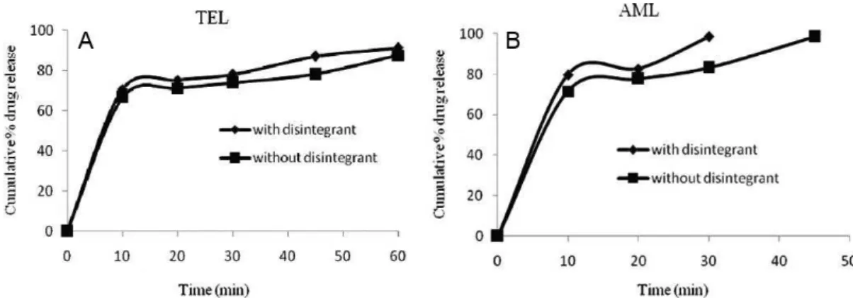
FIGURE 5- Comparison of dissolution proiles of TEL/AML tablets manufactured with and without a disintegrant.(A) Proile for
TEL and (B) for AML.
conditions and checking the dissolution behavior in the presence of the proposed dissolution test conditions. The effect of tablet hardness (5 kg/cm2vs. 8 kg/cm2 ) and the
disintegrant (with disintegrant vs. without disintegrant) are
shown in Figures 4 and 5, revealing that dissolution rate of
both drugs was slightly faster for tablets with less hardness and with a disintegrant. Therefore, this dissolution method has the ability to discriminate changes in the composition
and manufacturing process. The dissolution proile data
were also compared mathematically using the similarity
it factor (f2).
If f2 is less than 50, then two dissolution profiles are considered dissimilar. The similarity increases as the f2 value increases above 50 and approaches 100 (FDA 1997). The similarity factor f2 was calculated from the
dissolution proile, using ive points for telmisartan and
three points for amlodipine besylate. Among these, one
Table III lists the f2 values used for the comparison of the
dissolution proiles for each of the process parameters evaluated. These results conirm that the dissolution test
procedure has discriminating power regarding variation in the composition and manufacturing process.
Method validation
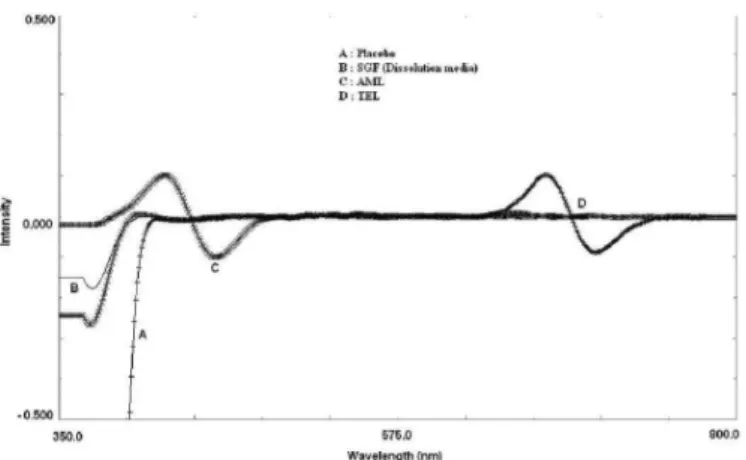
Specificity
The synchronous luorescence derivative spectra of
the placebo, TEL and AML in the dissolution medium are
shown in Figure 6. Perusal of the igure shows that there was no interference from the excipients in the tablets and
dissolution medium with the derivative response of either of the drugs (TEL and AML) at their respective analytical
wavelengths. Therefore, the proposed method is speciic.
Linearity
The linearity was evaluated by the least square
regression method. The responses for TEL at 675 nm were found to be linear in the concentration range of 4-14 µg/mL, with a correlation coeficient (r) value of 0.997. Similarly, the responses for AML at 458 nm were linear in the concentration range of 1-6 µg/mL, with a correlation coeficient (r) value of 0.999. The results indicate a good
linear relationship between the derivative response and
concentration. Linearity plots are shown in Figure 7.
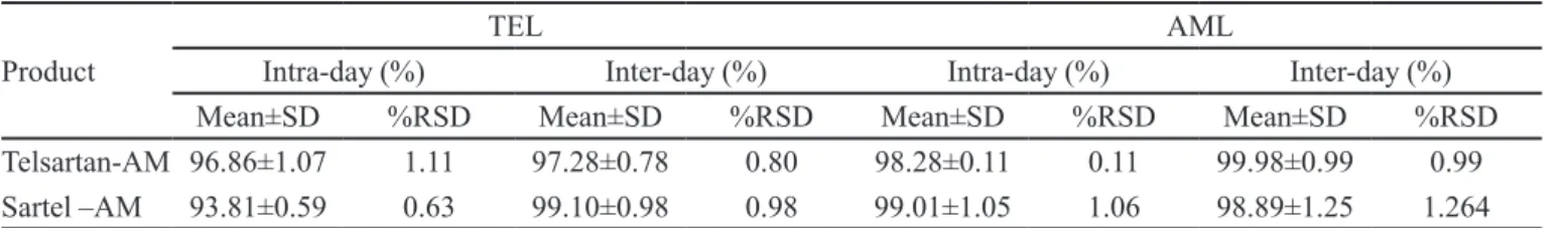
Precision
The percent relative standard deviation values for intra and inter day precision studies were found to be less
than 2 and there was no signiicant difference observed
between the intra- and inter-day values, which indicates that the proposed method was reproducible and precise. The results are reported in Table IV.
Accuracy
Accuracy shows the agreement between the standard value and the observed value. The accuracy results of the proposed dissolution method are reported in Table V. Percent recovery was from 97.0-99.0% and percent
relative standard values were less than 2; this signiies
that the dissolution method is accurate for its intended use.
CONCLUSION
The discriminating and dissolution test developed and validated for TEL/AML tablets was considered
satisfactory. The best conditions optimized for dissolution
testing for TEL/AML tablets were: 900 mL of simulated
gastric fluid (SGF without enzymes), a paddle-type
apparatus, stirring speed of 75 rpm, a temperature of 37
ºC±0.5 and collection time of 60 min. The dissolution
studies for TEL and AML combination tablets in the proposed dissolution media were found to be better, because of their discriminating power, utility as a surrogate of the gastrointestinal tract environment (biologically relevant). In addition, these studies provide information for pharmaceutical researchers involved in the development of new biorelevant dissolution media and predicting the in vivo performance of poorly soluble drugs. The
TABLE III - Similarity factor (f2) for dissolution proiles of tablets
with different parameters
f2 Value Process
parameter
Tablets with hardness of 5 kg/ cm2 vs. 8 kg/cm2
Tablets containing disintegrant vs. no
disintegrant
TEL 44.47 63.14
AML 46.14 49.21
FIGURE 6 - Synchronous first order derivative spectrum of placebo (A), blank dissolution medium (B), AML (C) and TEL (D).
TABLE IV - Precision data for the proposed dissolution method
Product
TEL AML
Intra-day (%) Inter-day (%) Intra-day (%) Inter-day (%)
Mean±SD %RSD Mean±SD %RSD Mean±SD %RSD Mean±SD %RSD
Telsartan-AM 96.86±1.07 1.11 97.28±0.78 0.80 98.28±0.11 0.11 99.98±0.99 0.99
Sartel –AM 93.81±0.59 0.63 99.10±0.98 0.98 99.01±1.05 1.06 98.89±1.25 1.264
TABLE V - Results for accuracy for the proposed dissolution method
Analyte % level of recovery Amount (mg)
Added Recovered %Recovery %RSD
TEL 80 32 31.56 98.62 1.10
100 40 38.82 97.05 0.89
120 48 47.52 99.0 1.24
AML 80 4 3.90 97.5 1.02
100 5 4.85 97.0 0.98
120 6 5.89 98.16 0.85
results obtained from validation show that the proposed dissolution method was scientifically sound. These advantages encourage the routine use of the developed dissolution method in the quality control analysis of TEL and AML in tablet dosage forms.
ACKNOWLEDGMENTS
The authors are thankful to the management of Gokaraju Rangaraju College of Pharmacy for providing necessary facilities and infrastructure to carry out this research work.
REFERENCES
AMIDON, G.L.; LENNERNÄS, H.; SHAH, V.P.; CRISON, J.R. A theoretical basis for a biopharmaceutic drug classiication:
the correlation of in vitro drug product dissolution and in vivo bioavailability. Pharm. Res., v.12, n.3, p.413-420, 1995.
ANDRADE-EIROA, A.; DE-ARMAS, G.; ESTELA, J.M.; CERDA, V. Critical approach to synchronous spectroluorimetry. Trends Anal. Chem., v.29, n.8, p.885-901, 2010.
ANTHONY, C.M.; DAVID, M.O.; BRIAN, W. Clarke’s analysis of drugs and poisons. 3.ed. London: Pharmaceutical Press,
2004. p.629, 1601.
BASAVAIAH, K.; RAGHU, M.S.; VINAY, K.B. Simple and rapid spectrophotometric assay of levocetirizine in pharmaceuticals through charge-transfer complexation using chloranilic acid and 2,3-dichloro-5,6-dicyanoquinone as π-acceptors. Bull. Chem. Soc. Ethiop., v.26, n.3,
p.319-328, 2012
BELAL, F.; EL-BRASHY, A.; EL-ENANY, N.; TOLBA, M. Conventional and irst derivative synchronous luorometric
determination of ethamsylate in pharmaceutical preparations
and biological luids. Application to stability studies. J. Floresec., v.21, n.4, p.1371-1384, 2011.
DRESSMAN, J.B.; AMIDON, G.L.; REPPAS, C.; SHAH,
V.P. Dissolution testing as prognostic tool for oral drug absorption: immediate release dosage forms. Pharm. Res.,
v.15, n.1, p.11-22, 1998. .
EI-WASSEF, D.R.; EI-SHEEBINY D.T.; ABU-EI-ENEIN M.A.; EI-ASHRY S.M. Simultaneous determination of labetalol and furosemide by irst derivative synchronous spectroluorimetry. J. Fluoresc, v.19, n.5, p.817-828, 2009.
FOOD AND DRUG ADMINISTRATION. FDA. Guidance
for industry: immediate release solid dosage forms: in vitro dissolution testing and in vivo bioequivalence
GALIA, E.; NICOLAIDES, E.; HORTER, D.; LÖBENGER, R.; REPPAS, C.; DRESSMAN, J.B. Evaluation of various
dissolution media for predicting in vivo performance of class I and II drugs. Pharm. Res., v.15, n.5, p.698-705, 1998.
GOMEZ-HENS, A. Modern aspects of luorimetry as applied
to clinical chemistry. Pure Appl. Chem., v.63, n.8,
p.1083-1088, 1991.
HE, Z.; ZHONG, D.; CHEN, X.; LIU, X.; TANG, X.; ZHAO,
L. Development of a dissolution medium for nimodipine tablets based on bioavailability evaluation. Eur. J. Pharm. Sci., v.21, n.4, p.487-491, 2004.
INTERNATIONAL CONFERENCE ON HARMONIZATION.
ICH. Harmonized tripartite guideline. Validation of
analytical procedures: text and methodology Q2B. Rockville: ICH,1996. p.1-10.
KOTTAI, R.; SANKHLA, A.; GUPTA, S.H.; SMITH, A.A.;
MANAVALAN, R. Development and validation of a reversed phase HPLC method for simultaneous determination of amlodipine and telmisartan in pharmaceutical dosage form. J. Appl. Chem. Res., v.12, p.43-52, 2010.
KULKARNI, A.P.; SHAHNAWAZ, M.; ZAHEER, Z.;
DEHGHAN, M.H.G. Development and validation of a
dissolution method for pioglitazone tablets. Dissolut. Technol., v.19, n.4, p.36-45, 2012.
MENEGOLA, J.; STEPPE, M.; SCHAPOVAL, E.E. Dissolution
test for citalopram in tablets and comparison of in vitro
dissolution proiles. Eur. J. Pharm. Biopharm., v.67, n.2,
p.524-530, 2007.
MHASKE, R.A.; GAROLE, D.J.; MHASKE, A.A.;
SAHASRABUDHE, S. RP-HPLC method for simultaneous determination of amlodipine besylate, valsartan, telmisartan,
hydrochlorothiazide and chlorthalidone: application to
commercially available drug products. Int. J. Pharm. Sci. Res., v.3, n.1, p.141-149, 2012.
MARK, H.; WORKMAN JR., J. Derivative in spectroscopy.
Part I - The behavior of the derivative. Spectroscopy, v.18,
n.4, p.32-33, 2003.
OLIVEIRA, E.; AIEVEDO, R; BONEILIO, R.; OLIVEIRA, D.B.; REBEIRO, G.P.; ARAVJO, M.B. Dissolution test
optimization for meloxicam in the tablet pharmaceutical
form. Braz. J. Pharm. Sci., v.45, n.1, p.67-73, 2009.
PANIKUMAR, A.D.; SUNITHA, G.; VENKAT RAJU, Y.; SATHESH BABU, P.R.; SUBRAHMANYAM, C.V.S.
Development of biorelevant and discriminating method
for dissolution of efavirenz and its formulations. Asian J. Pharm. Clin. Res., v.5, n.3, p.220-223, 2012.
PANIKUMAR, A. D.; SUNITHA, G.; VENKAT RAJU, Y.; SATHESH BABU, P.R.; SUBRAHMANYAM, C.V.S.
Development of dissolution test method for drotaverine hydrochloride/mefenamic acid combination using derivative spectrophotometry. Trop. J. Pharm. Res., v.12, n.2, p.227-232, 2013.
PRATAP, Y.P.; MANISH, A.R.; SWATI, U.K.; RESHMA,
B.K. Simultaneous spectrophotometric estimation of amlodipine besylate and telmisartan in tablet dosage form. Der Pharmacia Chemica, v.4, n.2, p.725-730, 2012
RAYMOND, C.R.; PAUL, J.S.; SIAN, C.O. Handbook of
pharmaceutical excipients. 2.ed. Chicago, London: Pharmaceutical Press, American Pharmacists Association,
2007. p.132, 385,725.
SONI, T.; NAGDA, E.; GANDHI, T.; CHOTAI, N.P.
Development of discriminating method for dissolution of aceclofenac marketed formulations. Dissolut. Technol.,
v.15, n.2, p.31-34, 2008.
VIGNADUZZO, S.E.; CASTELLANO, P.; KAUFMAN,
T.S. Development and validation of a dissolution test for
meloxicam and pridinol mesylate from combined tablet
formulation. India J. Pharm. Sci., v.72, n.2, p.197-203, 2010.
UNITED STATES PHARMACOPEIA.USP 30: The National
formulary: NF 25: by authority of the United States
Pharmacopeia Convention prepared by the council of
experts and its expert committees. Rockville: United States
Pharmacopeia Convention,2007.v.2.p.1092.
ZONGYUN, H.; RUBEN, L.; ROBERT, F.; ANNE-FRANCOISE, A.; ALYSON, S.; DENIS, S. Development
of a single in-vitro dissolution method for a combination trilayer tablet formulation of clopidogrel and pravastatin. Dissolut. Technol., v.18, n.2, p.12-19, 2011.
Received for publication on 21st May 2013