Neuronal Cell Fate Specification by the
Convergence of Different Spatiotemporal
Cues on a Common Terminal Selector
Cascade
Hugo Gabilondo1☯, Johannes Stratmann2☯, Irene Rubio-Ferrera1, Irene Millán-Crespo1,
Patricia Contero-García1, Shahrzad Bahrampour2, Stefan Thor2*, Jonathan Benito-Sipos1
*
1Departamento de Biología, Universidad Autónoma de Madrid, Cantoblanco, Madrid, Spain,2Department of Clinical and Experimental Medicine, Linköping University, Linköping, Sweden
☯These authors contributed equally to this work. *[email protected](ST);[email protected](JBS)
Abstract
Specification of the myriad of unique neuronal subtypes found in the nervous system depends upon spatiotemporal cues and terminal selector gene cascades, often acting in sequential combinatorial codes to determine final cell fate. However, a specific neuronal cell subtype can often be generated in different parts of the nervous system and at different stages, indicating that different spatiotemporal cues can converge on the same terminal selectors to thereby generate a similar cell fate. However, the regulatory mechanisms underlying such convergence are poorly understood. The Nplp1 neuropeptide neurons in theDrosophilaventral nerve cord can be subdivided into the thoracic-ventral Tv1 neurons and the dorsal-medial dAp neurons. The activation of Nplp1 in Tv1 and dAp neurons depends upon the same terminal selector cascade:col>ap/eya>dimm>Nplp1. However, Tv1 and dAp neurons are generated by different neural progenitors (neuroblasts) with differ-ent spatiotemporal appearance. Here, we find that the same terminal selector cascade is triggered byKr/pdm>grnin dAp neurons, but byAntp/hth/exd/lbe/casin Tv1 neurons. Hence, two different spatiotemporal combinations can funnel into a common downstream terminal selector cascade to determine a highly related cell fate.
Author Summary
A fundamental challenge in developmental neurobiology is to understand how the great diversity of neuronal subtypes is generated during nervous system development. Neuronal subtype cell fate is established in a stepwise manner, starting with spatial and temporal cues that confer distinct identities to neural progenitors and trigger expression of terminal selector genes in the early-born neurons. Terminal selectors are those that determine the final neuronal subtype cell fate. Intriguingly, similar neuronal subtypes can be generated
a11111
OPEN ACCESS
Citation:Gabilondo H, Stratmann J, Rubio-Ferrera I, Millán-Crespo I, Contero-García P, Bahrampour S, et al. (2016) Neuronal Cell Fate Specification by the Convergence of Different Spatiotemporal Cues on a Common Terminal Selector Cascade. PLoS Biol 14 (5): e1002450. doi:10.1371/journal.pbio.1002450
Academic Editor:William A. Harris, University of Cambridge, UNITED KINGDOM
Received:October 16, 2015
Accepted:April 1, 2016
Published:May 5, 2016
Copyright:© 2016 Gabilondo et al. This is an open access article distributed under the terms of the
Creative Commons Attribution License, which permits unrestricted use, distribution, and reproduction in any medium, provided the original author and source are credited.
Data Availability Statement:All relevant data are within the paper and its Supporting Information files
Funding:The funders had no role in study design, data collection and analysis, decision to publish, or preparation of the manuscript. This work was funded by Swedish Research Council (VR-NT; 621-2010-5214;
www.vr.se) to ST, Wallenberg Foundation
(KAW2012.0101;www.wallenberg.com/kaw/) to ST,
Swedish Cancer Foundation (120531;www.
by different progenitors and under the control of different spatiotemporal cues; thus, we wondered how such convergence is achieved. To address this issue, we have decoded the specification of two highly related neuropeptide neurons, which are generated at different locations and time-points in theDrosophilanervous system. We find that two different combinations of spatiotemporal cues, in two different neural progenitors, funnel onto the same terminal selector gene, which in turn activates a shared regulatory cascade, ultimately resulting in the specification of a similar neuronal cell subtype identity.
Introduction
During nervous system development, vast numbers of different neuronal subtypes are gener-ated, and understanding the process of cell fate specification remains a major challenge. Studies have shown that establishment of distinct neuronal identities requires complex cascades of reg-ulatory information, starting from spatial and temporal selector genes [1] and feeding onward to terminal selector genes [2,3], often acting in combinatorial codes to dictate final and unique cell fate [4–6]. One particularly intriguing regulatory challenge pertains to the generation of highly related neuronal subtypes in different regions of the central nervous system (CNS). Examples are plentiful and include e.g., various groups of dopaminergic and serotonergic neu-rons in the mammalian CNS [7,8], as well as neuropeptide-producing neurons in many sys-tems [9,10]. The appearance of highly related neurons in different regions and at distinct developmental time-points clearly indicates that different spatial and temporal cues can con-verge to trigger the same terminal selector code, to thereby trigger a similar final cell fate. How-ever, the underlying mechanisms are unclear.
In the developingDrosophilaventral nerve cord (VNC), two distinct sets of neurons selec-tively express the neuropeptide Nplp1: dAp and Tv1. Both subtypes express the LIM-homeo-domain transcription factor Apterous (Ap; mammalian Lhx2a/b) and the transcription co-factor Eyes absent (Eya; mammalian Eya1-4). dAp neurons constitute a dorsal-medial set of bilateral neurons running the length of the ventral nerve cord, while Tv1 neurons are located ventrolaterally in the three thoracic segments (Fig 1A and 1B). Both dAp and Tv1 project axons ipsilaterally and anteriorly, and join a common Ap fascicle [11,12]. While it is possible that other aspects of their cell fate are different, their common neuropeptide expression and axonal projections suggest that dAp and Tv1 can be grouped into a highly related, if not identi-cal, neuronal subtype. A number of regulatory genes and pathways acting in the specification of the Tv1 neurons have been elucidated [6,11,13–20]. These studies reveal that Tv1 cell fate depends upon a feedforward cascade in which spatial cues, provided by Hox and Hox cofactor input (Antp, Exd and Hth), and temporal cues, provided by the temporal factor Cas, activate a
col!ap/eya!dimmterminal selector cascade. This selector cascade ultimately results in the
activation of Nplp1 neuropeptide expression. dAp neurons depend upon the samecol!ap/
eya!dimmterminal selector cascade as Tv1. However, dAp neurons are not restricted to
tho-racic segments, but rather are distributed throughout the VNC (Fig 1A and 1B). In addition, they are born at an earlier stage than Tv1 [12]. Furthermore, while Tv1 is generated by NB5-6T, the lineage that generates dAp is unknown [6]. Not surprisingly, the upstream spatial and temporal cues that trigger the terminal selector cascade in the Tv1 neuron do not affect the dAp cells [14,17]. Thus, the dAp and Tv1 cells represent a unique scenario for addressing how neurons generated by different neuroblasts (NBs) and with different spatial and temporal regu-lators can activate the identical terminal selector cascade to ultimately dictate a highly related, if not identical, neuronal subtype identity.
26172fcf4eb029fa6ec7da6901432ea0/?vgnextoid= 264ecb2b1890f310VgnVCM1000001d04140aRCRD) to JBS.
Competing Interests:The authors have declared that no competing interests exist.
Here, we identify the NB generating the dAp neurons as NB4-3 and find that these dAp neurons are generated during an earlier time in development than Tv1, dictated by the tempo-ral factors Kr and Pdm. Hence, the same terminal selector cascade (col!ap/eya!dimm) is
triggered by distinct spatial and temporal cues in two different neuroblasts. Additionally, we find two crucial and specific factors refining the action of those spatiotemporal selectors: the GATA factor Grain (Grn), acting in dAp neurons, and the Ladybird early factor (Lbe), in Tv1 neurons. Thus, thecol!ap/eya!dimmterminal selector cascade is triggered by the Cas/Exd/
Hth/Antp/Lbe spatiotemporal code in NB5-6T, but by the Kr/Pdm/Grn code in NB4-3. These results demonstrate that distinct spatiotemporal combinatorial codes can converge onto a com-mon terminal selector cascade. Because the generation of highly related neurons in different regions of the CNS and at distinct time-points represents a common feature of many animal systems, the regulatory logic outlined here is likely to be widespread.
Results
dAp is Generated by NB4-3 in an Early Temporal Window
Previous work demonstrated that the terminal selector cascade composed bycol!ap/
eya!dimmis critical for the Nplp1 terminal cell fate both in dAp and Tv1 neurons, and can
trigger this fate broadly in the CNS when combinatorially misexpressed [6,11,13–20]. Tv1 neu-rons have been most extensively studied, and their NB origin is well understood [14,17]. They are generated at the end of the NB5-6T lineage, under a Castor (Cas) temporal window and through a type 0 division mode (Fig 1A) [14,21]. dAp neurons arise from a distinct, previously unknown NB lineage. Thus, we began by identifying the progenitor NB that gives rise to dAp
Fig 1. dAp and Tv1 neurons in theDrosophilaVNC.(A) Lateral view of early embryonicDrosophilaCNS, showing NB5-6T in the three thoracic segments (red) and the NB that gives rise to the dAp cells (green). Model of NB5-6T lineage, with the Tv1 neuron (red/blue). (B) Model of late embryonicDrosophilaVNC (air-filled trachea [AFT] stage), depicting the Ap clusters in the thoracic segments and the dAp cells located in the segments T1-A10.
neurons, utilizing sets of markers that identify most, if not all, of the 30 NBs generated in each hemisegment [22–26]. Eya expression commences in dAp at St13, and using Eya together with a number of NB markers, we found that dAp neurons are generated by NB4-3 (Fig 2A–2I). To follow the development of this lineage, we made use of acolenhancer that drives reporter expression selectively in the dAp neuron, as well as in the NB4-3 and parts of the lineage (Fig 2J and 2K). NB4-3 is known to delaminate at St late 11 [25], and we can observe the lineage usingcol-GFPfrom this stage and onward. We mapped the expression of the temporal genes, and as anticipated from the early birth of dAp, evident by Eya expression, we did not find expression of the late temporal factor Cas (S3A Fig). We did not observe expression of the early temporal factor Kr (Fig 2L–2N). Because bothhbandKrmutants affect dAp specification (see below), we envision that Hb and Kr are expressed in the NB4-3 prior to the onset of col-GFP. One of the two“middle”temporal factors Pdm1 (Nubbin [Nub], which together with Pdm2 we collectively refer to as Pdm1/2) was, however, expressed in several cells in the NB4-3 lineage (col-GFPcells) (Fig 2Q). When Col and Eya are turned on, we can identify Nub expres-sion specifically in the early dAp neuron itself at St13, to subsequently be downregulated at St15 (Fig 2Q and 2R). Using anti-phospho-Ser10 on Histone 3 (pH3), we were able to monitor cell divisions in the NB4-3 lineage, which revealed that dAp is born by a ganglion mother cell (GMC) and is hence generated in a type I proliferation window (Figs2KandS3B). In order to unambiguously show that dAp comes from a type I lineage, we analyzed the dAp neuron in a
sanpodo(spdo) mutant background. Corroborating the notion deduced with the pH3 analysis,
we observe two dAp neurons in that mutant background (S3B Fig). Therefore, dAp is born in a type I proliferation window.
In a screen for regulators and specific markers of dAp cell fate, we identified the GATA factor Grain (Grn) as being expressed in the dAp cell (see below), and could hence use agrn-lacZ
reporter to map the NB4-3 lineage. We found thatgrn-lacZexpression is concomitant with Pdm and hence precedes Col, being turned on in the GMC that generates the dAp cell (Fig 2O). We further observedgrn-lacZexpression in dAp neurons at all later embryonic stages (Fig 2P).
In summary, we map the origin of dAp to NB4-3 and find that it is born in the middle of this lineage. At the stage when NB4-3 generates the GMC that in turn will divide to generate the dAp neuron, it expresses Pdm and Grn (Fig 2O and 2Q). Hence, dAp and Tv1 are lineage-unrelated neurons, generated in different temporal windows, mid versus late, and during two different proliferation modes, type I versus type 0.
The Early Born dAp Neurons Depend upon Early Temporal Genes
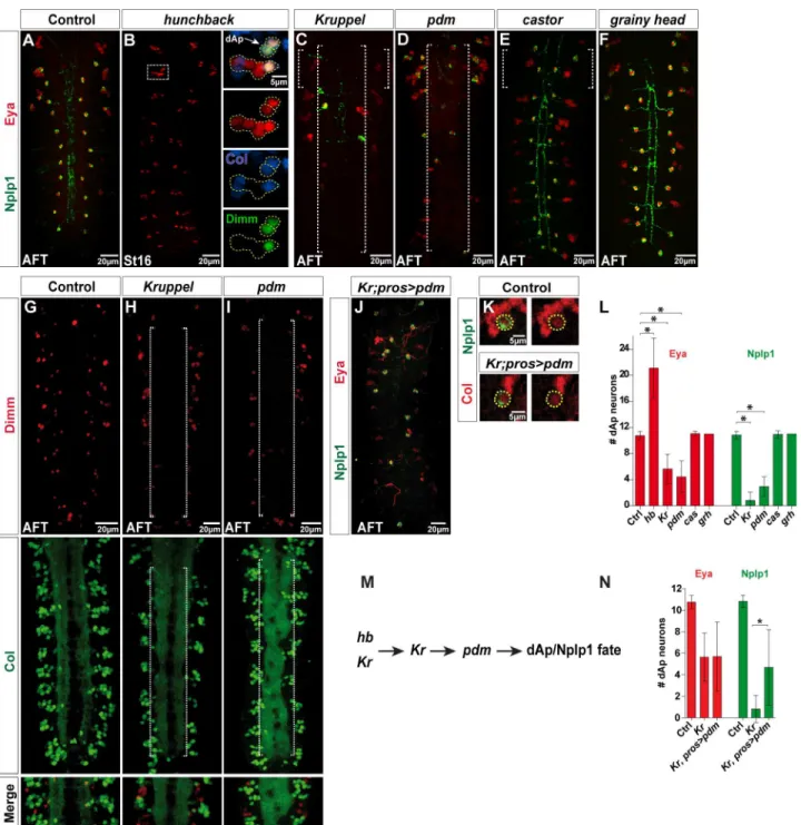
Having identified that dAp is born early in NB4-3, we next tested the expression of Nplp1/Eya markers in mutants for the temporal genes. In the early temporal mutanthb, we observed an apparent duplication of dAp neurons, evident by Col, Eya, and Dimm expression (Fig 3Band
S2). InKrmutants we found a reduction of Col, Eya, Dimm, and Nplp1 expressing cells (Fig 3C, 3H and 3L). As anticipated from the expression of Pdm in the GMC generating the dAp cells, and in the dAp cells themselves, we also observed loss of dAp neuron markers inpdm
cell death mutantDf(3R)H99, which removes all embryonic cell death [29]. We did not, how-ever, note any rescue of dAp cell in these double mutants (S7A and S7B Fig). We conclude that dAp neurons, which are born in an early temporal window, depend upon the early temporal geneshb,Kr, andpdmfor their specification. In contrast, Tv1 neurons, which are born late, depend upon the late temporal genecas.
grain
is Necessary for dAp but not for Tv1 Specification
The distinct NB origin and spatiotemporal generation of dAp and Tv1 demonstrates that two different sets of spatiotemporal inputs can converge upon the same terminal selector cascade (col!ap/eya!dimm), which triggers Nplp1 expression. Although the temporal factors are
selectively expressed at different points of the lineage development, they are broadly spatially expressed in most NBs during neurogenesis [27,28]. Hence, we predicted the existence of addi-tional upstream spatially-defining regulatory genes acting with theKrandpdmtemporal genes. In order to identify such additional upstream cues, we analysed a number of mutants for changes in Nplp1 expression in dAp but not in Tv1 cells or vice-versa (seeMaterials and Meth-ods). In the case of the dAp neurons, one mutant identified in this survey wasgrain(grn), which encodes a GATA transcription factor known to be dynamically expressed in the devel-oping VNC [30]. Our expression mapping of NB4-3 revealed expression ofgrnlacZin the NB at StE11, in the GMC at StL11, and in early dAp cells from St14 and onward to St16 (Figs2O, 2P
andS4A). Addressing the function ofgrn, we found that severalgrnallelic combinations all dis-played complete loss of Col, Eya,aplacZ, Dimm, and Nplp1 expression in dAp, but not in Tv1 neurons (Fig 4A–4GandS1 Data). To determine if dAp neurons undergo cell death ingrn
mutants, we expressed the cell death blocker p35. This did not, however, result in rescue of dAp cells (S7C Fig). Thus, thegrnmutant analysis indicates thatgrnis an early factor, acting upstream ofcol, in the dAp specification cascade. Strikingly,grnis not involved in triggering this cascade in the Tv1 neuron (Figs4A–4D,S4A and S4B).
grain
Acts at an Early Stage of dAp Specification
colis a critical determinant of early dAp neuron identity [6], and we find thatgrnacts upstream ofcol. Thus, we next addressed whether all of thegrnfunctions in the dAp specification are
rule out certain NBs as progenitors of the dAp cell. The dAp cell is positive forhkb-lacZand Msh, which indicates that NB4-3 is the progenitor of dAp cells. (J) NB staining for Msh shows overlap with Dpn and the col-dAp-GFP enhancer, which indicates that the NB generating the dAp cell, is NB4-3. (K) Expression of GFP, Pros, Dpn, and pH3 at St11 and St12 shows a pH3/Pros positive and Dpn negative cells (St11 thick yellow dashed circle) overlapping with the GFP expression from thecol-dAp-GFPenhancer construct, which suggests that the dAp cell is born in a type I division. (L) GFP, Kr, and Dpn expression at different stages (yellow dashed circles). At StE11, Dpn and GFP are expressed in the NB, but Kr is not detectable. At St11, the NB 4-3 lineage progresses (based on the GFP signal from the enhancer construct), Dpn is still detectable, but Kr is not expressed. By StL11, two strong GFP positive cells are detectable and GFP expression is evident in a cell in the previous NB location, but without a Dpn signal. (M) GFP, Kr, and Eya expression at St12, St13, and St14 shows that one of the cells expressing strong GFP is the dAp cell and turns on Eya expression by stage 13 (thick yellow dashed circle). (N) Kr is not expressed in the dAp cells at stage 16 (yellow dashed circles). (O) Costaining for GFP andβgal of thecol-dAp-GFPenhancer together withgrn-lacZ, shows thatgrain, one of the critical factors for the dAp specification is expressed in the NB4-3 lineage (yellow dashed circles). (P) Staining for Eya andβgal shows thatgrnis expressed in the dAp cells by stage 14 (yellow dashed circles). (Q) GFP (col-dAp-GFP), Col, and (Nub) Pdm expression at different stages in the NB4-3 lineage reveals that GFP is detectable prior to endogenous Col expression at StL11. At this stage, Nub (Pdm) starts being expressed in the two strong GFP positive cells (thick yellow dashed circles). At StM13 the GFP signal remains strongly expressed; furthermore, Col and Nub (Pdm) are expressed robustly. Of note, Col and Nub (Pdm) expression overlaps in one of the strong GFP positive cells (thick yellow dashed circles). At St14, Col expression is still strong, whereas Nub (Pdm) expression is downregulated (thick yellow dashed circles); Col, GFP, Nub (Pdm) expressing cell is the dAp cell. (R) Nub (Pdm) is not expressed in the dAp cells at stage 15 (yellow dashed circles). (S) GFP (col-dAp-GFP), Col, and Eya show an overlap in the dAp cell at St14. (T) Part of the lineage model of the NB 4–3. At StE11, Kr was not expressed in the NB (L). Still, we find Grn and Nub expression in the NB and the daughter cells. Together with the positive pH3 staining, prior to birth of the dAp cell, this model suggests that the dAp cell is born in a type I division mode by St12 and subsequently activates Col, Eya, and by later stages Dimm and Nplp1. Genotypes: (A)mirr-lacZ. (B)unpg-lacZ. (C)ind-lacZ. (D, E)OregonR. (F)en-lacZ. (G)OregonR. (H)hkb-lacZ/+. (J)col-dAp-GFP;col-dAp-GFP. (L, M, Q, and S)col-dAp-GFP;col-dAp-GFP. (N)OregonR. (O and P)col-dAp-GFP/+; col-dAp-GFP/grn-lacZ. (R)ap-Gal4, UAS-mRFP/CyO.
Fig 3. Early temporal genes are critical for dAp specification.(A–F) Expression of Eya and Nplp1 in control and temporal mutants, at St16 or AFT. (B) In hbmutants (boxed area), we observe two dorsal Ap cells (yellow dotted circles), which both express Eya, Col, and Dimm. Quantification of Nplp1 positive dAp cells inhbmutants fails sincehbmutants do not develop into stage AFT at which Nplp1 is expressed. (C and D) BothKrandpdmmutants show decreased numbers of Eya and Nplp1 expressing dAp cells (long dotted brackets). (E and F) Eya and Nplp1 expression incasandgrhmutants is not affected. (G–I) Dimm and Col expression shows a loss of both factors in the dAp cells inKrandpdmmutants (long dotted brackets). (J) Cross-rescue ofKr mutants byUAS-pdmfrompros-Gal4does not rescue Eya expression in dAp cells, but can partially rescue Nplp1 expression. (K) Col and Nplp1 expression in dAp cells of control andKrmutants expressingpdmfrompros-Gal4shows thatpdmcan restore the Col and Nplp1 expression inKrmutants. (L) Quantification of Eya and Nplp1 positive dAp neurons in temporal mutants (n>10; asterisk denotesp<0.05; Student´s two-tailedttest; seeS1 Data). (M) Genetic model of the dAp specification cascade, showing that the early temporal genesKrandpdmact to specify the dorsal Ap cell fate. (N) Quantification of Eya and Nplp1 positive dAp neurons in (n>10 VNC; asterisk denotesp<0.05; Student´s two-tailedttest; seeS1 Data). Numbers of Nplp1 positive dAp neurons inKrmutants expressingpdmare significantly increased compared toKrmutants. Genotypes: (A)OregonR. (B)hbP1,hbFB. (C, H)Kr1,KrCD. (D, I) Df(2L)ED773. (E)casΔ1/ casΔ1. (F)grhIM/grhIM. (G)OregonR. (J, and K)Kr1,KrCD;pros-Gal4/UAS-nub.
Fig 4.grainis critical for dAp specification.(A, B) Eya and Nplp1 expression in VNCs at stage AFT. Ingrn mutants, Eya and Nplp1 expression in dAp cells is almost completely lost (long dotted bracket). (C-F) Expression of Dimm, Col andβgal (aprK568) in control andgrnmutants. Ingrnmutants all three markers are
strongly downregulated, specifically in dAp cells (long dotted brackets). In contrast, expression in Tv1 cells is unperturbed. (G) Quantification of Nplp1 and Eya expressing dAp cells in control andgrnmutant VNCs (n>10 VNCs; asterisks denote significant difference ingrnmutants compared to control;p<0.05, Student´s two-tailedttest; seeS1 Data). Genotypes: (A)OregonR. (B)grn7L12/grnSPJ9. (C, E)aprK568/+. (D, F) aprK568/+; grnSPJ9/grn7L12.
mediated bycol. To this end, we re-expressedcolingrnmutants fromGal4drivers with differ-ent temporal onset:pros-Gal4at St10 andelav-Gal4at St12 [17,21]. We found robust re-appearance of dAp neurons, showing both the Eya and Nplp1 markers, when we used either
thepros-Gal4orelav-Gal4drivers (Fig 5B, 5D and 5G). As anticipated from previous studies
[6], expression ofUAS-colfrom eitherpros-Gal4orelav-Gal4also triggered a number of ectopic Eya/Nplp1 cells (Fig 5A and 5C). In a reciprocal experiment we tried to cross-rescue dAp cell fate incolmutants by expressinggrnfromelav-Gal4orpros-Gal4. We did not, how-ever, find any rescue of dAp cell specification in these cross-rescues (Fig 5E, 5F and 5H;S5 Fig
andS1 Data).
Together, these results suggest that the main, if not the only, role ofgrnin dAp cells is to trigger the expression ofcol, setting in motion the cascade of regulatory events that culminate with the dAp specification.
grain
Acts Downstream of the
Kr
and
pdm
Temporal Genes
Our lineage and expression analyses indicated thatgrnacts downstream of theKrandpdm
temporal genes, and that its primary role is to triggercolexpression. To further test this notion, we drove the expression ofgrninKrandpdmmutants. In both cases, we found partial rescue of the dAp neurons (Fig 6A, 6B and 6E). Next, we expressedcolinKrandpdmmutants and observed rescue of dAp neurons in both experiments (Fig 6C, 6D and 6FandS1 Data). Misex-pression ofUAS-colagain triggered a number of ectopic Eya/Nplp1 cells (Fig 6C and 6D).
These findings indicate that dAp cell fate is specified by aKr/pdm>grn>colcascade, in
which the function ofKr/pdmis to activategrn, and the function ofgrnis to activatecol. How-ever, the partial rescue ofKrbygrnsuggests thatKrmay be involved in a feedforward manner to regulatecol.
ladybird early
Delimits the Broad Action of the Spatio-temporal Cues
Responsible for the Tv1 Specification
Having identifiedgrnas a key spatial regulator upon which the temporal factors act to specify a dAp fate, we attempted to find a counterpart ofgrnin Tv1 fate specification. Recently we per-formed a large-scale forward genetic screen looking for genes critical for Tv4/FMRFa cell fate which resulted in the identification of additional genes controlling NB5-6T development [31]. One of the mutants identified in this genetic screen, by its loss ofFMRFa-EGFPexpression, was mapped toladybird early(lbe) (mammalianLbx1/2). This EMS allele,lbe12C005, has a non-sense mutation at amino acid 29 (a likely null allele) [31], and was placed over deletion
Df(lbl-lbe)B44to avoid genetic background problems (hereafter referred to aslbemutants). Inlbe
mutants, we observe a complete loss of Eya, Dimm, and Nplp1 (Fig 7A and 7B). Strikingly, we find thatlbedoes not affect dAp neurons (Fig 7A, 7B, 7M and 7N).
In order to further characterize the loss-of-function phenotype oflbe, we analysed the expression of other key regulators acting during Ap cluster specification. Inlbemutants, we observed normal expression of the sub-temporal factor Nab (Fig 7C and 7D) [14]. However, we observed complete loss of Col, Eya, Dimm, and Nplp1 expression (Fig 7A–7D,7G and 7H). The loss of Col expression inlbemutants prompted us to reciprocally address Lbe expression incolmutants. We observed normal Lbe expression as well as normal Nab expression incol
p<0.05, Student´s two-tailedttest). (H) Quantification of Eya and Nplp1 positive dAp cells incolmutants withgrnmisexpression shows thatgrndoes not rescue thecolmutant phenotype (n = 7 VNCs forpros>grn, Eya cell quantification; for all others, n>10 VNCs; asterisks denotep<0.05, Student´s two-tailed ttest; seeS1 Data). Genotypes: (A)UAS-col/pros-Gal4. (B)UAS-col/pros-Gal4; grnSPJ9/grn7L12. (C)elav-Gal4/UAS-col. (D)elav-Gal4/UAS-col; grnSPJ9/ grnSPJ9. (E)col1/col1,UAS-grn; pros-Gal4/+. (F)col1/col1,UAS-grn; elav-Gal4/+.
doi:10.1371/journal.pbio.1002450.g005
Fig 6. Cross-rescue reveals thatKrandpdmact upstream ofgrnandcol.(A–D) Eya and Nplp1 expression in cross-rescue ofKrandpdmmutants with eitherUAS-colorUAS-grnmisexpressed frompros-Gal4, at stage AFT. (A) While the number of Eya expressing dAp neurons inKrmutants is not restored by misexpression ofgrn, (B) inpdmmutants misexpression ofgrnresults in partial rescue in numbers of dAp neurons expressing both Eya and Nplp1. (C) Cross-rescue ofKrwithcolcan fully rescue the mutant phenotype and results in ectopic numbers of dAp neurons expressing both Eya or Nplp1. (D) Cross-rescue ofpdmwithcolcan fully rescue thepdmmutant phenotype with respect to Eya and Nplp1 positive dAp neurons, and results in ectopic Eya and Nplp1 expression. (E, F) Quantification of Eya and Nplp1 positive dAp neurons from the different rescue experiments (n = 9 VNCs forKr; pros>grn, n = 8 VNCs for pdm; pros>col. For all others n>10 VNCs; asterisk denotesp<0.05, Student´s two-tailedttest; seeS1 Data). Genotypes: (A)Kr1,KrCD; pros-Gal4/
UAS-grn. (B)Df(2L)ED773; pros-Gal4/UAS-grn. (C)Kr1,KrCD; pros-Gal4/UAS-col. (D)Df(2L)ED773; pros-Gal4/UAS-col.
Fig 7.lbeis critical for Ap cluster formation and Tv1 specification.(B) Eya, Dimm, and Nplp1 expression in control VNCs at stage AFT, showing that Eya is expressed in all four neurons of the Ap clusters and the dAp cells. Dimm is expressed in two out of four Ap cluster cells and the dAp cells, while Nplp1 is expressed in one neuron (Tv1 cell) of each Ap cluster and the dAp cells. (C) Inlbemutants Eya, Dimm and Nplp1 expression is lost in the Ap clusters, whereas their expression in dAp cells is unchanged. (D–E) GFP (lbe(K)-EGFP), Dpn, Col, and Nab expression in NB5-6T at St14 showing that Col
expression is lost inlbemutants while Nab expression is unaffected. (F, G) GFP, Dpn, Lbe, and Nab expression in NB5-6T at St14 in control andcolmutants, showing that Lbe expression is not affected incolmutants. (H, I) GFP, Dimm, Nplp1, and Eya expression in the Ap cluster at stage AFT in control, reveals loss of Eya, Dimm, and Nplp1 inlbemutants. (J, K) GFP, Lbe, Eya, and Nab expression in the Ap cluster at St16 reveals no effect on Lbe expression incol mutants. (L, M) GFP, Lbe, Col, Dpn expression in NB5-6T at St12 and St13 reveals that Lbe is expressed prior to the onset of Col expression. (N, O) Eya, Col, Dimm, and Nplp1 expression at St17 and AFT in the dAp cells of control andlbemutants, revealing no difference in cell fate specification with respect to Nplp1 expression. Genotypes: (B)OregonR. (C)lbe12C005/Df(lbl-lbe)B44. (D, L, M)lbe(K)-EGFP. (E)lbe(K)-EGFP/+;lbe12C005/Df(lbl-lbe)B44. (F) lbe(K)-EGFP/TTG homozygous. (G)col1/col3;lbe(K)-EGFP/+.
Ap cluster determinants and is critical for the activation of thecol!ap/eya!dimmterminal
selector cascade.
lbe
Acts in a Feedforward Manner with
col
in the Tv1 Specification
Cascade
lberegulatescol, but is this the only role thatlbeplays, or does it play multiple roles, perhaps acting on targets downstream ofcol? To address this, we attempted to cross-rescuelbeusing
elav-Gal4drivingUAS-col. First, as a control, we rescuedlbemutants withUAS-lbe, and, as
anticipated, this resulted in rescue of thoracic lateral Eya/Dimm/Nplp1 cells (Fig 8A and 8B). Next, we attempted to cross-rescuelbewithUAS-col, but did not observe any thoracic lateral Eya/Dimm/Nplp1 cells (Fig 8C). These results indicate thatlbeplays roles in addition to acti-vatingcol, perhaps acting downstream together withcol. To test this idea, we misexpressedlbe
andcolalone, and compared this to the effects of combinatorial misexpression. We noted that each gene alone could trigger ectopicaplacZ/Eya/Dimm/Nplp1 expression. However, their com-binatorial action was striking, with vast numbers of ectopicaplacZ/Eya cells (Fig 8D–8G). Inter-estingly, only a subset of ectopicaplacZ/Eya cells co-expressed Dimm/Nplp1, which may be explained by the fact thatlbeandcolare also critical for the Ap cluster Tv2 and Tv3 cell fate: non-neuropeptide expressing interneurons. Finally, we addressed whetherlbeis regulated by other Tv1 upstream regulators, and stained for Lbe incas,hthandAntpmutants. This revealed no effects on Lbe expression in any of these three mutants (S1A–S1F Fig). Reciprocally, we testedlbemutants for expression of Cas, Hth, and Antp, but did not observe any effects (S1G–
S1J Fig).
These results demonstrate thatlbeacts in parallel to the four other Ap cluster upstream determinants, and acts in a feedforward manner, first activatingcoland subsequently acting withcolto activate Ap/Eya/Dimm/Nplp1 (Fig 8H).
Discussion
A number of previous studies have addressed the final steps of neuronal specification with regards to neuropeptide expression in single neuronal lineages inDrosophila[6,11,13,14,17,20,
33–36]. However, there are many examples inDrosophilaof neurons in diverse locations expressing the same neuropeptide [10]. The current study addresses the mechanism by which different upstream cues are integrated to trigger neuropeptide, Nplp1, expression in two spa-tially and temporally unrelated cells: the Tv1 and dAp cells. We find that the late-born Tv1 cell requires an interplay in the NB5-6T lineage of late temporal selector gene input fromcas
together with spatial input fromAntp,lbe,hth, andexdto activatecol; a key trigger gene for the Nplp1 terminal selector cascade (Fig 9). In contrast, the early-born dAp cell requires an input of the early temporal selectorsKrandpdm, together with the GATA factorgrn, to activatecol
in NB4-3 (Fig 9). Oncecolis activated in either Tv1 or dAp cell, an identical feed-forward ter-minal selector cascade plays out downstream ofcolto activate the Nplp1 expression. Hence, the more restricted expression ofgrnandlbeacts to refine the broader spatiotemporal cues, triggering a highly restricted terminal selector code, which is initiated by Col expression. Thus,
colcould be viewed as a genetic integrator of different spatiotemporal input.
Logical Basis for the Different Spatiotemporal Selector Inputs
similar lines, Tv1 cells are born late in NB5-6T and depend upon the late temporal selector Cas, while dAp cells are born early-middle and hence depend upon Kr and Pdm.
In NB5-6T, Col is triggered by a combinatorial code of spatiotemporal selectors (cas Antp,lbe,
hth, andexd) that to some extent explain its selective expression. However, Col expression is in itself fairly broad and highly dynamic in the developing VNC [6], and hence its expression can-not explain the highly restricted expression of Ap/Eya. However, herelbeplays a secondary role, as it acts withcolto activate Ap/Eya expression. Hence, the highly selective expression oflbe, in only a few row 5 NBs, and its feedforward action withcolcombine to refine the action ofcol.
In the case of dAp and NB4-3, we are likely still missing additional upstream and feedfor-ward regulators. First, although Grn contributes to refine the action of Kr/Pdm, the specific activation of expression of Kr, Pdm, and Grn is still not restricted enough to explain the specific triggering of Col in NB4-3. Second, as mentioned above, Col itself is also broadly expressed and needs additional factors to refine its role in NB4-3. Thus, we envision the existence of addi-tional upstream factors in the dAp genetic cascade.
Expanding Steps of Coherent Feedforward Loops
Expression analysis in mutant and misexpression backgrounds will most often help to place two regulators, X and Y, in relationship to each other. If X expression is not lost in Y mutants,
Fig 8.lbeis critical for Ap cluster formation and Tv1 specification.(A–C) Expression of Eya, Dimm, and Nplp1 in control and inlbemutants rescued with UAS-lbe, or cross-rescued withUAS-col, driven fromelav-Gal4. Rescue oflbemutants by misexpression ofUAS-lbecan rescue the mutant phenotype in the Ap clusters with respect to Nplp1 expression and gives rise to more Eya positive cells. In contrast, the cross-rescue oflbeby misexpression ofUAS-colfails to rescue the Ap cluster expression of Eya, Dimm, and Nplp1. (D–G) Expression of Eya,βgal (aprK568), Dimm, and Nplp1. Single misexpression oflbeorcol
results in some ectopic Eya,βgal (aprK568), Dimm, and Nplp1 expression. Co-misexpression oflbeandcolresults in a dramatic increase of ectopic Eya,βgal
(aprK568), Dimm, and Nplp1 expression. (H) Model of a potential feed-forward cascade for the Nplp1 specification.lbeboth activatescoland potentially feeds
forward on downstream targets such aseyaandap. Genotypes: (A)OregonR. (B)UAS-col/+;lbe12C005/Df(lbl-lbe)B44,elav-Gal4. (C)UAS-lbe/+;lbe12C005/Df
(lbl-lbe)B44,elav-Gal4. (D)OregonR. (E)aprK568/+;elav-Gal4/UAS-lbe. (F)aprK568/+;elav-Gal4/UAS-col.(G)UAS-col/aprK568;elav-Gal4/UAS-lbe.
doi:10.1371/journal.pbio.1002450.g008
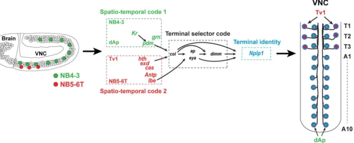
Fig 9. Illustration summarizing the findings.(Left) Lateral view of the early developingDrosophilaembryonic CNS depicting the thoracic NB5-6T, which generates the Tv1 cells, and the NB4-3, which generates the dAp cells. (Middle-right) Our results reveal that the critical terminal selector genecolis activated by different spatio-temporal selector genes acting in the two different NB lineages. In the NB5-6Tcolis activated by the late temporal genecas, together with Hox input, viaAntp/hth/exd, andlbe. In NB4-3colis activated by the early temporal genesKrandpdmand the GATA genegrn. Downstream ofcol, the Nplp1 activation cascade in the NB5-6T and NB 4–3 lineages is near identical and acts to specify the related cell fate of the dAp and Tv1 cells. In the NB5-6T lineage, we identified two new players involved in the Nplp1 cell fate specification:lbewhich activatescoland feeds forward ontoapandeya, andKr, which shows a late onset in the Tv1 cell to maintaincolexpression. Hence, different spatiotemporal selector genes acting in cells of a different developmental history triggers a common terminal selector cascade via the key entry point genecol.
but Y expression is lost in X mutants, one would propose that X regulates Y. However, to address whether or not the only thing X does is to regulate Y, we employ two other approaches: cross-rescue and combinatorial misexpression. The cross-rescue can show, for example, that dAp cells can be fully rescued ingrnmutants bycolre-expression, while in con-trast, Tv1 cells cannot be rescued inlbemutants bycolre-expression. Regarding combinato-rial misexpression, we observe a striking combinatocombinato-rial misexpression effect oflbe/colwhen compared to either gene alone. Such cross-rescue and co-misexpression experiments prompts us to postulate a direct linear and non-feedforward regulation ofcolbygrnin dAp cells. In contrast, for Tv1 cells we propose feedforward regulation oflbeoncol, and subse-quently withcolonap/eya. Such loops, i.e.,X!YX!Z, are denoted coherent feedforward
loops, and are common inEscherichia coliand yeast gene regulatory networks [37]. Coherent feedforward loops act as regulatory timing devices and allow for gene X to carry different reg-ulatory output (or meaning) at successive developmental time-points.colis a salient example of this; its transient expression in NB5-6T triggers an initial“generic”Ap/Eya interneuron cell fate in the four Ap cluster neurons, while its maintained expression (specifically in Tv1) acts to propagate the terminal selector cascade that ultimately results in the activation of Nplp1 [6,14].
Coherent feedforward loops have also been identified during nervous system development in other animals, includingCaenorhabditis elegans[38,39]. With regards to neuropeptide cell specification, we now find increasingly longer loops; five steps between Kr and Nplp1 in dAp cells, and ranging in developmental time from St10 to late embryonic stage. The presence of coherent feedforward loops has not been extensively tested in vertebrate systems, primarily because cross-rescue and multiple combinatorial misexpression experiments are technically challenging in these systems. But it is tempting to speculate that coherent feedforward loops are extensively utilized by more complex systems, and that the number of regulatory levels in these loops may increase with evolutionary complexity.
Materials and Methods
Fly Stocks
lbe12C005[31].Df(lbl-lbe)B44,UAS-lbe, andladybird earlyfragment K drivinglacZ(referred to
aslbe(K)-lacZ) (provided by C. Jagla) [32].lbe(K)-EGFP[40].elav-Gal4(provided by A.
DiA-ntonio) [41].prospero-Gal4(F. Matsuzaki, Kobe, Japan).casΔ1andcasΔ3(provided by W. Odenwald) [42].UAS-nls-myc-EGFP(referred to asUAS-nmEGFP) [11].col1,col3[43] and
UAS-col(provided by A. Vincent) [44].hkb5953(referred to ashkblacZ) [45].UAS-apand
apmd544(referred to asapGal4)[46].aprK568(referred to asaplacZ) [47].UAS-grn-HA(#F001916; provided by FlyORF).grhIM[48].hbP1,hbFBandKr1,KrCD[27],unpg1912-r37= unpg-lacZ (pro-vided by C.Q. Doe) [23].Antp12(provided by F. Hirth) [49].ind-lacZanden-lacZ(provided by H. Reichert).grn-lacZ,grn7L12,grnSPJ9,UAS-grn(provided by J. Castelli-Gair Hombría).
col-dAp-GFPwas generated by inserting a genomic fragment from thecolgene into the vector
pEGFP.attB (provided by K. Basler and J. Bischof) and generating transgenes by PhiC31 trans-genic integration (BestGene Inc, California, United States).
From Bloomington Drosophila Stock Center:Antp25(BL#3020).Df(2L)ED773(removes bothnubandPdm2; BL#7416).mirr-lacZ(mirrB1-12; BL#30023).elavC155=elav-Gal4
(BL#458).elav-Gal4(BL#8765).hth5E04(BL#4221).Df(3R)Exel6158(BL#7637; referred to as
Exploratory Screen to Study Nplp1 Specification
The following transcription factor mutants were scored for changes in Nplp1 expression, with-out any apparent effects:escargot (esg),shuttle craft (stc),elbow/No ocelli (el/noc),rotund (rn),
eagle (eg),kruppel homolog (kr h),knirps (kni),schnurri (shn),klumpfuss (klu),zfh2,dachshund
(dac),defective proventriculus (dve),seven up (svp),vein (vn),beadex (bx),scribbler (sbb).
Immunohistochemistry
Primary antibodies were: Guinea pig Deadpan (1:1,000) (provided by J.B. Skeath). Rabbit a-ß-Gal (1:5,000; ICN-Cappel, Aurora, Ohio, US). Rabbit a-GFP (1:500; Molecular Probes, Eugene, OR, US). Guinea pig Col (1:1,000), guinea pig Dimm (1:1,000), chicken a-proNplp1 (1:1000), and rabbit a-proFMRFa (1:1,000). Rat a-Grh (1:1,000). Rabbit a-Cas (1:250) (provided by W. Odenwald). Rat mAb a-GsbN (1:10) (provided by R. Holmgren). Mouse a-Nubbin (referred to in the figure as Nub [Pdm]; 1:20) (provided by Steve Cohen). Mouse mAb a-Dac dac2–3 (1:25), mAb a-Antp (1:10), mAb a-Pros MR1A (1:10), mAb a-Eya 10H6 (1:250) (Developmental Studies Hybridoma Bank, Iowa City, Iowa, US). Guinea pig anti-Odd (1:500); guinea pig anti-Runt (both provided by M. Ruiz and D. Kosman). Rat a-Msh (1:500) (provided by Z. Paroush) [51].
Confocal Imaging and Data Acquisition
Zeiss LSM 700 or Zeiss META 510 Confocal microscopes were used for fluorescent images; confocal stacks were merged using LSM software or Adobe Photoshop. Statistic calculations were performed in Graphpad prism software (v4.03). To address statistical significance, Stu-dent'sttest or nonparametric Mann-Whitney U test or Wilcoxon signed rank test, in the case of non-Gaussian distribution of variables, was used. Images and graphs were compiled in Adobe Illustrator.
Supporting Information
S1 Data. Data underlying Figs3L, 3N,4G,5,6E, 6FandS5D. (XLSX)
S1 Fig.lbeacts in parallel withAntp,cas, andhth.(A–F) GFP/βgal, Col, Nab, and Lbe expres-sion in the NB5-6T at St14 in control andAntp,cas, andhthmutants.Antpandhthmutants show loss of Col expression, while Lbe expression is not affected. (D)casmutants show, in addition to a negative Col expression, a loss of Nab expression, sincecasregulatesnabvia the sub-temporal genesqz. Lbe expression is however not affected. (G-J) Staining against Antp, Cas, and Hth at St14 in NB5-6 of control andlbemutants shows that neither of these three fac-tors are affected inlbemutants. Genotypes: (A)lbe(K)-GFP. (B)lbe(K)-GFP/+;Antp25/Antp12.
(C)lbe(K)-GFP. (D)lbe(K)-GFP/+;casΔ1/casΔ3. (E)lbe(K)-lacZ. (F)lbe(K)-lacZ/+;hth5E04/
hthDf. (G, I)lbe(K)-GFP. (H, J)lbe(K)-GFP/+;lbe12C005/Df(lbl-lbe)B44. (TIF)
S2 Fig. Origin of extra dAp cells inhbmutant background.Co-staining for GFP and Eya of
thecol-dAp-GFPenhancer (to visualize the NB4-3 from which dAp cell originated) in (A)
con-trol and (B) hb mutant background. Both the bonafide dAp and the supernumerary one are GFP positive. Thus, the supernumerary dAp generated inhbmutant originate from the NB4-3. Genotypes:col-dAp-GFP/+; hbP1,hbFB.
S3 Fig. Castor is not expressed in the NB4-3 early lineage and dAp is generated in a type I division mode.(A and B) Co-staining for GFP and Cas of thecol-dAp-GFPenhancer (to visu-alize the NB4-3 lineage from which dAp cell originated) at Stage 12 and 13 to analyze the expression of Cas in the NB4-3 lineage when it is generating the dAp neuron. Cas is not expressed in the NB4-3 early lineage (C) Co-staining for GFP, Dimm, and Eya of the col-dAp-GFPenhancer (to visualize the NB4-3 lineage from which dAp cell originated) inspodomutant background. Additional dAp cell express GFP, Eya, and Dimmed in Spodo mutant. Genotypes:
(A)col-dAp-GFP;col-dAp-GFP. (B)col-dAp-GFP/+;spdo6104/spodo6104.
(TIF)
S4 Fig. Grn expression in the NB5-6 lineage at two different stages.(A) Co-staining forβgal of thegrn-lacZand Eya to analyze the expression ofgrn-lacZat the Ap cluster at St16. We do not findgrn-lacZexpression in the NB5-6 lineage at St 16. (B) Co-staining for GFP of thelbe
(K)-GFP(to visualize the NB5-6 lineage) together withβgal for thegrn-lacZconstruct at Stage
15 to analyze the expression ofgrn-lacZin the NB5-6 lineage. We do not findgrn-lacZ expres-sion in the NB5-6 lineage at St 15. Genotypes: (A)grn-lacZ/+. (B)lbe(K)-GFP/ grn-lacZ. (TIF)
S5 Fig. Overexpression ofgrn.(A-C) Co-staining for Eya, Nplp1 and Col in (A) control, (B)
pros-Gal4>UAS-grn, and (C)elav-Gal4>UAS-grngenetic background. Overexpression ofgrn
is not able to induce ectopic dAp neurons. (D) Quantification of Nplp1 and Eya expressing dAp cells in control,prospero-Gal4>UAS-grn, andelav-Gal4>UAS-grngenetic background
VNCs (n = 7 VNCs forpros>grnfor Eya cell quantification; for all others, n>10 VNCs;
aster-isks denotep<0.05, Student´s two-tailedt-test; seeS1 Data) Genotypes: (A)OregonR. (B).
prospero-Gal4/UAS-grn. (C)elav-Gal4/UAS-col.
(TIF)
S6 Fig. Origin of supernumerary Eya cells incolandlbeco-misexpression.(A, B) Co-stain-ing forβgal, Dimm, Eya, and GsbN of themirror-lacZconstruct in (A) control and (B)
elav-Gal4>UAS-col,UAS-lbegenetic background. White dotted lines represent the Gsbn
compart-ment whereas magenta dotted lines represent the Mirr compartcompart-ment. Supernumerary Eya cells generated byUAS-col,UAS-lbeco-misexpression originate from lineages generated by NBs in row 5 (Gsbn) as well from lineages generated by NBs in row 1, 2 and 3 (mirr-lacZ). Genotypes:
(A)mirr-lacZ/ UAS-col; UAS-lbe. (B)elav-Gal4;; mirr-lacZ/ UAS-col,UAS-lbe.
(TIF)
S7 Fig.Kr,pdm, andgrnare not required for dAp cell survival.(A–C) Co-staining for Dimm and Eya inKr,pdm, andgrnmutants, in which cell death has been impaired byDf(3R) H99or by expression of cell death blockerUAS-p35. dAp cells are lost in mutants and not restored by cell death impairment.
(TIF)
Acknowledgments
Author Contributions
Conceived and designed the experiments: HG JS ST JBS. Performed the experiments: HG JS IRF IMC PCG SB. Analyzed the data: HG JS ST JBS. Wrote the paper: ST JBS.
References
1. Allan DW, Thor S. Transcriptional selectors, masters, and combinatorial codes: regulatory principles of neural subtype specification. Wiley interdisciplinary reviews Developmental biology. 2015; 4(5):505– 28. Epub 2015 Apr 8. doi:10.1002/wdev.191PMID:25855098.
2. Hobert O. Regulatory logic of neuronal diversity: terminal selector genes and selector motifs. Proceed-ings of the National Academy of Sciences of the United States of America. 2008; 105(51):20067–71. Epub 2008/12/24. doi:10.1073/pnas.0806070105PMID:19104055; PubMed Central PMCID: PMC2629285.
3. Wenick AS, Hobert O. Genomic cis-regulatory architecture and trans-acting regulators of a single inter-neuron-specific gene battery in C. elegans. Developmental cell. 2004; 6(6):757–70. Epub 2004/06/05. doi:10.1016/j.devcel.2004.05.004PMID:15177025.
4. Sharma K, Sheng HZ, Lettieri K, Li H, Karavanov A, Potter S, et al. LIM homeodomain factors Lhx3 and Lhx4 assign subtype identities for motor neurons. Cell. 1998; 95(6):817–28. PMID:9865699
5. Thor S, Andersson SG, Tomlinson A, Thomas JB. A LIM-homeodomain combinatorial code for motor-neuron pathway selection. Nature. 1999; 397(6714):76–80. PMID:9892357
6. Baumgardt M, Miguel-Aliaga I, Karlsson D, Ekman H, Thor S. Specification of Neuronal Identities by Feedforward Combinatorial Coding. PLoS Biol. 2007; 5(2):295–308.
7. Bjorklund A, Dunnett SB. Dopamine neuron systems in the brain: an update. Trends in neurosciences. 2007; 30(5):194–202. Epub 2007/04/06. doi:10.1016/j.tins.2007.03.006PMID:17408759.
8. Gaspar P, Lillesaar C. Probing the diversity of serotonin neurons. Philosophical transactions of the Royal Society of London. 2012; 367(1601):2382–94. Epub 2012/07/25. doi:10.1098/rstb.2011.0378 PMID:22826339; PubMed Central PMCID: PMC3405676.
9. Hokfelt T, Broberger C, Xu ZQ, Sergeyev V, Ubink R, Diez M. Neuropeptides—an overview. Neuro-pharmacology. 2000; 39(8):1337–56. Epub 2000/05/20. PMID:10818251.
10. Park D, Veenstra JA, Park JH, Taghert PH. Mapping peptidergic cells in Drosophila: where DIMM fits in. PLoS ONE. 2008; 3(3):e1896. PMID:18365028. doi:10.1371/journal.pone.0001896
11. Allan DW, Pierre SE, Miguel-Aliaga I, Thor S. Specification of Neuropeptide Cell Identity by the Integra-tion of Retrograde BMP Signaling and a Combinatorial TranscripIntegra-tion Factor Code. Cell. 2003; 113 (1):73–86. PMID:12679036.
12. Lundgren SE, Callahan CA, Thor S, Thomas JB. Control of neuronal pathway selection by the Dro-sophilaLIM homeodomain geneapterous. Development (Cambridge, England). 1995; 121:1769–73.
13. Allan DW, Park D, St Pierre SE, Taghert PH, Thor S. Regulators acting in combinatorial codes also act independently in single differentiating neurons. Neuron. 2005; 45(5):689–700. PMID:15748845.
14. Baumgardt M, Karlsson D, Terriente J, Diaz-Benjumea FJ, Thor S. Neuronal subtype specification within a lineage by opposing temporal feed-forward loops. Cell. 2009; 139(5):969–82. PMID:
19945380. doi:10.1016/j.cell.2009.10.032
15. Benveniste RJ, Thor S, Thomas JB, Taghert PH. Cell type-specific regulation of the Drosophila FMRF-NH2 neuropeptide gene by Apterous, a LIM homeodomain transcription factor. Development (Cam-bridge, England). 1998; 125(23):4757–65.
16. Hewes RS, Park D, Gauthier SA, Schaefer AM, Taghert PH. The bHLH protein Dimmed controls neuro-endocrine cell differentiation in Drosophila. Development (Cambridge, England). 2003; 130(9):1771– 81. PMID:12642483.
17. Karlsson D, Baumgardt M, Thor S. Segment-specific neuronal subtype specification by the integration of anteroposterior and temporal cues. PLoS Biol. 2010; 8(5):e1000368. PMID:20485487. doi:10.1371/ journal.pbio.1000368
18. Miguel-Aliaga I, Thor S. Segment-specific prevention of pioneer neuron apoptosis by cell-autonomous, postmitotic Hox gene activity. Development (Cambridge, England). 2004; 131(24):6093–105. PMID:
15537690.
19. van Meyel DJ, O'Keefe DD, Thor S, Jurata LW, Gill GN, Thomas JB. Chip is an essential cofactor for apterous in the regulation of axon guidance in Drosophila. Development (Cambridge, England). 2000; 127(9):1823–31. PMID:10751171.
21. Baumgardt M, Karlsson D, Salmani BY, Bivik C, MacDonald RB, Gunnar E, et al. Global programmed switch in neural daughter cell proliferation mode triggered by a temporal gene cascade. Developmental cell. 2014; 30(2):192–208. Epub 2014/07/30. doi:10.1016/j.devcel.2014.06.021PMID:25073156.
22. Bossing T, Udolph G, Doe CQ, Technau GM. The embryonic central nervous system lineages of Dro-sophila melanogaster. I. Neuroblast lineages derived from the ventral half of the neuroectoderm. Devel-opmental biology. 1996; 179(1):41–64. PMID:8873753.
23. Doe CQ. Molecular markers for identified neuroblasts and ganglion mother cells in the Drosophila cen-tral nervous system. Development (Cambridge, England). 1992; 116(4):855–63. PMID:1295739.
24. Prokop A, Technau GM. The origin of postembryonic neuroblasts in the ventral nerve cord of Drosoph-ila melanogaster. Development (Cambridge, England). 1991; 111(1):79–88. PMID:1901786.
25. Schmid A, Chiba A, Doe CQ. Clonal analysis of Drosophila embryonic neuroblasts: neural cell types, axon projections and muscle targets. Development (Cambridge, England). 1999; 126(21):4653–89. PMID:10518486.
26. Schmidt H, Rickert C, Bossing T, Vef O, Urban J, Technau GM. The embryonic central nervous system lineages of Drosophila melanogaster. II. Neuroblast lineages derived from the dorsal part of the neu-roectoderm. Developmental biology. 1997; 189(2):186–204. PMID:9299113.
27. Isshiki T, Pearson B, Holbrook S, Doe CQ. Drosophila neuroblasts sequentially express transcription factors which specify the temporal identity of their neuronal progeny. Cell. 2001; 106(4):511–21. PMID:
11525736.
28. Kambadur R, Koizumi K, Stivers C, Nagle J, Poole SJ, Odenwald WF. Regulation of POU genes by castor and hunchback establishes layered compartments in the Drosophila CNS. Genes & develop-ment. 1998; 12(2):246–60. PMID:9436984.
29. White K, Grether ME, Abrams JM, Young L, Farrell K, Steller H. Genetic control of programmed cell death in Drosophila. Science (New York, NY. 1994; 264(5159):677–83. PMID:8171319.
30. Garces A, Thor S. Specification of Drosophila aCC motoneuron identity by a genetic cascade involving even-skipped, grain and zfh1. Development (Cambridge, England). 2006; 133(8):1445–55. PMID:
16540509.
31. Bivik C, Bahrampour S, Ulvklo C, Nilsson P, Angel A, Fransson F, et al. Novel Genes Involved in Con-trolling Specification of Drosophila FMRFamide Neuropeptide Cells. Genetics. 2015; 200(4):1229–44. Epub 2015/06/21. doi:10.1534/genetics.115.178483PMID:26092715; PubMed Central PMCID: PMC4574234.
32. De Graeve F, Jagla T, Daponte JP, Rickert C, Dastugue B, Urban J, et al. The ladybird
homeobox genes are essential for the specification of a subpopulation of neural cells. Developmental biology. 2004; 270(1):122–34. PMID:15136145.
33. Benito-Sipos J, Estacio-Gomez A, Moris-Sanz M, Baumgardt M, Thor S, Diaz-Benjumea FJ. A genetic cascade involving klumpfuss, nab and castor specifies the abdominal leucokinergic neurons in the Dro-sophila CNS. Development (Cambridge, England). 2010; 137(19):3327–36. Epub 2010/09/09. doi:10. 1242/dev.052233PMID:20823069.
34. Benito-Sipos J, Ulvklo C, Gabilondo H, Baumgardt M, Angel A, Torroja L, et al. Seven up acts as a tem-poral factor during two different stages of neuroblast 5–6 development. Development (Cambridge, England). 2011; 138(24):5311–20. PMID:22071101.
35. Lundell MJ, Hirsh J. eagle is required for the specification of serotonin neurons and other neuroblast 7–3 progeny in the Drosophila CNS. Development (Cambridge, England). 1998; 125(3):463–72. PMID:
9425141.
36. Miguel-Aliaga I, Thor S, Gould AP. Postmitotic specification of Drosophila insulinergic neurons from pioneer neurons. PLoS Biol. 2008; 6(3):e58. PMID:18336071. doi:10.1371/journal.pbio.0060058 37. Alon U. Network motifs: theory and experimental approaches. Nature reviews Genetics. 2007; 8
(6):450–61. Epub 2007/05/19. doi:10.1038/nrg2102PMID:17510665.
38. Etchberger JF, Flowers EB, Poole RJ, Bashllari E, Hobert O. Cis-regulatory mechanisms of left/right asymmetric neuron-subtype specification in C. elegans. Development (Cambridge, England). 2009; 136(1):147–60. Epub 2008/12/09. doi:10.1242/dev.030064PMID:19060335; PubMed Central PMCID: PMC2685964.
39. Johnston RJ Jr., Copeland JW, Fasnacht M, Etchberger JF, Liu J, Honig B, et al. An unusual Zn-finger/ FH2 domain protein controls a left/right asymmetric neuronal fate decision in C. elegans. Development (Cambridge, England). 2006; 133(17):3317–28. PMID:16887832.
41. DiAntonio A, Haghighi AP, Portman SL, Lee JD, Amaranto AM, Goodman CS. Ubiquitination-depen-dent mechanisms regulate synaptic growth and function. Nature. 2001; 412(6845):449–52. PMID:
11473321.
42. Mellerick DM, Kassis JA, Zhang SD, Odenwald WF. castor encodes a novel zinc finger protein required for the development of a subset of CNS neurons in Drosophila. Neuron. 1992; 9(5):789–803. PMID:
1418995.
43. Crozatier M, Valle D, Dubois L, Ibnsouda S, Vincent A. Head versus trunk patterning in the Drosophila embryo; collier requirement for formation of the intercalary segment. Development (Cambridge, England). 1999; 126(19):4385–94. PMID:10477305.
44. Vervoort M, Crozatier M, Valle D, Vincent A. The COE transcription factor Collier is a mediator of short-range Hedgehog-induced patterning of the Drosophila wing. Curr Biol. 1999; 9(12):632–9. PMID:
10375526.
45. Bhat KM. The patched signaling pathway mediates repression of gooseberry allowing neuroblast speci-fication by wingless during Drosophila neurogenesis. Development (Cambridge, England). 1996; 122 (9):2921–32. PMID:8787765.
46. O'Keefe DD, Thor S, Thomas JB. Function and specificity of LIM domains in Drosophila nervous sys-tem and wing development. Development (Cambridge, England). 1998; 125(19):3915–23.
47. Cohen B, McGuffin ME, Pfeifle C, Segal D, Cohen SM. apterous, a gene required for imaginal disc development inDrosophilaencodes a member of the LIM family of developmental regulatory proteins. Genes Dev. 1992; 6:715–29. PMID:1349545
48. Nusslein-Volhard C, Wieschaus E, Kluding H. Mutations affecting the pattern of the larval cuticle in Dro-sophila melanogaster. Development. 1984; 193:267–82. PMID:FBrf0041708.
49. Abbott MK, Kaufman TC. The relationship between the functional complexity and the molecular organi-zation of the Antennapedia locus of Drosophila melanogaster. Genetics. 1986; 114(3):919–42. Epub 1986/11/01. PMID:3098627; PubMed Central PMCID: PMC1203021.
50. Campos-Ortega JA, Hartenstein V. The embryonic development ofDrosophila melanogaster. New York: Springer-Verlag; 1985.