Abstract
Submitted: September 26, 2016 Modiication: November 16, 2016 Accepted: January 9, 2017
Effect of photodynamic therapy and
non-thermal plasma on root canal
illing: analysis of adhesion and sealer
penetration
Objective: The aim of this study was to evaluate the effect of photodynamic therapy (PDT) and non-thermal plasma (NTP) on adhesion and sealer penetration in root canals. Material and Methods: Sixty single-rooted premolars were used. The teeth were prepared using a crown-down technique. NaOCl and EDTA were used for irrigation and smear layer removal, respectively. The root canals were divided into three groups: control, PDT, and NTP. After treatments, the roots were illed using gutta-percha and either AH Plus (AHP) or MTA Fillapex (MTAF) sealers. Samples were sectioned at 4, 8, and 12 mm from the apex (1-mm slices)and analyzed by the push-out bond strength test (adhesion) and confocal laser scanning microscopy (sealer penetration). Data were statistically evaluated using Kruskal-Wallis, Dunn’s, and Spearman’s tests. Results: Regarding AHP, bond strength was similar in the NTP group and in the control group, but signiicantly lower in the PDT group. As to MTAF, both therapies showed lower values than the control group. In the confocal analysis of AHP, maximum and mean penetration, and penetrated area were statistically higher in the control group than in the PDT and NTP groups. Penetrated perimeter was similar among groups. Regarding MTAF, all parameters yielded better results in the NTP than in the control group. The PDT and control groups showed similar results except for penetrated area. Conclusion: PDT and plasma therapy affected the adhesion and sealer penetration of root canals illed with AH Plus and MTA Fillapex and there is no positive correlation between adhesion and sealer penetration.
Keywords: Confocal microscopy. Photochemotherapy. Plasma gases. Root canal illing materials.
Marilia MENEZES1
Maíra PRADO1
Brenda GOMES2
Heloisa GUSMAN3
Renata SIMÃO1
1Universidade Federal do Rio de Janeiro, Departamento de Engenharia Metalúrgica e de Materiais,
Rio de Janeiro, RJ, Brasil.
2Universidade Estadual de Campinas, Faculdade de Odontologia de Piracicaba, Departamento de
Odontologia Restauradora, Área de Endodontia, Piracicaba, SP, Brasil.
3Universidade Federal do Rio de Janeiro, Departamento de Clínica Odontológica, Área de
Endodontia, Rio de Janeiro, RJ, Brasil. Corresponding address:
Introduction
The basic requirements for root canal treatment are effective chemomechanical preparation and
three-dimensional obturation of the root canal system1. The
complexity of the root canal system, with isthmuses,
ramiications, and dentinal tubules, makes it impossible
to eliminate microorganisms from root canals during
preparation24. In addition to the routinely used chemical
substances and instruments, other technologies have
been proposed to promote antimicrobial activity in the root canal system, such as photoactivated disinfection
and non-thermal plasma7,15,20,27,28.
Root canal obturation is a very important step
for a successful treatment. The use of gutta-percha with various root canal sealers is the most common
obturation method. AH Plus sealer (Dentsply Maillefer,
Ballaigues, Switzerland) is a resin-based sealer widely
used for root canal illing due its acceptable physical
properties, low solubility and disintegration, apical
sealing ability, good adhesion, antimicrobial action,
and good biological properties2. However, studies
have demonstrated AH Plus higher cytotoxic effects compared to MTA-based sealer30. MTA Fillapex (Angelus
Dental Solutions, Londrina, PR, Brazil) is a calcium
silicate-based root canal sealer that contains salicylate
resin, diluting resin, natural resin, bismuth oxide, nanoparticulate silica, and MTA. It was developed to
utilize the good features of MTA; relatively high levels
of biocompatibility, antimicrobial activity, and sealing
ability have been reported for this material1.
Adhesion and penetration are two important aspects
to be considered in sealer selection. Adhesion of an
endodontic sealer is deined as its capacity to adhere
to root canal walls and promote the union of gutta-percha cones to each other and to the dentin2,26. Sealer
penetration into dentinal tubules is also a required
feature, as it can improve the connection between
sealer and dentin13. The penetration ability of root canal
illing materials with antibacterial effect into dentinal
tubules may also help avoiding colonization by residual
bacteria and root canal reinfection1,6.
Studies have shown that bond strength and sealer penetration may be affected by the pretreatment of
root canal walls and by the type of sealer used2,11,21.
Regarding the effects of auxiliary technologies used
for root canal disinfection, photoactivated disinfection does not adversely affect the bond strength of AH Plus
to dentin, but it has a negative effect on MTA Fillapex
sealer17,18.
This study assessed the effects of photodynamic therapy (PDT) and non-thermal plasma (NTP) on
adhesion and sealer penetration in root canals illed with
AH Plus and MTA Fillapex and the correlation between
adhesion and sealer penetration.
Material and Methods
Specimen preparation
Sixty straight single-rooted premolar teeth were
used. Teeth with a fully formed apex were selected,
whereas roots with resorption defects, fractures, or
open apices were excluded. Crowns were sectioned below the cemento-enamel junction so that the lengths
of all roots were adjusted to 14 mm using a low-speed
diamond saw (Isomet; Buehler Ltd, Lake Bluff, IL,
USA) under water cooling. Patency of each root canal
was checked using a size 10 K-ile (Dentsply Maillefer,
Ballaigues, Switzerland) and working length (WL) was
established at 1 mm short of the apex. All teeth had
their apices sealed with utility wax (Technew, Rio de
Janeiro, RJ, Brazil) to prevent low through them.
Cleaning and shaping were performed with a
crown-down technique, using Miltex nickel-titanium rotary
instruments (Integra® Miltex®, York, PA, USA). The
following sequence was used: 35/.10 to prepare the
middle-coronal third. The sequence used in the apical
third was: 20/.03; 15/.05; 22/.04; 25/.04; 20/.06; and
20/.07. All iles reached the WL. Canals were irrigated
with 1 mL of 5.25% sodium hypochlorite (Mil Fórmulas,
Rio de Janeiro, RJ, Brazil) between each ile change.
The smear layer was removed after instrumentation
with 3 mL of 17% EDTA (Maquira Indústria de Produtos Odontológicos Ltda, Londrina, PR, Brazil), 1 mL per
minute. Thereafter, the roots were irrigated with 1
mL of distilled water to remove EDTA, followed by 1
mL of sodium hypochlorite. Finally, the root canals
were lushed with 5 mL of distilled water and dried
with medium paper points (Endo Points, Manacapuru,
AM, Brazil). The teeth were divided into three groups
(n=20): control (no employment of auxiliary technology used for root canal disinfection), PDT, and NTP.
Photodynamic therapy
For photodynamic therapy (PDT), after being
prepared as described above, the root canals were
was then stirred with a sterile #15 K-ile (Dentsply
Maillefer, Ballaigues, Switzerland) and allowed to stand for 2 min in the root canal (pre-irradiation time). A diode
laser (Twin laser, MMOptics, São Carlos, SP, Brazil) was
used as a radiation source with total power of 100 mW
and wavelength of 660 nm. Optical iber was initially
inserted up to the WL, and spiral movements, from
apical to coronal, were performed to allow for adequate
distribution of light throughout the root canal. Total
irradiation time was 90 s, resulting in an energy of 8 J for each sample, as described by Oliveira, et al.19
(2015).
Plasma therapy
A non-thermal atmospheric pressure plasma jet (Plasma Pen™, PVA Tepla America, Corona, CA, USA)
and a mixture of helium and oxygen (98% He and 2%
O2, White Martins, Rio de Janeiro, RJ, Brazil) was used.
The gas pressure was kept at 6 bar and 1000 V was applied to generate plasma.
During treatment, the distance between the tip of
the plasma jet and the sample was approximately 5
mm. The teeth were exposed to the plasma for 1 min.
Root canal illing
In control and experimental groups (after
photodynamic or plasma therapy), all roots were
immediately illed with gutta-percha cones (medium,
Microtipped, Endo Points, Manacapuru, AM, Brazil)
and AH Plus (Dentsply, Petropolis, RJ, Brazil) or MTA
Fillapex (Angelus, Londrina, PR, Brazil) sealers, a total
of 6 subgroups (n=10), i.e., control AH Plus, control MTA Fillapex, PDT AH Plus, PDT MTA Fillapex, NTP AH
Plus, and NTP MTA Fillapex.
For confocal laser scanning microscopy, each
sealer was luorescently labeled by adding rhodamine
B (Sigma-Aldrich, St. Louis, Missouri, USA) at an
approximate ratio of 0.1 w/w%20. Both sealers were
mixed according to the manufacturer’s instructions. A
gutta-percha cone covered with sealer was introduced into the root canal. Another medium cone was further
used as accessory until the entire length of the
root canal was illed. A #45 McSpadden condenser
(Dentsply Maillefer, Ballaigues, Switzerland) was then used. The plugger was advanced apically up to 4 mm
from the apical stop and slowly removed. Afterwards,
the plugger was removed slowly whilst being pushed
softly against one side of the canal. Roots were sealed with provisional restorative material (Cavitec, Caitech
Produtos Odontológicos, Rio do Sul, SC, Brazil).
Specimens were kept in an incubator at 37°C and 100% humidity for 2 days.
Push-out test
Each root was horizontally sectioned with a
slow-speed water-cooled diamond saw (Buehler Isomet 2000, Lake Bluff, IL, USA) at 4, 8, and 12 mm from
the apex14 to produce 1-mm thick slices for each root
region (apical, middle, and coronal).
Loading was performed using an electromechanical machine (EMIC DL200MF, São José dos Pinhais, PR,
Brazil) at a crosshead speed of 0.5 mm/min until bond
failure occurred. Three tips with different diameters
were used for load application in the push-out test in the different thirds (0.76 mm for cervical third, 0.60 mm for
middle third, and 0.40 mm for apical third). Debonding
values (maximum load) were used to calculate the
push-out strength in megapascals (MPa), according to the following formula:
Push-out bond strength (MPa) =
Maximum load (N)
Adhesion area (mm2)
The adhesion area was calculated by using the
following formula: A=π(R + r)[(h2+(R-r)2]0.5
where π=3.14, R is the coronal side radius, r is the
apical side radius, and h is the thickness of the slice. The thickness of each slice was measured using
a digital caliper (Vonder, Curitiba, PR, Brazil) and
the coronal and apical radii were measured using a
stereoscope (Leika MZ75, Meyer Instruments, Houston, TX, USA) and IM50 software (Leika IM50 Image
manager, Houston, TX, USA).
Confocal microscopy
After the push-out test, the remaining gutta-percha was removed and the sections were polished manually
with wet 1200-, 2400- and 4000-grit silicon carbide
(SiC) abrasive paper (Carbimet Disc Set, Buehler, Lake
Bluff, IL, USA). For each abrasive paper, the sections were polished for 1 min.
Specimens were mounted onto glass slides and
examined under a confocal laser scanning microscope
(Leica Microsystems GmbH, Mannheim, Germany) using a 5x objective (Leica Microsystems GmbH, Mannheim,
Germany). Absorption and emission wavelengths for
rhodamine-B were set to 540 and 590 nm. Images from
Image analysis was performed using Adobe
Photoshop (Adobe Systems Incorporated, San Jose, CA, USA). Six images were compiled to create an image
with the whole tooth. First, confocal microscopy images
of the slice were chosen (Figure 1A). Then, the image
of the slice captured with the stereoscope was selected
(Figure 1B), creating the inal image (Figure 1C).
To calculate maximum penetration, measurements
of the penetration areas were recorded on the slice
(Figure 1D) and maximum penetration was registered (green line) (Figure 1E). For average penetration
depth, four points were selected (Figure 1F) and
sealer penetration was registered (green line) (Figure
1G). Total perimeter of the root canal (Figure 1H) was measured and the penetrated perimeter registered
(green line) (Figure 1I). As to the penetrated area,
irst the total area of the slice (Figure 1J) was assessed
and then the sealer penetrated area was calculated (Figure 1K)5,8.
Statistical analysis
The Kolmogorov-Smirnov test was used to assess
the normality of data. Since data were not normally
distributed, the Kruskal-Wallis test was used for general
comparison and Dunn’s test for pairwise comparison. The Spearman’s test was used to correlate data. The
signiicance level was set at 5%.
Results
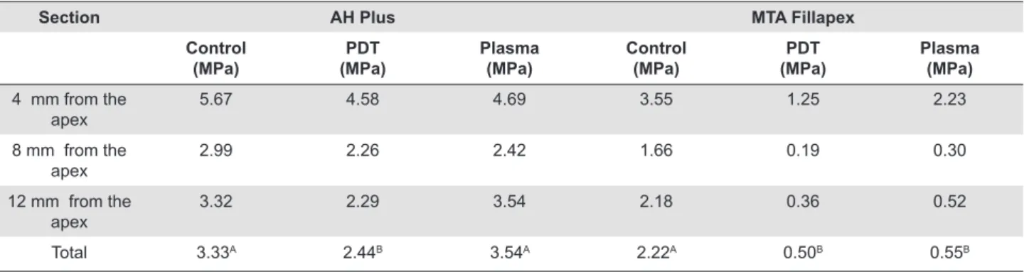
Table 1 shows the median bond strength values
(MPa) for both sealers. Plasma therapy results were similar to the control group, whereas PDT presented
signiicantly low strength when AH Plus was used.
Conversely, both therapies showed lower bond strength
than the control group using MTA Fillapex.
Figure 2 and 3 show representative confocal images
of the different groups for AH Plus and MTA Fillapex
Section AH Plus MTA Fillapex
Control (MPa)
PDT (MPa)
Plasma (MPa)
Control (MPa)
PDT (MPa)
Plasma (MPa)
4 mm from the apex
5.67 4.58 4.69 3.55 1.25 2.23
8 mm from the apex
2.99 2.26 2.42 1.66 0.19 0.30
12 mm from the apex
3.32 2.29 3.54 2.18 0.36 0.52
Total 3.33A 2.44B 3.54A 2.22A 0.50B 0.55B
A, B Comparison between groups of the same sealer (Statistical analysis on the row). Different letters indicate statistically signiicant values Table 1- The median bond strength values (MPa) for both sealers
sealers, respectively.
Table 2 shows median values of maximum and mean sealer penetration depth, penetrated perimeter
(%), and penetrated area (%) for the AH Plus sealer.
Overall, the control group presented statistically higher
values than the PDT and the plasma therapy groups for maximum penetration, mean penetration, and
penetrated area, which did not differ among groups.
Regarding penetrated perimeter, the groups showed
similar values. When the segments were separately evaluated and compared with the overall results, those
obtained for the coronal and middle thirds (8 and 12
mm from the apex) were similar to the overall analysis.
There was no difference among groups for the apical third. After comparing the different segments, the
apical third showed similar or lower values compared
to the middle and coronal thirds.
Regarding MTA Fillapex, plasma therapy showed better results for all parameters than did the control
group. PDT group values were similar to the control
group for maximum penetration, mean penetration,
and penetrated perimeter and similar to the plasma therapy group for penetrated area. The sealer applied
to the apical third did not show differences among
groups. After comparing the different segments, the
values for the apical third were similar or lower when compared to the other groups.
In the push-out test and confocal analysis, the
Spearman’s test showed no positive correlation
between bond strength and sealer penetration.
Figure 2- Representative images of confocal for AH Plus sealer
Group Maximum depth of penetration (µm)
Mean depth of penetration (µm)
Penetrated perimeter (%)
Penetrated area (%)
Total Control 1045.95A 594.87A 82.99A 17.76A
PDT 613.04B 389.77B 98.95A 11.18B
Plasma 581.25B 398.85B 100.00A 14.95B
4 mm from the apex (apical)
Control 632.71Ab* 357.23Ab* 61.80Ab* 14.10Ab*
PDT 541.90Aa• 284.95Ab• 58.18Ab• 12.02Aa•
Plasma 505.57Aa♣ 295.17Aa♣ 83.96Ab♣ 16.36Aa♣
8 mm from the apex (middle)
Control 1192.77Aa* 574.44Aa* 82.49Aa* 19.06Aa*
PDT 684.18Ba• 436.32Aa• 99.37Aa• 11.25Ba•
Plasma 543.41Ba♣ 417.02Aa♣ 100.00Aab♣ 15.28ABa♣
12 mm from the apex (coronal)
Control 1154.93Aa* 750.78Aa* 100.00Aa* 25.42Aa*
PDT 641.80Ba• 418.91Ba• 100.00Aa• 9.18Ba•
Plasma 664.50Ba♣ 448.43Ba♣ 100.00Aa♣ 9.90Ba♣
(A,B) Comparison between groups; (a,b) comparison between segments in the same group (control*, PDT•, plasma♣)
Table 2- Confocal analysis for AH Plus sealer
Group Maximum depth of
penetration (µm)
Mean depth of penetration (µm)
Penetrated perimeter (%)
Penetrated area (%)
Total Control 1,380.47B 823.44B 82.86B 32.58B
PDT 1,398.63B 927.50B 91.85A 40.61B
Plasma 1,645.36A 1,290.03A 100.00A 61.13A
4 mm from the apex (apical)
Control 997.51Aa* 662.23Aa* 75.53Aa* 20.73Aa*
PDT 1,147.36Aa• 576.71Aa• 68.02Aa• 18.76Aa•
Plasma 1,268.46Ab♣ 885.12Ab♣ 95.54Aa♣ 42.48Ab♣
8 mm from the apex (middle)
Control 1,545.46Aa* 937.72Ba* 96.31Aa* 41.94Ba*
PDT 1,910.25Aa• 1,163.26ABa• 91.16Aa• 49.39ABa•
Plasma 1,884.52Aa♣ 1,350.20Aa♣ 100.00Aa♣ 67.06Aa♣
12 mm from the apex (coronal)
Control 1,471.29Aa* 868.09Ba* 88.47Ba* 30.99Ba*
PDT 1,454.64Aa• 921.83Aa• 99.05ABa• 46.29ABa•
Plasma 1,886.04Aa♣ 1,457.29Aa♣ 100.00Aa♣ 63.63Aa♣
(A,B) Comparison between groups; (a,b) comparison between segments in the same group (control*, PDT•, plasma♣)
Table 3- Confocal analysis for MTA Fillapex sealer
Discussion
PDT and plasma therapy have been proposed as auxiliary therapy in chemomechanical preparation
due to their antimicrobial activity7,15,20,27,28. Both
technologies create reactive oxygen species, causing
serious damage to microorganisms through irreversible oxidation of cell components12,19,25. The effects of these
therapies have been studied in different periods of
time7,12,15,20,25,27,28. Although these technologies have
shown favorable results concerning their antimicrobial activity, little is known about their impact on adhesion
and sealer penetration.
In the present study, PDT was applied for 90 s.
This period was chosen because it was the minimum period found in the literature that antimicrobial activity
was veriied by the same parameters employed in the
present study12,19,25.
A mixture of helium and oxygen (98% He and 2%
O2) was applied for 60 s for its antimicrobial properties.
Also, this mixture has non-thermal characteristics
acting at room temperature and causing no damage to periapical and periodontal tissues. Additionally, short
periods are clinically favorable.
Flow and adhesion are essential properties when
choosing the proper endodontic sealer. Flow allows adequate penetration of the sealer into the dentinal
tubules and may favor contact and coninement of
microorganisms to the dentinal tubules, providing
better antiseptic action. Adequate low ability allows illing irregularities, isthmuses, and accessory canals.
thus aiding stability of the illing mass and preventing
microleakage24. The present study evaluated the effect
of PDT and plasma therapy on adhesion and sealer
penetration using two sealers – a resin-based sealer
(AH Plus) and an MTA-based sealer (MTA Fillapex).
Sealers were also compared for each third and adhesion and sealer penetration were correlated.
With respect to AH Plus, plasma therapy adhesion
was similar to the control group, while PDT yielded
signiicantly lower values. An explanation for the poor
results of PDT would be the possible interference/
remnants of the photosensitizing agent on the dentin
surface. On the other hand, plasma therapy was used
in dry root canals and had no inluence on adhesion. This result contradicts the indings of Ok, et al.17,18
(2013,2014), who verified that photoactivated
disinfection did not adversely affect bond strength
of AH Plus to the root canal dentin. Different results can be associated with different laser systems,
photosensitizing agents, and with the segments
selected for the push-out test. Regarding the use of
plasma, any study had previously evaluated its effect on adhesion, not allowing comparisons with data from
the literature.
PDT and plasma showed low bond strength values
for MTA Fillapex compared to the control group, showing the negative effect of these therapies on this
sealer adhesion. Our results are consistent with those
of Ok, et al.17 (2013), who veriied that photoactivated
disinfection adversely affected bond strength of MTA Fillapex. According to these authors, this might have
occurred due the type of photosensitizing agent used.
AH Plus showed higher bond strength than MTA
Fillapex, in line with Sagsen, et al.23 (2011). An
explanation for the poor adhesion of MTA Fillapex
is that the apatite formed by MTA and
phosphate-buffered saline may be deposited within collagen ibrils,
promoting controlled mineral nucleation on dentin, seen as the formation of an interfacial layer with
tag-like structures22,23. Low bond strength of MTA Fillapex
could be due to the low adhesion capacity of these
tag-like structures22,23. Additionally, throughout the
experiment MTA Fillapex showed to be quite friable.
However, Assmann, et al.4 (2012) found similar bond
strength comparing AH Plus and MTA Fillapex.
Sealers were manipulated in association with rhodamine. Bitter, et al.5 (2009) associated rhodamine
with cements and observed that bond strength values
were not affected by rhodamine, values were similar
to those reported in the literature. The same occurred
in the present study, bond strength values found here were similar to those reported in the literature4,14.
AH Plus sealing ability was statistically higher in
the control group than in the PDT and plasma therapy
groups regarding maximum penetration, mean penetration, and penetrated area, and both treatments
did not differ between themselves. Regarding
penetrated perimeter, all groups showed similar
values. However, a different behavior was found for MTA Fillapex. Here, plasma therapy had better results
for all parameters than the control group. Maximum
penetration, mean penetration, and penetrated
perimeter were similar in the PDT and control groups, with similar results for penetrated area in the plasma
therapy group. Different results can be related to sealer
composition and its interaction with the dentin surface,
as well as to different viscosity16.
MTA Fillapex penetration was better than that of
AH Plus, possibly due to low viscosity and high low
ability of the former3,16. These results are in accordance
with previous studies10,16, however, other studies found
similar results when comparing MTA Fillapex and AH
Plus9,24.
The use of the two sealers in the apical segment
did not show differences among groups. The other thirds results were similar to the overall analysis.
This difference can be associated with the penetration
depth of PDT and plasma. Regarding PDT, the presence
of vapor lock may have hindered the action of the photosensitizing agent29. In plasma therapy, anatomical
limitations due to the distance between plasma pen and
apical third may have prevented the action of plasma
on the apical third.
The Spearman’s test did not show a positive
correlation between adhesion and penetration
parameters in any of the sealers studied. Thus, it was
veriied that while both are important characteristics
and the key to root canal filling success2,6,13,26,30,
good adhesion is not directly correlated with good
penetration of AH Plus and MTA Fillapex sealers.
Conclusion
PDT and plasma therapy affected the adhesion and
sealer penetration in root canals illed with AH Plus and
MTA Fillapex. Moreover, no positive correlation between
and MTA Fillapex.
Acknowledgements
This study was supported by Brazilian agencies
Faperj, Capes (PNPD) & CNPq. The authors deny any
conlicts of interest related to this study.
References
1- Akcay M, Arslan H, Durmus N, Mese M, Capar ID. Dentinal tubule
penetration of AH Plus, iRoot SP, MTA illapex, and guttalow bioseal root canal sealers after different inal irrigation procedures: a confocal
microscopic study. Lasers Surg Med. 2016;48:70-6.
2- Alfredo E, Silva SR, Ozório JE, Souza-Neto MD, Brugnera-Júnior A, Silva-Sousa YT. Bond strength of AH Plus and Epiphany sealers on root dentine irradiated with 980 nm diode laser. Int Endod J. 2008;41:733-40.
3- Amoroso-Silva PA, Guimarães BM, Marciano MA, Duarte MA, Cavenago BC, Ordinola-Zapata R, et al. Microscopic analysis of the quality of obturation and physical properties of MTA Fillapex. Microsc Res Tech. 2014;77:1031-6.
4- Assmann E, Scarparo RK, Böttcher DE, Grecca FS. Dentin bond
strength of two mineral trioxide aggregate-based and one epoxy resin-based sealers. J Endod. 2012;38:219-21.
5- Bitter K, Paris S, Pfuertner C, Neumann K, Kielbassa AM. Morphological and bond strength evaluation of different resin cements to root dentin. Eur J Oral Sci. 2009;117:326-33.
6- Bouillaguet S, Shaw L, Barthelemy J, Krejci I, Wataha JC. Long-term sealing ability of pulp canal sealer, AH-Plus, GuttaFlow and Epiphany. Int Endod J. 2008;41:219-26.
7- Du T, Ma J, Yang P, Xiong Z, Lu X, Cao Y. Evaluation of antibacterial effects by atmospheric pressure nonequilibrium plasmas against
Enterococcus faecalis bioilms in vitro. J Endod. 2012;38:545-9. 8- Gharib SR, Tordik PA, Imamura GM, Baginski TA, Goodell GG. A confocal laser scanning microscope investigation of the epiphany obturation system. J Endod. 2007;33:957-61.
9- Kok D, Rosa RA, Barreto MS, Busanello FH, Santini MF, Pereira JR, et al. Penetrability of AH plus and MTA Fillapex after endodontic treatment and retreatment: a confocal laser scanning microscopy study. Microsc Res Tech. 2014;77:467-71.
10- Kuçi A, Alaçam T, Yavaş O, Ergul-Ulger Z, Kayaoglu G. Sealer
penetration into dentinal tubules in the presence or absence of smear layer: a confocal laser scanning microscopic study. J Endod. 2014;40:1627-31.
11- Leal F, Simão RA, Fidel SR, Fidel RA, Prado M. Effect of inal irrigation
protocols on push-out bond strength of an epoxy resin root canal sealer to dentin. Aust Endod J. 2015;41:135-9.
12- Liu CT, Wu CJ, Yang YW, et al. Atomic oxygen and hydroxyl radical generation in round helium-based atmospheric-pressure plasma jets by various electrode arrangements and its application in sterilizing
Streptococcus mutans. IEEE Trans Plasma Sci. 2014;42:3830-6. 13- Mamootil K, Messer HH. Penetration of dentinal tubules by endodontic sealer cements in extracted teeth and in vivo. Int Endod J. 2007;40:873-81.
14- Mokhtari H, Rahimi S, Forough Reyhani M, Galledar S, Mokhtari Zonouzi HR. Comparison of push-out bond strength of gutta-percha to root canal dentin in single-cone and cold lateral compaction techniques with AH Plus sealer in mandibular premolars. J Dent Res Dent Clin Dent Prospects. 2015;9:221-5.
15- Ng R, Singh F, Papamanou DA, Song X, Patel C, Holewa C, et al. Endodontic photodynamic therapy ex vivo. J Endod. 2011;37:217-22. 16- Nikhil V, Bansal P, Sawani S. Effect of technique of sealer agitation on percentage and depth of MTA Fillapex sealer penetration: a comparative
in-vitro study. J Conserv Dent. 2015;18:119-23.
17- Ok E, Ertas H, Saygili G, Gok T. Effect of photodynamic therapy on
bond strength of root canal illing. J Endod. 2013;39:1428-30.
18- Ok E, Ertas H, Saygili G, Gok T. Effect of photo-activated disinfection on bond strength of three different root canal sealers. Eur J Dent. 2014;8:85-9.
19- Oliveira BP, Aguiar CM, Câmara AC, Albuquerque MM, Correia
AC, Soares MF. The eficacy of photodynamic therapy and sodium hypochlorite in root canal disinfection by a single-ile instrumentation
technique. Photodiagnosis Photodyn Ther. 2015;12:436-43.
20- Pan J, Sun K, Liang Y, Sun P, Yang X, Wang J, et al. Cold plasma therapy of a tooth root canal infected with Enterococcus faecalis bioilms in vitro. J Endod. 2013;39:105-10.
21- Prado M, Simão RA, Gomes BP. Effect of different irrigation protocols on resin sealer bond strength to dentin. J Endod. 2013;39:689-92 22- Reyes-Carmona JF, Felippe MS, Felippe WT. Biomineralization ability and interaction of mineral trioxide aggregate and white portland cement
with dentin in a phosphate-containing luid. J Endod. 2009;35:731-6. 23- Sagsen B, Ustün Y, Demirbuga S, Pala K. Push-out bond strength of
two new calcium silicate-based endodontic sealers to root canal dentine. Int Endod J. 2011;44:1088-91.
24- Silva RV, Silveira FF, Horta MC, Duarte MA, Cavenago BC, Morais IG, et al. Filling effectiveness and dentinal penetration of endodontic sealers: a stereo and confocal laser scanning microscopy study. Braz Dent J. 2015;26:541-6.
25- Simoncelli E, Barbieri D, Laurita R, Liguori A, Stancampiano A,
Viola L, et al. Preliminary investigation of the antibacterial eficacy of a
handheld Plasma Gun source for endodontic procedures. Clin Plasma Med. 2015;3:77-86.
26- Sousa-Neto MD, Silva Coelho FI, Marchesan MA, Alfredo E, Silva-Sousa YT. Ex vivo study of the adhesion of an epoxy-based sealer to human dentine submitted to irradiation with Er:YAG and Nd:YAG lasers. Int Endod J. 2005;38:866-70.
27- Souza LC, Brito PR, Oliveira JC, Alves FR, Moreira EJ, Sampaio-Filho HR, et al. Photodynamic therapy with two different photosensitizers as a supplement to instrumentation/irrigation procedures in promoting intracanal reduction of Enterococcus faecalis. J Endod. 2010;36:292-6. 28- Sun K, Yang X, Ye G, Pan H, Wang J. Evaluation of two different cold plasma treatments on root canal infected with Enterococcus faecalis bioilms. Hua Xi Kou Qiang Yi Xue Za Zhi. 2013;31:195-8.
29- Tay FR, Gu LS, Schoeffel GJ, Wimmer C, Susin L, Zhang K, et al. Effect of vapor lock on root canal debridement by using a side-vented needle for positive-pressure irrigant delivery. J Endod. 2010;36:745-50. 30- Zhang W, Li Z, Peng B. Ex vivo cytotoxicity of a new calcium