Original article
Blockade of the Naloxone-induced Aversion in Morphine-conditioned Wistar
Rats by L-Arginine Intra-central Amygdala
1
Mahnaz Rahimpour, *2 Manizheh Karami, 1Sara Karimi, 3 Abbas Haghparast, 4 Mohammad Reza Jalali, 5 Farzaneh Sabouni
Abstract
Objective(s)
Single injection of naloxone, a selective antagonist of morphine, prior to the drug conditioning testing was used to investigate on morphine dependence.
Materials and Methods
Conditioning to morphine (2.5-10 mg/kg, s.c.) was established in adult male Wistar rats (weighing 200-250 g) using an unbiased procedure. Nitric oxide agents were microinjected into the central amygdala prior to naloxone-paired place conditioning testing.
Results
The results showed that morphine produced a significant dose-dependent place preference in animals. Naloxone (0.1-0.4 mg/kg, i.p.) injections pre-testing of the response to morphine (7.5 mg/kg, s.c.) caused a significant aversion at the higher doses (0.4 mg/kg, i.p.). This response was reversed by microinjection of L-arginine (0.3-3 µg/rat, intra-central amygdala) prior to naloxone on the day of the testing. The response to L-arginine was blocked by pre-injection of NG-nitro-L-arginine methyl ester (L-NAME) (intra-central amygdala).
Conclusion
A single injection of naloxone on the test day of morphine place conditioning may simply reveal the occurrence of morphine dependence in rats, and that the nitric oxide in the central amygdala most likely plays a key role in this phenomenon.
Keywords: Amygdala, Morphine dependence, Naloxone, Nitric oxide
1- MS student, Department of Biology, Faculty of Basic Sciences, Shahed University, Tehran, Iran 2-Department of Biology, Faculty of Basic Sciences, Shahed University, Tehran, Iran
*Corresponding author: Tel: +98-21-5121-2626; Fax: +98-21-5121-2601; email: [email protected] 3- Neuroscience Research Center, Shahid Beheshti University of Medical Sciences, Tehran, Iran
Introduction
Much evidence indicates that the chronic usage of morphine produces physical and psychological dependence characterized by the expression of withdrawal symptoms upon cessation of the drug (1-5). Morphine is also implicated in reward (6-8) because of enhanced dopamine release in shell of accumbens by doubling of the firing rate of dopamine projecting neurons from the ventral tegmental area (9). Place conditioning is a simple and an effective method to assess the rewarding properties of drugs (10-17). It has been demonstrated that morphine-induced place preference is mediated by mu-opioid receptors (13, 14, 18-20) to which binding of the opioids, enhances the level of extracellular dopamine in the amygdala (7, 8, 21).
Naloxone, a selective antagonist is able to neutralize the opioid effect by competing for the same receptor sites (19). Some authors have notified that infusion of naloxone into the ventral tegmental area or periaqueductal gray blocks the acquisition of morphine place preference (22). In contrast, systemic administration of naloxone potentiated the expression of morphine-conditioned place preference (CPP) in experimental animals (23). Moreover, injection of naloxone in morphine-dependent Swiss-Webster mice has shown a significant increase in mu-opioid receptor expression during opioid withdrawal in some brain areas including amygdala (24). This antagonist has been shown to precipitate the withdrawal symptoms after a short-term infusion or even a single dose injection both in humans and animals (25).
Nitric oxide, a chemical of great importance (26) and a main retrograde neurotransmitter (27) is produced postsynaptically in response to the activation of central excitatory amino acids. Nitric oxide plays a role in the regulation of behavior (28), and is implicated in the actions of opiates (29). Morphine stimulates the release of nitric oxide in amygdala through a sensitive route for the naloxone and NG-nitro-L-arginine methyl ester (L-NAME) (17), the fact demonstrating that the nitric oxide in limbic system modulates the morphine-induced psychological dependence. Despite this evidence, little is known about the
mediation of the psychological dependence which is supposed to be induced by a limited period of morphine therapy used to establish a place preference by the drugs of abuse. Therefore, in the present study, we tried to examine the morphine conditioning in the task paired with a single pretesting injection of naloxone to address the drug dependence more deeply, and investigating the possibility of the nitric oxide production in the central amygdala in the process of morphine dependence in Wistar rats.
Materials and Methods
Subjects
Adult male Wistar rats weighing 200-250 g (Pasteur Institute, Tehran, Iran) were housed in standard plastic cages in groups of 2 in a
controlled colony room (temperature 21±3 °C). They were maintained on a 12 hr
light/dark cycle (lights on at 07.00 a.m.) with food and water ad libitum. The experiments were carried out during the light phase of the cycle. Each animal was tested once. Six to eight animals were used per groups which were killed by overdose of chloroform at the end of each experiment. All experiments were done in accordance with the National Institutes of Health Guide for the Care and use of laboratory animals approved by the local ethical committee of Shahed University (Document No: 7941).
Drugs
Morphine sulphate (Temad, Co., Tehran, Iran) was prepared freshly in sterile 0.9% NaCl solution, and injected subcutaneously (s.c.). The intraperitoneal (i.p.) injection volume of naloxone hydrochloride (Tolid-Daru Co., Tehran, Iran) was 1 ml/kg for all groups. Vehicle was 0.9% physiological saline. L-Arginine (Sigma Chemical Co., USA) and NG-nitro-L-arginine methyl ester (L-NAME; Research Biochemical Inc., USA) were also prepared freshly in sterile 0.9% NaCl solution.
Nitric oxide agents, L-arginine and/or L-NAME were bilaterally injected into the
organization of Iran) at the dose of 100 mg/kg i.p. was used to anesthetize the experimental animals.
Stereotaxic surgery
The animals were anesthetized and placed in a stereotaxic apparatus, with the incisor bar set at approximately 3.3 mm below horizontal zero to achieve a flat skull position. An incision was made to expose the rat skull. Two holes were drilled in the skull at stereotaxic coordinates: AP= -2.12 mm posterior to bregma, and L= ±4.1 mm according to the atlas of Paxinos and Watson (30). Two guide cannulae (21-Gauge) were inserted into the holes. For animals receiving bilateral injections into the central amygdala, the guide cannulae were lowered 6 mm below bregma through the holes drilled at the desired coordinates. The guide cannulae were anchored with a jeweler’s screw, and the incision was closed with dental cement. After surgery, dummy inner cannulae inserted into the guide cannulae and left in the place until injections were made. All animals were allowed to being recovered for 1 week before the beginning of the behavioral testing.
Intra-central amygdala injection
The animals were gently restrained by hand; the dummy cannulae were removed from the guide cannulae. Drugs were directly injected into the nucleus through guide cannulae by using injection needles (27-Gauge) connected by polyethylene tubing (0.3 mm internal diameter) to a 5.0 µl glass Hamilton syringe. The injection needles projected a further 1.8 mm ventral to the tip of the guides. The injection volume was 1.0 µl for all groups that was made over a 30 sec period; and the injection cannulae were left in the guide cannulae for an additional 60 sec to facilitate the diffusion of the drugs.
Conditioning place preference apparatus and paradigm
Place Conditioning Apparatus
A two compartment conditioned place preference (CPP) apparatus (30×60×30 cm) was used in these experiments. Place
conditioning was conducted using an unbiased program, with a little change to the previously described design (17, 31, 32): the apparatus was divided into two equal-sized compartments. In the middle of the apparatus a removable wall was inserted. Both compartments were completely colored white but striped black differently (vertical vs. horizontal). The compartments were also distinguishable by texture and olfactory cues. To provide the tactile difference between the compartments, one of the compartments was floored smooth vs. the other compartment which was grinded. A drop of natural aqueous rose extract was placed at the corner of the compartment equipped with a textured floor, to provide the olfactory difference between the compartments. In this apparatus, rats displayed no consistent preference for one of the compartments, confirming that this procedure was unbiased. All experiments were recorded using an Ethovision system equipped with a video camera which was located 120 cm above the apparatus. In addition, the video files were then reviewed by an observer who was blind to the experiments.
Conditioning paradigm
The conditioning paradigm consisted of three phases.
Pre-conditioning (familiarization) phase
On day 1, animals received one habituation session. They were placed in the middle line of the apparatus and allowed free access to the entire apparatus for 15 min, while the removable wall was raised 12 cm above the floor. The time spent by rats in each compartment was recorded by using the Ethovision system and by an observer who was blind to the treatments after reviewing the video files.
Conditioning phase
09.00 am and 03.00 pm). Control groups received saline (1 ml/kg, s.c.) twice a day with a 6 hr interval. The duration of all conditioning sessions was 45 min. During these sessions the removable wall was inserted along the seam separating the two compartments. For each drug dose, animals were randomly assigned into groups of 8 rats. Then, the groups were injected with drug and confined in a compartment for drug pairing (drug-paired side). The drug treatment for half of the groups was paired with one compartment, while for the other half it was paired with the other compartment (13, 16, 17, 33). The presentation order of morphine and saline were counterbalanced for each drug dose.
Post-conditioning (testing) phase
Test sessions were carried out on day 5, one day after the last conditioning session, in a morphine-free state. Each animal was tested only once. For testing, the removable wall was raised 12 cm above the floor and each control animals was allowed free access to both compartments of the apparatus for 15 min. The time spent in both compartments was then assessed. The time spent in the drug-paired compartment on testing day minus that of spent in the same compartment on day of the familiarization, representing the score of change in place preference (in sec), was expressed as mean ± SEM.
Experimental design
Induction and assessment of morphine place conditioning
The effects of administration of morphine (2.5, 5, 7.5 and 10 mg/kg; s.c.) on induction of place conditioning in animals were determined. Each different doses of morphine or saline was injected in a 3-day schedule of conditioning task as described in detail in section 2.4. The time spent in the
drug-paired compartment on the test day (post-conditioning phase) minus that of spent
in the same compartment on the day of the familiarization (pre-conditioning phase) was calculated to assess the CPP induction. Each drug dose was tested in 8 animals which were examined only once. Animals were tested in a
morphine-free state which may eliminate the influence of morphine-induced motor effects on the response (16, 22, 34).
Effects of naloxone, an antagonist of mu-opioid receptor, in a morphine conditioning procedure
In this set of experiments, single injections of different doses of naloxone (0.1-0.4 mg/kg, i.p.) were done on the test day (post-conditioning phase), 10 min before the morphine place conditioning test. The time spent in the drug-paired compartment on the test day minus that of spent in the same compartment on the pre-conditioning day (familiarization day) was calculated to assess the response induction by the drugs. Animals were tested in a morphine-free state.
Effects of intra-central amygdala nitric oxide agents either alone or in combination with single injection (i.p.) of naloxone in the morphine conditioning task
To determine the effects of different doses of intra-central amygdala L-arginine or L-NAME (0.3-3 µg/rat) on morphine-CPP, alone or in combination with naloxone, the agents were bilaterally administered 1-2 min before the morphine place conditioning test or injection of naloxone on the test (post-conditioning) day. These agents (0.3-3 µg/rat, intra-central amygdala) were pre-administered (1-2 min) to naloxone before morphine response testing to determine the interaction between the drugs. Naloxone (0.4 mg/kg, i.p.) was injected 10 min prior to the morphine (7.5 mg/kg, s.c.) place conditioning test. Control groups were simply received saline (1 µl/rat intra-central amygdala or 1 ml/kg, i.p.).
Histological verification
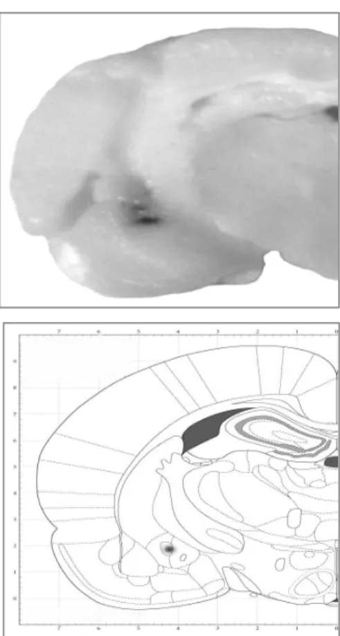
before sectioning. Brain slices were taken through the brain areas of cannulae placements, and the cannulae placements were verified (Figure 1) using the atlas of Paxinos and Watson (2005). Data from rats with injection sites located outside the appropriate area were excluded from the statistical analyses.
Statistical analysis
All results were expressed as mean±SEM (standard error of mean). In order to compare the changes in place preference (sec) obtained in all groups (vehicle and experimental groups), one- and/or two-way analysis of variance (ANOVA) followed by appropriate post-hoc analysis (Tukey’s test) for multiple comparisons were used, as needed. P-values less than 0.05 (P< 0.05) were considered to be statistically significant.
Results
Histological verification of microinjection sites in the central amygdale
Figure 1 reveals the injection site intra-central amygdala after administration of 1 µl of a methylene blue solution by using the same injection set up as used for the drugs.
Dose-response of morphine in conditioning place preference paradigm
Figure 2 shows the effect of different doses of morphine (2.5-10 mg/kg, s.c.) in CPP paradigm in Wistar rats. Administration of morphine resulted in a significant response in comparison with the saline group (F4,35=
3.258; P< 0.05). The opioid induced a meaningful preference to the place dose-dependently. In view of the results, morphine at the dose of 7.5 mg/kg (s.c.) was used for the subsequent studies.
Effect of naloxone on the expression of morphine response in the place conditioning procedure
Figure 3 shows the effect of single injection of different doses of naloxone (0.1-0.4 mg/kg, i.p.) prior to morphine response. Pre-testing administration of naloxone in experimental animals, which were morphine (7.5 mg/kg,
s.c.) injected, resulted in a significant effect as compared to the control group in morphine-CPP. The two-way ANOVA indicated the interaction (Fdrug(1,70)=7.383, P< 0.05;
Fdose(4,70)=2.221, P< 0.05: Fdrug*dose(4,70)=2.997,
P< 0.05). Further analysis demonstrates that despite the fact that the antagonist did not induce any response alone; the narcotic drug, when injected on day of the test in morphine-administered animals, reversed the effect of morphine effective dose in a dose- dependent manner. In view of the results, naloxone at the highest dose (0.4 mg/kg, i.p.) was used for the subsequent studies.
Effect of nitric oxide production in the central amygdala on the naloxone-modulated morphine response in the place conditioning paradigm
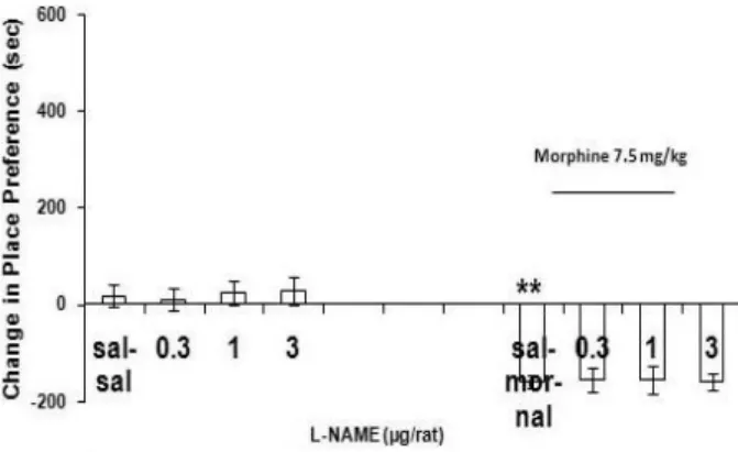
Pre-testing injection of single doses of L-arginine or L-NAME (0.3, 1.0 and 3.0 µg/rat,
intra-central amygdala) resulted no significant effect in the animals which were simply injected saline (1 ml/kg, s.c.), during the conditioning procedure (Figure 4 and Figure 5). On the other hand, bilateral injection of L-arginine but not L-NAME (0.3, 1.0 and 3.0 µg/rat, intra-central amygdala), prior (1-2 min) to the injection of naloxone (0.4 mg/kg, i.p.) before testing of morphine response (7.5 mg/kg, s.c.), resulted in a significant effect (Figure 5) compared to the control group. Analysis by two-way ANOVA revealed the interaction between drugs (Fdrug(1,56)= 0.891, P> 0.05; Fdose(3,56)= 7.750, P<
0.0001: Fdrug*dose(3,56)=9.245, P< 0.0001). Further
Figure 1. A Cannulae placements in central amygdala evidenced by ink injection in a volume of 1 µl/rat by using the same set up as used for intra-central amygdala injection of drugs (AP: -2.12).
B: Verification from atlas of Paxinos and Watson (2005).
Figure 2. Dose response curve for morphine-induced conditioned place preference in opioid-naive male
Wistar rats. Morphine (2.5-10 mg/kg) or saline (1 ml/kg) was given subcutaneously (s.c.) in a 3-day
schedule of an unbiased conditioning paradigm. The control group received saline (1 ml/kg, s.c.), twice daily for 3 days. Data are expressed as mean of change in place preference±SEM. Change in place preference is defined as the time spent in the drug-paired place on day of testing minus that spent in the same place during pre-conditioning.
Tukey-Kramer post hoc analysis showed the differences:
*P< 0.05, and **P< 0.01 difference to control (sal-sal).
Figure 3. Naloxone response curve in morphine treated male rats. Naloxone (0.1-0.4 mg/kg) was given intraperitoneally (i.p.) on day of the testing of place conditioning to morphine (7.5 mg/kg, s.c.). The rats were first injected with naloxone, and after 10 min they were tested in a morphine-free state. Control group simply received saline (1 ml/kg, i.p.) pre-testing. Data are expressed as mean of change in conditioning±SEM. **P< 0.01 difference compared to the negative control (sal-sal).
##P< 0.01 difference compared to the positive control (sal-mor).
+++P< 0.001 difference compared to the respective dose groups according to post hoc measurements.
Figure 4. Response curve induced by single injection of L-arginine (0.3-3 µg/rat, intra-central amygdala) pre-testing, in the control task or prior to naloxone injection before morphine response testing. 10 min after saline (1 ml/kg, i.p.) or naloxone injection (0.4 mg/kg, i.p.) the rats were tested in a morphine-free state. Data are expressed as mean of change in conditioning±SEM. **P< 0.01 difference compared to saline control (sal-sal)
# P< 0.05, and ## P< 0.01 differences compared to positive control (sal-mor-nal)
Figure 5. Response curve induced by single injection of L–NAME (0.3-3 µg/rat, intra-central amygdala) pre-testing in the control task or prior to naloxone before morphine response testing. 10 min after saline (1 ml/kg, i.p.) or naloxone injection (0.4 mg/kg, i.p.) the rats were tested in a morphine-free state. Data are expressed as mean of change in conditioning±SEM.
**P< 0.01 difference compared to saline control (sal-sal).
Figure 6. Response curve induced by prior injection of L-NAME (0.3-3 µg/rat, intra-central amygdala) prior to L-arginine before the injection of naloxone (0.4 mg/kg, i.p.) on day of testing of morphine response. 10 min after naloxone injection, the rats were tested in a morphine-free state according to the schedule of conditioning paradigm. The control group was injected with saline (1 µg/rat, intra-nucleus or 1 ml/kg, i.p.) with respect to the way of injection of the desired drug prior to testing. Data are expressed as mean of change in conditioning±SEM.
**P< 0.01 difference compared to control (sal).
Discussion
In this study, morphine (2.5, 5, 7.5 and 10 mg/kg, s.c.) induced a significant conditioned dose-dependent place preference in male Wistar rats. This effect may reflect the role of the mu- and delta- opioid receptors (20, 35) as well as several neuronal systems (13) including glutamate and GABAB receptors
(36) in the mediation of the morphine
rewarding in rats.
Injection of naloxone (0.1-0.4 mg/kg, i.p.) before the morphine (7.5 mg/kg, s.c.)-CPP test in the post-conditioning phase (test day), induced a significant place aversion. It indicates an opioid system involvement (37) in the morphine dependence in a place preference paradigm measured by an unbiased task with no pre-conditioning preference. In accordance, previous studies have shown an aversive effect of naloxone by using either a biased (38) or a balanced biased procedure with no strong preconditioning preferences (39, 40). Our results, for the first time, may indicate that the style of the protocol (biased vs. unbiased) designed to analyze the dependence on morphine in the conditioning, by using a single injection of the antagonist prior to opioid response, does not play a role in displaying of the drug dependency. In addition, it seems that the same subtypes of the receptors might be involved in the observed interaction. In contrast, a reinforcing property of naloxone in the process has been reported in those cases in which the intraperitoneal route of the administration of the antagonist was chosen in the protocol (41). Other studies also indicated that high doses of naloxone inhibit the expression of morphine place conditioning in mice (3, 42). In a previous survey on acute morphine dependence, the magnitude of naloxone potency to precipitate morphine withdrawal is shown to be depended to morphine dose (43). Although there is no molecular evidence to elucidate the mechanism governing the effect observed in the present study, it might lead in a conclusion that this property of naloxone is mediated through the activation of the same subtype opiate receptors (40) which are involved in the conditioning to the drugs of abuse. Other explanation might be that different subtypes of opioid receptors are involved in this effect since the role of kappa-opioid receptors has been demonstrated both in the development of physical and psychological dependence on morphine (44) and the precipitation of the withdrawal signs by naloxone as well (45). It is recently noted that both mu and GABAA
morphine treatment (46). There is also evidence expressing that the central amygdala is involved in place aversion induced by naloxone in single-dose morphine-treated rats (47). Previous studies proposed the role of several neurotransmitter systems in the expression of morphine place conditioning (13, 14).
Nitric oxide also participates in morphine-induced CPP (17, 48-51). The role of the nitric oxide at the central amygdala in morphine CPP has already been proposed by this laboratory (17), but, role of nitric oxide system in the central amygdala in naloxone place aversion was evaluated in the present work for the first time. A repeated morphine injection (3-sessions) was used to associate between primary unconditioned properties of the environment and the conditioned stimulus (13). But, Ishida et al (2008) used a single-session morphine treatment to survey on the acute dependence. Although single injection of the L-arginine (0.3-3 µg/rat, intra-central amygdala) as a nitric oxide precursor showed no significant effect in the control task, present observations demonstrated that injection of L-arginine before naloxone injection produces a significant effect on naloxone-induced responses. The data may reflect that nitric oxide as a candidate for modulating morphine dependence in the central amygdala participates in the acute (psychological) dependence on morphine which was exhibited
in the avoidance of the conditioned place because of interaction between naloxone and morphine. L-NAME (0.3-3 µg/rat, intra-central amygdala) blocked this effect when microinjected in combination with L-arginine. However, sole injection of L-NAME did not show any significant effect compared to the control groups in morphine-induced CPP. Therefore, it can be suggested that L-arginine may cause an activation of the nitric oxide system in central amygdala which consequently interacts with naloxone to present the dependence on the opioid in the naloxone-paired morphine conditioning test. It can be supported by the finding that different neural systems mediate morphine reward and the spontaneous withdrawal signs in the acute dependence (15).
Although, the role of the opioid system in the acute dependency is not obviously defined to date, nevertheless, this work may clearly indicate that both opioid and non-opioid mechanisms modulate this effect, the fact that the nitric oxide, a highly active neurotransmitter, in the central amygdala may mediate the pharmacological effects of the morphine and naloxone at the molecular level, remains to be studied.
Acknowledgment
This study was supported in part by Research Deputy of Shahed University (document no; 7941 for the research project), Tehran, Iran.
References
1. Gutstein HB, Akil H. Opioid analgesics. In: Hardman JG, Limbird LE, Goodman Gilman A, editors. Goodman and Gilman’s the Pharmacological Basis of Therapeutics.10th ed. New York: McGraw-Hill; 2001.p.569-619. 2. Karami M, Zarrindast MR. Morphine sex-dependently induced place conditioning in adult Wistar rats. Eur J
Pharmacol 2008; 582:78-87.
3. McClung CA. The molecular mechanisms of morphine addiction. Rev Neurosci 2006; 17: 393-402.
4. Watanabe T, Nakagawa T, Yamamoto R, Maeda A, Minami M, Satoh M. Involvement of glutamate receptors within the central nucleus of the amygdala in naloxone-precipitated morphine withdrawal-induced conditioned place aversion in rats. Jpn J Pharmacol 2002; 88: 399-406.
5. Watanabe T, Nakagawa T, Yamamoto R, Maeda A, Minami M, Satoh M. Involvement of noradrenergic system within the central nucleus of the amygdala in naloxone-precipitated morphine withdrawal-induced conditioned place aversion in rats. Sychopharmacol (Berl) 2003; 170: 80-88.
6. Ikemoto S. Dopamine reward circuitry: two projection systems from the ventral midbrain to the nucleus accumbens-olfactory tubercle complex. Brain Res Rev 2007; 56:27-78.
7. Spanagel R, Weiss F. The dopamine hypothesis of reward: past and current status. Trends Neurosci 1999; 22:521-527.
8. Wise RA. Drug-activation of brain reward pathways. Drug Alcohol Depend 1998; 51:13-22. 9 Di Chiara G, North RA. Neurobiology of opiate abuse. Trends Pharmacol Sci 1992; 13:185-193.
13. Tzschentke TM. Measuring reward with the conditioned place preference (CPP) paradigm: update of the last decade. Addict Biol 2007; 12:227-462.
14. Tzschentke TM. Measuring reward with the conditioned place preference paradigm: A comprehensive review of drug effects, recent progress and new issues. Prog Neurobiol 1998; 56:613-72.
15. Vargas-Perez H, Ting-A-Kee R, van der Kooy D. Different neural systems mediate morphine reward and its spontaneous withdrawal aversion. Eur J Neurosci 2009; 29:2029-2034.
16. Wise RA. The role of reward pathways in the development of drug dependence. Pharmacol Ther 1987: 35:227-263.
17. Zarrindast MR, Karami M, Sepehri H, Sahraei H. Influence of nitric oxide on morphine-induced conditioned place preference in the rat central amygdala. Eur J Pharmacol 2002; 453: 81-89.
18. Crain SM, Shen KF. Ultra-low concentrations of naloxone selectively antagonize excitatory effects of morphine on sensory neurons, thereby increasing its anti-nociceptive potency and attenuating tolerance/dependence during chronic co-treatment. Proc Natl Acad Sci USA 1995; 92:10540-10544.
19. Wang H-Y, Friedman E, Olmstead MC, Burns LH. Ultra-low-dose naloxone suppresses opioid tolerance, dependence and associated changes in mu-opioid receptor-G protein coupling and Gbetagamma signaling. Neuroscience 2005; 135:247-261.
20. Zheng-xiong XI, Stein EA. Gabaergic mechanisms of opiate reinforcement. Alcohol 2002; 37:485-494. 21. Di Chiara G, Imperato A. Drugs abused by humans preferentially increase synaptic dopamine concentrations in
the mesolimbic system of freely moving rats. Proc Natl Acad Sci USA 1988; 85:5274-5278.
22. Olmstead MC, Franklin KB. Development of a conditioned place preference: effects of lesions of various CNS sites. Behav Neurosci 1997; 111:1313-23.
23. Neisewander JL, Pierce RC, Bardo MT. Naloxone enhances the expression of morphine-induced conditioned place preference. Psychopharmacology (Berl) 1990; 100:201-205.
24. Diaz SL, Barros VG, Antonelli MC, Rubio MC, Balerio GN. Morphine withdrawal syndrome and its prevention with baclofen: Autoradiographic study of mu-opioid receptors in prepubertal male and female mice. Synapse 2006; 60:132-140.
26. Fukuto JM, Mayer B. The enzymology of nitric oxide synthase. In: Feelisch M, Stamler JS, editors. Methods in nitric oxide research. New York: John Wiley & Sons Ltd; 1996. p. 147-157.
27. Garthwaite J. Glutamate, nitric oxide and cell-cell signaling in the nervous system. Trends Neurosci 1991; 14:60-67.
28. Moncada S, Palmer RMJ, Higgs EA. Nitric oxide: physiology, pathophysiology, and pharmacology. Pharmacol Rev 1991; 43:109-142.
29. Kivastik T, Rutkauskaite J, Zharkovsky A. Nitric oxide synthesis inhibition attenuates morphine-induced place preference. Pharmacol Biochem Behav 1996; 53:1013-1015.
30. Paxinos G, Watson CR. The Rat Brain in Stereotaxic Coordinates. 5th ed. San Diego: Elsevier Academic Press; 2005.
31. Haghparast A, Azizi P, Hassanpour-Ezatti M, Khorrami H, Naderi N. Sub-chronic administration of AM251, CB1 receptor antagonist, within the nucleus accumbens induced sensitization to morphine in the rat. Neurosci Lett 2009; 467:43-47.
32. Karami M, Zarrindast MR, Sepehri H, Sahraei H. Role of nitric oxide in the rat hippocampal CA1 area on morphine-induced conditioned place preference . Eur J Pharmacol 2002; 449: 113-9.
33. Moaddab M, Haghparast A, Hassanpour-Ezatti M. Effects of reversible inactivation of the ventral tegmental area on the acquisition and expression of morphine-induced conditioned place preference in the rat. Behav Brain Res 2009; 198:466-471.
34. Overton DA. State-dependent learning produced by addicting drugs. In: Fisher S, Freedman, AM, editors. Opiate addiction: origin and treatments. Washington DC: Winston; 1973. p. 61-75.
35. Bie B, Zhu W, Pan ZZ. Rewarding morphine-induced synaptic function of delta-opioid receptors on central glutamate synapses. J Pharmacol Exp Ther 2009; 329:290-296.
36. Heinmiller A, Ting-A-Kee R, Vargas-Perez H, Yeh A, van der Kooy D. Tegmental pedunculopontine glutamate and GABA-B synapses mediate morphine reward. Behav Neurosci 2009; 123:145-155.
37. Skoubis PD, Matthes HW, Walwyn WM, Kieffer BL, Maidment NT. Naloxone fails to produce conditioned place aversion in mu-opioid receptor knock-out mice. Neuroscience 2001; 106:757-763.
38. Phillips AG, Le Paine FG. Reward produced by microinjection of (D-Ala2), Met5-enkephaline into the ventral tegmental area. Behav Brain Res 1982; 5:225-229.
39. Bozarth MA. Conditioned place preference. A parametric analysis using systemic heroin injections. In: Bozarth MA, editor. Methods of assessing the reinforcing properties of abused drugs. New York: Springer-Verlag;1987.p.241-273.
40. Mucha RF, van der Kooy D, O'Shaughnessy M, Bucenieks P. Drug reinforcement studied by the use of place conditioning in rat. Brain Res 1982; 243:91-105.
42. Zarrindast MR, Faraji N, Rostami P, Sahraei H, Ghoshouni H. Cross tolerance between morphine- and nicotine-induced conditioned place preferences in mic. Pharmacol Biochem Behav 2003; 74:363-369.
43. Schulteis C, Morse AC, Liu J. Repeated experience with naloxone facilitates acute morphine withdrawal: potential role for conditioning processes in acute opioid dependence. Pharmacol Biochem Behav 2003; 76:493-503.
44. Narita M, Funada M, Suzuki T. Regulations of opioid dependence by opioid receptor types. Pharmacol Ther 2001; 89:1-15.
45. Le Guen S, Gestreau C, Besson JM. Morphine withdrawal precipitated by specific mu, delta or kappa opioid receptor antagonists: a c-Fos protein study in the rat central nervous system. Eur J Neurosci 2003; 17:2425-2437. [46]. Skoubis PD, Matthes HW, Walwyn WM, Kieffer BL, Maidment NT. Naloxone fails to produce conditioned
place aversion in mu-opioid receptor knock-out mice. Neurosci. 2001; 106:757-63.
47. Ishida S, Shimosaka R, Kawasaki Y, Jin C, Kitamura Y, Araki H, et al. Involvement of the amygdala on place aversion induced by naloxone in single-dose morphine-treated rats. Yakugaku Zasshi (in Japanese) 2008; 128:395-403.
48. Cabral A, Ruggiero RN, Nobre MJ, Brandão ML, Castilho VM. GABA and opioid mechanisms of the central amygdala underlie the withdrawal-potentiated startle from acute morphine. Prog Neuropsychopharmacol Biol Psychiatry 2009; 33:334-344.
49. Manzanedo C, Aguilar MA, Rodriguez-Arias M, Minarro J. Sensitization to the rewarding effects of morphine depends on dopamine. Neuroreport 2005; 16:201-205.
50. Rezayof A, Zarrindast MR, Sahraei H, Haeri-Rohani A. Involvement of dopamine receptors of the dorsal hippocampus on the acquisition and expression of morphine-induced place preference in rats. J Psychopharmacol 2003; 17:415-423.