Pages: 264-268 DOI: 10.13057/biodiv/d160222
Short Communication:
Microfungal diversity on leaves of
Eusideroxylon zwageri, a threatened
plant species in Sarawak, Northern Borneo
A. LATEEF ADEBOLA1,2,♥, MUID SEPIAH1, MOHAMAD H. BOLHASSAN1, MANSOR WAN ZAMIR1 1
Department of Plant Science and Environmental Ecology, Faculty of Resource Science and Technology, Universiti Malaysia Sarawak, Kota Samarahan-94300, Sarawak, Malaysia. Tel./fax.: +60-146902928,
email: lateef.aa@unilorin.edu.ng 2 Department of Plant Biology, Faculty of Life Science, University of Ilorin, Nigeria
Manuscript received: 16 August 2015. Revision accepted: 27 September 2015.
Abstract. Adebola AL, Sepiah M, Bolhassan MH, Wan Zamir M. 2015. Microfungal diversity on leaves ofEusideroxylon zwageri, a threatened plant species in Sarawak, Northern Borneo. Biodiversitas 16: 264-268. A survey of the microfungal communities on green leaves and leaf litters of an endangered plant species,Eusideroxylon zwageriTeijsm. & Binn. (belian) was carried out for the first time. A total of 200 leaf segments were plated on both water agar and malt extract agar. 74 fungal species were identified from both leaf types with more fungal taxa found on the green leaves, with a Shannon diversity index of 3.85 compared to that on litters, 2.63 and the similarity between the microfungal communities on both leaf types was low with a Bray-Curtis similarity index of 0.366. The most dominant species on both leaf types includes Aphanocladium areanarum, Trichoderma koningii, Nectria sp., Chalara pteridina,
Hyphomycetes sp.3,hyalineMycelia sterilia, Circinotrichumsp., Phoma sp., Acremonium macroclavatum, Chaetopsina sp., Physarum
sp., Beltrania rhombica andColletotrichum acutatum.
Keywords:Endophytic, green leaves, leaf litters, new record, saprophytic
INTRODUCTION
General knowledge on the microfungal diversity and distribution is still inadequately understood. More studies have been done on fungal diversity and their spatial distribution in the temperate regions as compared to the tropics (Hawksworth 2001; Hawksworth and Rossman 1997). Many areas and substrates still remain unstudied in the world, most especially in the tropics and same applies to many plant species which are not yet studied for their associated microfungal communities. The most accepted fungal estimate of 1.5 million by Hawksworth (1991, 2001) was considered as too small by some authors (Cannon
1997; O’Brien et al. 2005) on the basis that the used plant
to fungus ratio of 1:6 used by Hawksworth, which assumed the plant diversity as 270,000, was too low, pointing out that there are about 300,000-320,000 plant species (Prance et al. 2000), 420,000 spp. (Govaerts 2001) and 117,734-575,320 spp. (Wortley and Scotland 2004). An important area of global fungal diversity which has been often overlooked is microfungi on vulnerable, threatened and endangered plant species. There is a wide gap of data on microfungi associated with many rare plant species, in terms of host-specific fungi and also fungal disease caused to the plants (Buchanan et al. 2002).
Eusideroxylon zwageriTeijsm. & Binn. (belian tree), is a typical case study of unstudied rare plants.E. zwageriis the only accepted species in the genusEusideroxylon which belongs to the family Lauraceae. This plant species, also called the Borneo Ironwood, is native to the Southeast
Asian forest and has been listed in the IUCN Global Red List of Threatened Species as an endangered species due to over logging and habitat destruction (IUCN 1998). To the best of our knowledge, no studies have been found in literature on microfungal communities on belian tree. At the plant family level (Lauraceae), comparatively few studies have been done on plant species belonging to the family Lauraceae, an example is the study done by (Paulus et al. 2006) onCryptocarya mackinnoniana from which 81 fungal taxa were identified using direct observation method
and on Chlorocardium rodiei by Cannon and Simmons
(2002) in which only 10 endophytic fungi were identified. This study aims at revealing the microfungal communities on green leaves and leaf litters ofE. zwageri
(belian tree) from Kubah National Park in Sarawak, Malaysia. The implications of this study will be far reaching in the understanding, protection and conservation of the belian tree.
MATERIALS AND METHODS
Sampling
samples were collected randomly under the belian tree, green leaves as just fallen green leaves and litters as already brown and weak leaves. The belian trees were very tall and it is practically impossible to detach a leaf from it. The samples collected were put in plastic bags, labeled and transported to the laboratory for processing.
Isolation of microfungi
Isolation of microfungi was based on the methods of Rakotoniriana et al. (2008) and Lateef et al. (2014) for endophytic fungi and saprobic fungi respectively, with some modifications. For endophytic fungi, the leaves were washed under running tap water to remove dust and debris adhering to them. The leaves were then cut into 1 cm2 with and without the midribs under aseptic conditions using a sterile scalpel. They were then surface sterilized with 70% ethanol for one minute, then in 10 % hydrogen peroxide (H2O2) solution for five minutes, rinsed with 70% ethanol for one minute and finally rinsed with deionized sterile distilled water five times to remove the sterilants and blotted on sterile filter paper to remove excess water. Five segments were plated separately on water agar (WA) and malt extract agar (MEA). The Petri dishes were sealed with parafilm and incubated at room temperature. Observation and isolation of the growing microfungi starts from the third day of incubation for MEA and four weeks for WA.
For saprophytic microfungi, the leaf samples were washed with double sterilized distilled water, cut into 1 cm2 into a 500 mL conical flask, washed again with sterile distilled water for 5 times and then blotted on sterilized filter paper. The leaf segments were then plated as done for the endophytic microfungi. A total of 200 leaf segments were plated, 100 each for green leaves and leaf litters. 20 replicate petridishes were used for the green leaves and leaf litters separately.
Frequencies of occurrence of each microfungi on the leaf segments were recorded. A Motic stereo microscope (MZ 168) and an Olympus compound microscope (CX-31) were used for monitoring of fungal reproductive structures and pictures were taken with a hand-held Samsung camera model ES91. Identification of the observed microfungi were made to genus level, and wherever possible, to species level.
Data analysis
The frequencies of occurrence of each microfungal taxa observed was calculated and the frequency of isolation was determined according to (Hata and Futai 1995; Osono 2008) as follows:
Freq. of isolation = Total no. of leaf segments a fungal taxa was present × 100 Total no. of leaf segments observed
The diversity indices such as Shannon and Simpson’s diversity indices as well as the Bray-Curtis similarity index were calculated using the software Estimates (Colwell 2013).
RESULTS AND DISCUSSION
The total of 74 taxa were identified from 200 leaf segments ofE. zwageri on WA and MEA, comprising 11 Ascomycetes, 55 anamorphic taxa, 3 Basidiomycetes, and
3 Zygomycetes. Non-sporulating mycelia (Mycelia
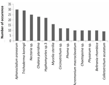
sterilia), both hyaline and melanized were also documented. 67 taxa were identified from green leaves while 20 taxa were from leaf litters (Table 1). The most dominant species observed from both leaf types were
Aphanocladium aranearum, Trichoderma koningii, Nectria
sp., Chalara pteridina, Hyphomycetes sp.3, Hyaline
Mycelia sterilia, Circinotrichum sp., Phoma sp., Acremonium macroclavatum, Beltrania rhombica, Chaetopsinasp. and Physarum sp. (Figure 1).
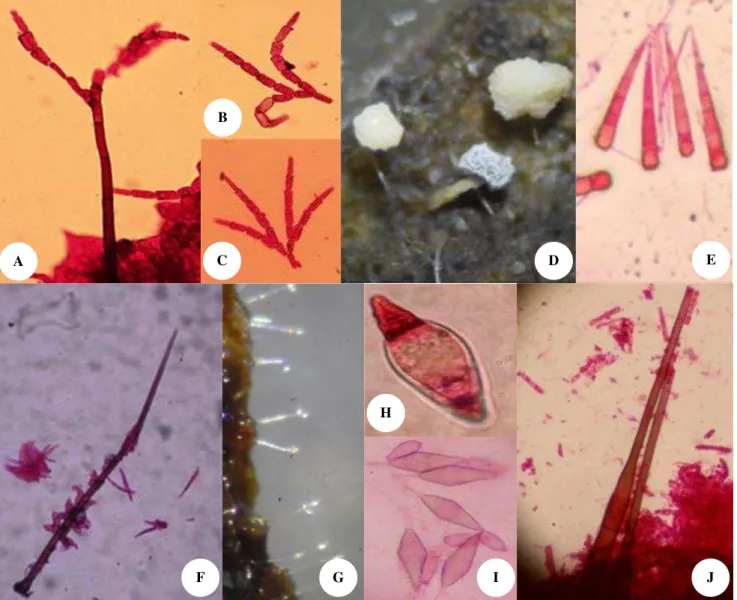
Other species identified includes Speiropsis
pedatospora (Figure 2.A-C.), Subulispora longirostrata
(Figure 2.E), Isthmotricladia sp., Fusarium solani, Cylindrocladium sp., Dactylaria obtriangularia and
Monacrosporium sp (Figure 2G-H). The Shannon and Simpson’s diversity indices were higher for the microfungal assemblage on green leaves, 3.85 and 35.93 respectively, than on leaf litters, 2.63 and 11.11 respectively (Tabel 2). This result indicates that leaves of belian support a high diversity of fungi when compared to that recovered from other endangered plant species (Sadaka and Ponge 2003; Shanthi and Vittal 2010; Goveas et al.
2011; Grbić et al. 2015). 41 endophytic fungal taxa were
identified from Coscinium fenestratum (Goveas et al. 2011)and 49 taxa from Nepetartanjensis (Grbić et al. 2015). Also, Sadaka and Ponge (2003) and Shanthi and Vittal (2010) identified 36 and 54 fungal taxa from
Quercus Rotundifolia andPavetta indica respectively. Green leaves are usually richer in nutrients than leaf litters (Lodge et al. 2014), thus supporting a more diverse species of microfungi. Green leaves of belian are thick and leathery in texture making it to last longer before decomposing. The high number of fungal taxa from leaves of belian tree altogether shows that this plant species supports the growth of many microfungal species.
Figure 1.Most dominant microfungal taxa on both green leaves and leaf litters ofEusideroxylon zwageri (belian)
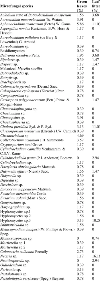
Table 1. Percent dominance of microfungi on green leaves and leaf litters ofEusideroxylon zwageri (belian)
Microfungal species
Green leaves (%)
Leaf litters (%)
Acladium state ofBotryobasidium consperum 1.56 3.68
Acremonium macroclavatumTs.Watan. 3.91 0
Aphanocladium aranearum (Petch) W. Gams 5.86 11.03
Aspergillus nomiusKurtzman, B.W. Horn & Hesselt.
1.17 0
Aureobasidium pullulans (de Bary & Löwenthal) G. Arnaud
1.17 0
Aureobasidiumsp. 0.39 0
Basidiomycetes 0.39 0.74
Beltrania rhombicaPenz. 1.95 3.68
Bipolarissp. 0.39 1.47
Bisporasp. 1.17 1.47
MelanizedMycelia sterilia 1.17 0
Botryodiplodiasp. 0.39 0
Botrytissp. 0.39 0
Brachyphorissp. 1.17 0
Calonectria pyrochroa (Desm.) Sacc. 0.39 0
Calosphaeria cyclospora (Kirschst.) Petr. 0.78 0
Camposporiumsp. 0.78 0
Ceriospora polygonacearum (Petr.) Piroz. & Morgan-Jones
0 1.47
Chaetendophragmiasp. 0.39 0
Chaetomiumsp. 0.78 0
Chaetopsinasp. 3.91 0
Chaetosphaeriasp. 0.39 0
Chalara pteridinaSyd. & P. Syd. 3.13 11.03
Chrysosporium merdarium (Ehrenb.) J.W. Carmich 0.39 0
Circinotrichumsp. 4.69 0
Colletotrichum acutatumJ.H. Simmonds 3.52 0
Cryptosporium tamiGrove 1.17 0
Cylindrocladium camelliaVenkataram. & C.S.V. Rame
0.39 0
Cylindrocladiella parva (P.J. Anderson) Boesew. 0 2.94
Cylindrocladiumsp. 1.17 0
Dactylaria obtriangulariaMatsush. 0.78 0
Didymella effusa(Niessl) Sacc. 1.56 1.47
Didymellasp. 0.39 0
Diplodia sp. 0.39 0
Drechslera sp. 0.39 0
Epicoccum nipponicumMatsush. 0.39 0
Fusarium merismoidesCorda 0.39 0
Fusarium solani (Mart.) Sacc. 1.56 0
Gonytrichum sp. 0.78 0
Harpographium sp. 1.17 0
Hyphomycetes sp.1 0.78 0
Hyphomycetes sp.2 1.56 0
Hyphomycetes sp.3 3.13 10.29
Isthmotricladia sp. 1.56 0
Metacapnodium juniperi (W. Phillips & Plowr.) Speg.
0.39 0
Monacrosporium sp. 0 0.74
Mortierella sp.1 0.39 0
Mortierellasp.2 1.17 0
Calonectria colhouniiPeerally 2.73 0
Nectria sp. 1.17 16.18
Neottiosporella sp. 0 2.94
Oidiodendron sp. 0.39 0
Periconia sp. 3.13 0
Pestalotiopsis sp. 0.78 0
Pestalotiopsis versicolor (Speg.) Steyaert 0.78 0
Phaeostalagmus sp. 0.39 0
Phoma sp. 0.78 7.35
Physarum sp. 3.91 0
Piricauda cochinensis (Subram.) M.B. Ellis 1.95 0
Rhinocladiella cristasporaMatsush. 3.52 0
Septonemasp. 0.78 0
Sordaria fimicola (Roberge ex Desm.) Ces. & De Not.
0.39 0
Speiropsis pedatosporaTubaki 1.17 0
HyalineMycelia sterilia 4.30 3.68
Subulispora longirostrataNawawi & Kuthub. 0.39 0
Subulispora procurvataTubaki 2.34 1.47
Tetrabrunneospora ellisiiDyko 0 1.47
Thielaviasp. 1.56 0
Thozetellasp. 0.78 0
Trichoderma koningiiOudem. 6.25 9.56
Trichosporiella sp. 1.95 0
Vermispora sp. 0 2.94
Veronaea indica (Subram.) M.B. Ellis 0.78 0
Volutella ciliata(Alb. & Schwein.) Fr. 0 4.41
Table 2. Diversity indices and Similarity index of the microfungal communties on green leaves and leaf litters of Eusideroxylon zwageri(belian)
Diversity index Green leaves Leaf litters
Number of isolates 256 132
Observed species 67 20
Number of Singletons 21 2
Number of Doubletons 12 6
ACE species estimate ± SD 83.09 20.14
Chao 1 species estimate ± SD 83.09 20.14
Shannon Index ± SD 3.85 2.63
Simpson Inv Index ± SD 35.93 11.11
Table 3.Similarity index of microfungi on green leaves and leaf litters ofEusideroxylon zwageri (belian)
Samples Index numbers
Species observed on green leaves 67 Species observed on leaf litters 20
Shared Species Observed 14
Sorensen Classic 0.322
Morisita-Horn 0.442
Bray-Curtis 0.366
Fourteen taxa were commonly identified from both green leaves and leaf litters with a Bray-Curtis similarity index of 0.366 (Table 3). Consequently, there were a total of 53 fungal taxa exclusively identified on green leaves while 7 taxa were from the leaf litters (Figure 3). There were more species exclusively on the green leaves than on the leaf litters which further suggests that the green leaves are more suitable for fungal growth. Furthermore,
Metacapnodium juniperi, Phaeostalagmus sp. and
Figure 2.Some microfungal diversity on green leaves and leaf litters ofEusideroxylon zwageri (belian). A.Speiropsis pedatospora, B -C. Conidia ofS. pedatospora, D. Physarum sp., E. Conidia ofSubulispora longirostrata, F.Circinotrichumsp.,G. Conidiophores of
Monacrosporium sp., H. Conidia ofMonacrosporiumsp., I. Conidia ofBeltraniasp., J.Chalara sp.
Figure 3. Number of microfungi exclusively observed only on green leaves and leaf litters ofEusideroxylon zwageri (belian)
usually frequently isolated from other plant species. Many rare fungal species have been identified on endangered plant species in New Zealand (Buchanan et al. 2002) and also in Sarawak (Hughes 1977). This occurrence signifies the microfungal loss which can be suffered with the extinction of such endangered plant species including belian.
Also, comparison of the fungal communities observed on belian leaves with other plant species in the family Lauraceae showed some similarity in their microfungal communities, in which Acremonium sp., Beltrania sp.,
Chaetopsis sp.,Chalara sp.,Dactylaria sp.,Pestalotiopsis
spp, Rhinocladiella cristaspora, Subulispora sp.,
Thozetella sp. and Volutella sp. have been recorded on leaves ofCryptocarya mackinnoniana (Paulus et al. 2006) as saprobic species. Colletotrichum acutatum have
previously been isolated from Chlorocardium rodiei
(Cannon and Simmons 2002) andCinnamomum bejolghota
A C
F G
H
I
E
J B
(Suwannarach et al. 2012) as an endophyte. Fusarium solani, Nectria sp., Oidiodendron sp., Periconia sp.,
Pestalotiopsis sp., Phoma sp. and Trichoderma sp. were
also recovered from Cinnamomum bejolghota
(Suwannarach et al. 2012). Other taxa recorded in this study are reported for the first time in the family Lauraceae.
The high demand for wood materials from belian and its slow growth, coupled with its habitat destruction makes it vulnerable to extinction. This study is the first report of microfungal communities on belian leaves in Sarawak. Green leaves supported a higher number of microfungi than the leaf litters. This observation contributes to the understanding of the biology ofE. zwageri and can be used to strengthen its conservation importance.
ACKNOWLEDGEMENTS
The first author is grateful to Universiti Malaysia Sarawak (UNIMAS) for the Zamalah scholarship awarded. We are also grateful to the Sarawak government and to Sarawak Forestry Co-operation (SFC) for permission to collect samples from the National Park.
REFERENCES
Buchanan P, Johnston P, Pennycook S, McKenzie E, Beever R. 2002. Data-deficient 2002, Rare and endangered fungi, Landcare Research. www.landcareresearch.co.nz/science/plants-animals-fungi/fungi/rare-and-endangered-fungi/data-deficient-2002 [Aug 8, 2015].
Cannon PF. 1997. Strategies for rapid assessment of fungal diversity. Biodiv Conserv 6: 669-680.
Cannon PF, Simmons CM. 2002. Diversity and host preference of leaf endophytic fungi in the Iwokrama Forest Reserve, Guyana. Mycologia 94: 210-220.
Colwell RK. 2013. EstimateS: Statistical Estimation of Species Richness
and Shared Species from Samples. Version 9 and earlier. User’s
Guide and Application. http://purl.oclc.org/estimates. [Aug 8, 2015]. Govaerts R. 2001. How many species of seed plants are there? Taxon 50:
1085-1090.
Goveas SW, Madtha R, Nivas SK, D’Souza L. 2011. Isolation of
endophytic fungi fromCoscinium fenestratum (Gaertn.) Colber. a red listed endangered medicinal plant. Bulg J Agric Sci 17: 767-772.
Grbić ML, Stupar M, Unković N, Vukojević J, Stevanović B, Grubišić D.
2015 May 9. Diversity of microfungi associated with phyllosphere of endemic Serbian plantNepeta rtanjensis Diklić & Milojević. Braz J Bot 38 (3): 597-603.
Hata K, Futai K. 1995. Endophytic fungi associated with healthy pine needles and needles infested by the pine needle gall midge,
Thecodiplosis japonensis. Can J Bot 73: 384-390.
Hawksworth DL. 1991. The fungal dimension of biodiversity magnitude, significance, and conservation. Mycol Res 98: 641-655.
Hawksworth DL. 2001. The magnitude of fungal diversity: the 1.5 million species estimate revisited. Mycol Res 105: 1422-1432.
Hawksworth DL, Rossman AY. 1997. Where are all the undescribed fungi? Phytopathology 87: 888-891.
IUCN. 1998. Eusideroxylon zwageri. Asian Regional Workshop on Conservation & Sustainable Management of Trees, Viet Nam, August 1996: The IUCN Red List of Threatened Species 1998: e.T31316A9624725. http://dx.doi.org/10.2305/IUCN.UK.1998. RLTS. T31316A9624725.en [Sep 25, 2015].
Lateef AA, Sepiah M, Bolhassan MH. 2014. Microfungi on green leaves and leaf litters from Santubong National Park, Sarawak. In: Microbes for the Benefits of Mankind, Proceedings of the International Conference on Beneficial Microbes (ICOBM), School of Industrial Technology, Universiti Sains Malaysia 2014, Sarawak.
Lodge DJ, Cantrell SA, González G. 2014. Effects of canopy opening and debris deposition on fungal connectivity, phosphorus movement between litter cohorts and mass loss. For Ecol Manag 332: 11-21.
O’Brien HE, Parrent JL, Jackson JA, Moncalvo J-M, Vilgalys R. 2005. Fungal community analysis by large-scale sequencing of environmental samples. Appl Environ Microbiol 71: 5544-5550. Osono T. 2008. Endophytic and epiphytic phyllosphere fungi ofCamellia
japonica: seasonal and leaf age-dependent variations. Mycologia 100: 387-391.
Paulus BC, Kanowski J, Gadek PA, Hyde KD. 2006. Diversity and distribution of saprobic microfungi in leaf litter of an Australian tropical rainforest. Mycol Res 110: 1441-1454.
Prance GT, Beentje H, Dransfield H, Johns R. 2000. The tropical flora remains undercollected. Ann Mo Bot Gard 87: 67-71.
Rakotoniriana EF, Munaut F, Decock C, Randriamampionona D, Andriambololoniaina M et al. 2008. Endophytic fungi from leaves of
Centella asiatica: occurrence and potential interactions within leaves. Antonie van Leeuwenhoek 93: 27-36.
Sadaka N, Ponge J-F. 2003. Fungal colonization of phyllosphere and litter of Quercus rotundifolia Lam. in a holm oak forest (High Atlas, Morocco). Biol Fertil Soils 39: 30-36.
Shanthi S, Vittal BPR. 2010. Fungi associated with decomposing leaf litter of cashew (Anacardium occidentale). Mycology 1: 121-129. Suwannarach N, Bussaban B, Nuangmek W, McKenzie EHC, Hyde KD,
Lumyong S. 2012. Diversity of endophytic fungi associated with
Cinnamomum bejolghota (Lauraceae) in Northern Thailand. Chiang Mai J Sci 39: 389-398.