Pais-Costa sR; aRaujo sLM; LiMa oat; Paes Ma; MaRtins sj. Laparoscopic left lateral segmentectomy for metachronic metastases of small intestine adenocarcinoma: a case report. J Coloproctol, 2011;31(4):387-392.
AbstRACt: Hepatectomy has been the standard treatment for metachronic metastases of non-colorectal (NCR) origin, mainly when the disease-free interval is more than two years. Laparoscopic hepatectomy has become the golden standard mainly for left side resections, due to lower morbidity, shorter hospital stay, early recovery and good cosmetic outcome. the authors report the case of a female patient with two metachronic metastases (ten years of disease-free survival), of non-colorectal origin (adenocarcinoma of small intestine), treated by laparoscopic left lateral segmentectomy (left hepatic lobectomy) with success. the postoperative progress was satisfactory. to date, the patient has presented no tumoral recurrence (six months of follow-up period). Laparoscopic left lateral segmentectomy can be satisfactorily performed in selected cases of hepatic metastasis. this approach presents low morbidity and good cosmetic result. The lack of alternative treatments and the poor prognosis of untreated cases have justiied surgical resection in order to increase overall survival. Nevertheless, this approach should be performed by hepatic surgery expertise teams trained on advanced laparoscopic procedures.
Keywords: laparoscopy; colorectal neoplasm; hepatectomy; neoplasm metastasis; liver neoplasm/surgery; liver neoplasm/secondary; survival rate.
Laparoscopic left lateral segmentectomy for metachronic metastases
of small intestine adenocarcinoma: a case report
seRgio Renato Pais-Costa1,seRgio Luiz MeLo aRaujo2, oLíMPia aLves teixeiRa LiMa3, MaRCio aLMeida Paes4, sandRo josé MaRtins5
1In Doctor’s Degree Program, Universidade Federal de São Paulo (UNIFESP) – São Paulo (SP), Brazil; Oncologic
Surgeon Physician, Hospital Santa Lucia – Brasília (DF), Brazil. 2Specialist in General Surgery; General Surgeon and Physician of the Hospital Santa Lucia – Brasília (DF), Brazil. 3Assistant Professor of Surgery at the Universidade de Brasília (UNB) – Brasilia (DF), Brazil; General Surgeon and Physician of the Hospital Santa Lucia – Brasília (DF), Brazil. 4Head of the Clinical Oncology Service of the Hospital de Base do Distrito Federal – Brasília (DF), Brazil; Oncologist at the Hospital Santa Lucia – Brasília (DF), Brazil. 5Adjunct Professor of Oncology at the Universidade de
Brasilia (UNB); Oncologist at the Hospital Santa Lucia – Brasília, (DF), Brazil.
Study carried out at the Hospital Santa Lucia – Brasília (DF), Brazil. Financing source: none.
Conlict of interest: nothing to declare.
Submitted on: 09/30/2010 Approved on: 10/21/2011
INtRODUCtION
Hepatectomy has been selected as the curative treatment of choice for metastases of colorectal (CR) origin. in a systematic review recently published by simmonds et al.1, the survival rates in 5 years in 16 se-ries, including R0 resection only, range from 15 to 67%, median of 30%. in the same study, postoperative mor-tality ranged from 0 to 6.6%, median of 3%. then, the
satisfactory results in the resection of non-colorectal (nCR) metastases. the results, when the nCR metasta-ses resection is performed in selected cametasta-ses, are similar to those observed in CR metastases resection, with rates in a 5-year survival ranging from 20 up to 45%8-13. in our community, the author of this study, in a prior study that compared CR and nCR metastases, observed si-milar survival, of around 20%, in a 5-year survival for the two groups7. Literature shows that this practice has been more freely adopted, especially when the disease
remains exclusively conined to the liver, limited to one
lobe, and mainly when the disease-free survival (dFs) between the primary tumor treatment and the distant le-sion presentation is more than two years8-13.
today, laparoscopic resection of metastases has become a reality, especially for lesions in the left he-patic lobe, as it is technically easier to be performed using this access. Laparoscopic hepatectomy (LH) offers many advantages in relation to the open surgery, such as lower morbidity, shorter hospital stay, reduced postoperative pain, reduced bleeding, reduced trans-fusion, early return to habitual activities, early feeding and good cosmetic outcome. Recent studies show that, in an oncologic perspective, LH does not show any difference in terms of recurrence, margins or survival when compared to the open technique if the principles of radicality are maintained14-29.
the authors describe the case of a young fema-le patient with two metachronic metastases (ten years of disease-free survival) in the left hepatic lobe (seg-ments ii-iii), originated from an adenocarcinoma of small intestine (terminal ileum), who was submitted to a laparoscopic left lateral segmentectomy (left he-patic lobectomy – segments ii+iii) with success. the postoperative progress was excellent, with six-month disease-free survival so far.
CAsE REPORt
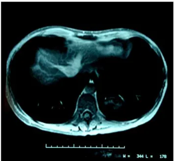
a 42-year-old female patient was submitted to a partial enterectomy in the terminal ileum (20-cm re-section) with primary entero-entero anastomosis due to an adenocarcinoma of small intestine (tnM sta-ging – t2n0M0) ten years before. no adjuvant treat-ment was performed at the time, with periodic exami-nations. in the routine exams, the abdominal control computed tomography showed two 3 cm metastases in the left hepatic lobe (segments ii-iii). the patient was in excellent conditions in general, with no sign of clinical or radiological extrahepatic dissemination. staging was complemented with magnetic resonan-ce of the abdomen, which showed these lesions only (Figure 1). a Pet-sCan (positron emission
tomo-graphy) was also performed, which conirmed an ex -clusively hepatic disease. the serum dosage of carci-noembryonic antigen was 41.2 ng/mL. as the patient
had two small metastases conined to the left lobe and
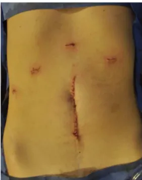
she presented a long disease-free period (ten years be-tween the enterectomy and the presentation of hepatic lesions), the surgical resection was proposed. the la-paroscopic procedure was suggested, which has been the medical team’s choice to treat lesions in the left hepatic lobe. Five trocars were used, and their sites are illustrated in Figure 2. in the abdominal cavity evaluation, no sign of distant dissemination was ob-served. Laparoscopic left lateral segmentectomy was then successfully performed, without interoccurren-ces, using the intrahepatic glissonian access techni-que, as described by Machado et al.29. no clamping of the liver hilum (Pringle maneuver) was performed. a hemostatic 10 mm LigaSure jaw (Valleylab, USA) for parenchyma sectioning and a white-loaded endoscopic stapler, of vascular type (Endogia 45 mm, Ethicon), for the left hilar elements and left hepatic vein.
the specimen was removed into an endo-bag, closed and without contamination, through a small median incision at the previous surgery site (Figures 2 and 3). the patient was fed on the same day (12 hours after the surgery). she did not receive blood, only dipyrone, as an analgesic substance. she presented good progress, without interoccurrences and was discharged from hospital on the third day af-ter the surgery. the histopathological exam showed only two lesions with metastatic adenocarcinoma and neoplasm-free margins. no adjuvant treatment was indicated, only periodic exams. six months after the surgery, the patient showed no symptoms and no disease recurrence.
DIsCUssION
the resection of nCR metastases has been con-sidered the standard curative treatment in selected
ca-ses. Several series have shown the beneit of resection
when compared to alternative palliative treatments, such as systemic chemotherapy. only resection can offer the real chance of healing or extended survival. However, the proper selection seems to be the main rationale of resection. then, factors such as: patient’s good conditions in general; proper nutritional and he-patic reserves; exclusively hehe-patic lesion; long disea-se-free survival between the primary tumor treatment and the presentation of metastases; and the nCR me-tastases etiology itself, seem to be the most important prognostic criteria for long-time survival observed in the literature7-13.
Laurent et al.12, when evaluating a series of 39 patients submitted to surgery due to nCR metastases with zero mortality, observed global survival of 35% in the 8-year period. only the disease-free survival (dFs) over 24 months was a positive prognostic fac-tor for long-term survival at the multivariate analysis (Mva). the primary tumor origin, such as gastrointes-tinal tract (git), genitourinary tract (gut) or others (sarcomas, breast, melanoma, etc.), was not a crite-rion of poor prognosis through the Mva. Weitz et al.9, in a study that analyzed a series of 141 patients from the Memorial sloan Kettering Cancer Center, in new York (zero mortality and 33% morbidity), observed that the main factors of good prognosis at the Mva were dFs over 24 months and primary tumor etiology
Figure 2. Sites of trocar utilization. Incision for specimen removal.
Figure 3. Left lateral sectorectomy. Note the two metastases in segments II-III (2 cm margin).
(tumor of reproductive system). However, Hemming et al.13, in toronto, Canada, evaluating a series a 37 patients whose postoperative mortality was also zero, reported 45% survival in a 5-year period; at the Mva,
no statistically signiicant difference was observed
ercolani et al.11, in an italian study that analyzed 83 cases of resection, observed 34% survi-val in a 5-year period, also with zero postoperative mortality, while morbidity was 20%. at the Mva, they found only the tumor volume as a prognostic factor for recurrence (metastasis volume larger than 125 cc), although the dFs over one year showed a
tendency towards that, but without signiicance due to the sample size. Speciically when the subgroup
of operated primary tumor was analyzed, a
statis-tically signiicant different was observed between
metastases from git versus gut tumors, sarcomas or even breast. For git tumors, survival was lower than in the others, with 17.3% in 3 years, and 8.4% in 5 years, respectively.
in our community, the author of this study, in a previously published case that analyzed ten patients submitted to resection of metachronic metastases of nCR (dFs over one year), observed 50% survival in a 3-year period, with zero mortality in the hepatec-tomies performed8. in another previous study of the same author, in which ten patients with nCR metas-tasis were compared to a similar contemporaneous cohort of 20 patients with nCR metastasis, all of them submitted to hepatic resection (all cases opera-ted by the same medical team), no statistically
signi-icant difference was observed for survival between
the two groups. the criteria of poor prognosis at the Mva in both groups (CR and nCR metastases) were: lymph node involvement (in hepatic hilum or prima-ry tumor) and more than one metastasis7.
Finally, in the study of greater number of ca-ses found in the literature (although multicenter and retrospective), adam et al.10, when evaluating 1,452 patients submitted to resection due to nCR metas-tases, observed 36% overall survival in a 5-year period (23% in 10-year survival), associated with 2.3% overall mortality and 21.5% morbidity. they found, at the Mva, the following factors of poor prognosis: age over 60 years, primary tumor that is not breast or melanoma cancer, squamous cells in the histological study (epidermoid carcinoma), dFs lower than one year, extra-hepatic disease, resec-tion with positive margin and larger hepatectomy. these authors also described a score, with a mathe-matical model, that can predict survival based on these prognostic factors. in addition, they sorted the
patients according to the Mva and evaluated their long-term survival. then, the patients were
classi-ied according to this score as: low risk (score 0–3,
46% survival in a 5-year period), medium risk (sco-re 4–6, 33% survival in a 5-year period) and high risk (score over 6, survival in a 5-year period lower than 10%).
When applying this mathematical model to this
case, the patient would be classiied as low risk, com -bined with her excellent nutritional condition, absence of comorbidities and good hepatic reserve, facts that
inluenced the selection of resection.
With the development in laparoscopic surgery in general, laparoscopic hepatectomy (LH) has been more freely indicated for the treatment of both benign and malign hepatic neoplasms. advantages, such as reduced intraoperative bleeding, lower morbidity, shorter hospital stay, early recovery and good cos-metic outcome, have been constantly observed14-29. For left-side resections, recently published control-led studies have suggested laparoscopy as the fa-vorite method especially of experienced surgeons in laparoscopic hepatic surgery15,16. this fact also
inluenced the decision on the access method used
in this study, as 20 LHs have been performed by the medical team (for both benign and malign tumors) in the last four years30.
on the other hand, regarding the treatment of malign neoplasm, recent studies have suggested that there is no difference between LH and open hepatec-tomy to treat metastases in portals, margins, local-systemic recurrence or long-term survival20,22,23,26,27. in our community, Machado et al.24, in a small series of four patients with CR metastasis submitted to LH,
have already demonstrated the beneit of LH, which
was performed with effectiveness and safety. in a re-view in the national literature, however, no case of LH used in the treatment of nCR metastasis has been found to date.
REsUmO: A hepatectomia tem sido o tratamento padrão para metástase de origem não colorretal (NCR) metacrônica, principal-mente quando o intervalo livre de doença é maior do que dois anos. A hepatectomia por laparoscopia tem se tornado padrão prin-cipalmente para as ressecções à esquerda, haja vista a menor morbidade, menor tempo de internação, reabilitação precoce e melhor resultado estético. Os autores relatam um caso de paciente com duas metástases metacrônicas (10 anos de sobrevida livre de doença), de etiologia não colorretal (adenocarcinoma de intestino delgado), tratada com segmentectomia lateral esquerda (lobectomia hepática esquerda) laparoscópica. Paciente apresentou boa evolução pós-operatória sem recidiva (seis meses de seguimento). segmentectomia lateral esquerda laparoscópica pode ser satisfatoriamente realizada em casos selecionados de metástases hepáticas, acarretando menor morbidade e melhor resultado estético. A falta de tratamentos alternativos e o prognóstico reservado nos casos de metástases NCR não operadas justiicam a ressecção com o objetivo de prolongar a sobrevida. No entanto, essa abordagem deve ser realizada por equipe es -pecializada em cirurgia hepática com treinamento em procedimentos laparoscópicos avançados.
Palavras-chave: laparoscopia; neoplasias colorretais; hepatectomia; metástase neoplásica; neoplasias hepáticas/cirurgia; neoplasias hepáticas/secundário; taxa de sobrevida.
REFERENCEs
1. simmonds PC, Primrose jn, Colquitt jL, garden oj, Poston gj, Rees M. surgical resection of metastases from colorectal cancer: a systematic review of published studies. Br j Cancer 2006;94:982-99.
2. Fong Y, Fortner j, sun RL, Brennan MF, Blumgart LH. Clinical score for predicting recurrence after hepatic resection for metastatic colorectal cancer: analysis of 1001 consecutive cases. ann surg 1999;230(3):309-21.
3. adam R. tratamento das metástases hepáticas do câncer colorretal. in: Correia MM, Mello elR, santos CeR (eds). Cirurgia do câncer hepatobiliar. Rio de janeiro: Revinter; 2003. p. 139-46.
4. Choi ea, Rodgers se, ahmad sa, abdalla eK. in: Feig BW, Berger dH, Fuhrman gM (eds.) the M.d. anderson surgical oncology Handbook. 4th ed. Philadelphia: Lippincott & Wilkins; 2006. p. 238-47.
5. Minagawa M, Makuuchi M, torzilli g, takayama t, Kawasaki s, Kosuge t, et al. extension of the frontiers of surgical indications in the treatment of liver metastases from colorectal cancer. ann surg 2000;231(4):487-99.
6. scheele j, altendorf-Hofmann a. surgical treatment of liver metastases. in: Blumgart LH, Fong Y (eds.) surgery of the liver and biliary tract. 3th ed. edinburgh: WB saunders; 2000. p. 1475-502.
7. Costa sRP, Horta sHC, Henriques aC, Waisberg j, speranzini MB. Hepatectomia para o tratamento de metástases colorretais e não-colorretais: análise comparativa em 30 casos operados. j Coloproctol 2009;29(2):216-25.
8. Costa sRP, Horta sHC, Miotto Mj, Costas MC, Henriques aC, speranzini M. Ressecção hepática para o tratamento de metástases não-colorretais e não-neuroendócrinas: indicações e resultados em 10 pacientes operados. einstein 2008;6(1):56-62. 9. Weitz j, .Blumgart LH, Fong Y, jarnargin WR, dangelica M,
Harrison Le, et al. Partial hepatectomy for metastases from noncolorectal, nonneuroendocrine carcinoma. ann surg 2005;241(2):269-76.
10. adam R, Chiche L, aloia t, elias d, salmon R, Rivoire M, et al. Hepatic resection for noncolorectal nonendocrine liver metastasis: analysis of 1452 patients and development of prognostic model. ann surg 2006;244(4):534-35.
11. ercolani g, grazi gL, Ravaioli M, Ramacciato g, Cescon M, varotti g, et al. the role of liver resection for noncolorectal, nonneuroendocrine metastases: experience with 142 observed cases. ann surg oncol 2005;12(6):1-8.
12. Laurent C, Rullier e, Feyle R, Masson B, saric j. Resection of noncolorectal and nonnneuroendocrine liver metastases: late metastases are the only chance of cure. World j surg 2001;25:1532-6.
13. Hemming aW, sielaff td, gallinger s, Cattral Ms, taylor BR, greig Pd, et al. Hepatic resection of noncolorectal nonneuroendocrine metastases. Liver transpl 2000;6(1):97-101. 14. ardito F, tayar C, Laurent a, Karoui M, Loriau j, Cherqui d.
Laparoscopic liver resection for benign disease. arch surg 2007;142(12):1188-93.
15. Campos RR, Hernández CM, Conesa aL, abellán B, Pérez PP, Paricio PP. La ressección laparoscópica de los segmentos del lóbulo izquierdo debe ser el abordaje inicial em centros com experiência. Cir esp 2009;85(4):214-21.
CONCLUsION
Laparoscopic left lateral segmentectomy (left he-patic lobectomy) can be satisfactorily performed in se-lected cases of hepatic metastasis, with lower morbi-dity and good cosmetic result. the lack of alternative
treatments and the poor prognosis of untreated cases
of NCR metastasis have justiied surgical resection in
16. Carswell Ka, sagias Fg, Murgatroyd B, Rela M, Heaton n, Patel ag. Laparoscopic versus open left lateral segmentectomy. BMC surgery 2009;9(14):1-9.
17. Cho jY, Han Hs, Yoon Ys, shin sH. outcomes of laparoscopic liver resection for lesions located in the right side of the liver. arch surg 2009;144(1):25-9.
18. Cugat e, Marco C. Cirugía laparoscópica del hígado. una opción madura? Cir esp 2009;85(4):193-5.
19. dulucq jL, Wintringer P, stabilini C, Berticelli j, Mahajna a. Laparoscopic liver resections: a single center experience. surg endosc 2005;19:886-91.
20. gagner M, Rogula t, selzer d. Laparoscopic liver resection:
beneits and controversies. Surg Clin North Am 2004;84:451-62.
21. gigot jF, gilneur d, azagra js, goergen M, Ceuterik M, Morino M, et al. auspices of the hepatobiliary and pancreatic section of the Royal Belgian society of surgery and the Belgian group for endoscopic surgery. Laparoscopic liver resection for malignant liver tumours: preliminary results of a multicenter european study. ann surg 2002;236(1):90-7. 22. Kofron aj, auffenberg Bs, Kung R, abecassis M. evaluation
of 300 minimally invasive liver resections at a single institution. ann surg 2007;246(3):385-94.
23. Lee KF, Cheung Ys, Chong Cn, tsang YYY, ng WWC, Ling e, et al. Laparoscopic versus open hepatectomy for liver tumours: a case control study. Hong Kong Med j 2007;13(6):442-8.
24. Machado MaC, Makdissi FF, almeida FaR, Luiz-neto M, Martins aCa, Machado MCC. Hepatectomia laparoscópica
no tratamento das metástases hepáticas. arq gastroenterol 2008;45(4):330-2.
25. zhang L, Chen Yj, shang Cz, zhang HW, Huang zj. total laparoscopic liver resection in 78 patients. World j gastroenterol 2009;15(45):5727-31.
26. Pilgrim CHC, to H, usatoff v, evans PM. Laparoscopic hepatectomy is a safe procedure for cancer patients. HPB 2009;11:247-51.
27. nguyen Kv, geller da. is laparoscopic liver resection safe and comparable to open liver resection for hepatocellular carcinoma? ann surg oncol 2009;16:1765-7.
28. Machado MaC, Makdissi FF, galvão FH, Machado MCC. intrahepatic glissonian approach for laparoscopic right segmental liver resections. am j surg 2008;196:e38-e42. 29. Machado MaC, Makdissi FF, surjan RC, Herman P, teixeira
aR, Machado MCC. Laparoscopic resection of left liver segments using intrahepatic glissonian approach. surg endosc 2009;23:2615-9.
30. Pais-Costa sR, araujo sLM, Lima aot, Chartuni a. Laparoscopic hepatectomy from hepatic tumors: a series of twenty resectable cases. ann oncol 2011;22(suppl 5): 67.
Correspondence to:
sergio Renato Pais-Costa sgas 710/910, sala 330