Disease: A Replication Study in the Spanish Population
Marta Conde-Jaldo´n1, Marco Antonio Montes-Cano1*, Jose´ Raul Garcı´a-Lozano1, Lourdes Ortiz-Ferna´ndez1, Norberto Ortego-Centeno2, Rocı´o Gonza´lez-Leo´n3, Gerard Espinosa4, Genaro Gran˜a-Gil5, Juan Sa´nchez-Burso´n6, Miguel Angel Gonza´lez-Gay7, Ana Celia Barnosi-Marı´n8, Roser Solans9,
Patricia Fanlo10, Mo´nica Rodrı´guez Carballeira11, Teresa Camps12, Santos Castan˜eda13, Javier Martı´n14, Marı´a Francisca Gonza´lez-Escribano1
1Servicio de Inmunologı´a, IBiS, H.U. Virgen del Rocı´o/CSIC/Universidad de Sevilla, Sevilla, Spain,2Servicio de Medicina Interna, H. Clı´nico San Cecilio, Granada, Spain,
3Servicio de Medicina Interna, H. U. Virgen del Rocı´o, Sevilla, Spain,4Servicio de Enfermedades Autoinmunes, H. Clinic, Barcelona, Spain,5Servicio de Reumatologı´a, CHU A Corun˜a, Spain,6Servicio de Reumatologı´a, H. de Valme, Sevilla, Spain,7Servicio de Reumatologı´a, H. Marques de Valdecilla, Santander, Spain,8Servicio de Medicina Interna, H. Torreca´rdenas, Almerı´a, Spain,9Servicio de Medicina Interna, H. Vall d’Hebron, Barcelona, Spain,10Servicio de Medicina Interna, H. Virgen del Camino, Pamplona, Spain,11Medicina Interna Unitat de Malalties Autoimmunes i Siste`miques Hospital Universitari Mu´tua Terrassa, Barcelona, Spain,12Servicio de Medicina Interna, H. Carlos Haya, Ma´laga, Spain,13Servicio de Reumatologı´a, H. de la Princesa, Madrid, Spain,14Instituto de Parasitologı´a y Biomedicina Lo´pez Neyra, CSIC, Granada, Spain
Abstract
Behc¸et’s disease (BD) is a multifactorial disorder associated with the HLA region. Recently, the ERAP1 gene has been proposed as a susceptibility locus with a recessive model and with epistatic interaction with HLA-B51. ERAP1 trims peptides in the endoplasmic reticulum to optimize their length for MHC-I binding. Polymorphisms in this gene have been related with the susceptibility to other immune-mediated diseases associated to HLA class I. Our aim was, the replication in the Spanish population of the association described in the Turkish population between ERAP1 (rs17482078) and BD. Additionally, in order to improve the understanding of this association we analyzed four additional SNPs (rs27044, rs10050860, rs30187 and rs2287987) associated with other diseases related to HLA class I and the haplotype blocks in this gene region. According to our results, frequencies of the homozygous genotypes for the minor alleles of all the SNPs were increased among patients and the OR values were higher in the subgroup of patients with the HLA-B risk factors, although differences were not statistically significant. Moreover, the presence of the same mutation in both chromosomes increased the OR values from 4.51 to 10.72 in individuals carrying the HLA-B risk factors. Therefore, although they were not statistically significant, our data were consistent with an association betweenERAP1 and BD as well as with an epistatic interaction betweenERAP1and HLA-B in the Spanish population.
Citation:Conde-Jaldo´n M, Montes-Cano MA, Garcı´a-Lozano JR, Ortiz-Ferna´ndez L, Ortego-Centeno N, et al. (2014) Epistatic Interaction ofERAP1and HLA-B in Behc¸et Disease: A Replication Study in the Spanish Population. PLoS ONE 9(7): e102100. doi:10.1371/journal.pone.0102100
Editor:Anna Carla Goldberg, Albert Einstein Institute for Research and Education, Brazil
ReceivedJanuary 29, 2014;AcceptedJune 14, 2014;PublishedJuly 14, 2014
Copyright:ß2014 Conde-Jaldo´n et al. This is an open-access article distributed under the terms of the Creative Commons Attribution License, which permits unrestricted use, distribution, and reproduction in any medium, provided the original author and source are credited.
Funding:This work was supported by Fondo de Investigaciones Sanitarias (10/1701), Fondos FEDER, Plan Andaluz de Investigacio´n (CTS-0197 and CTS-180), Red Enfermedades Inflamatorias y Reuma´ticas (RD08/0075/0013) and Consejerı´a de Salud de la Junta de Andalucı´a (0260/08). LOF is the recipient of a fellowship (FI11/ 00547). The funders had no role in study design, data collection and analysis, decision to publish, or preparation of the manuscript.
Competing Interests:The authors have declared that no competing interests exist. * Email: [email protected]
Introduction
Behc¸et disease (BD) is a multifactorial disorder with evidence of genetic contribution based on familial aggregation, predominance in patients with Mediterranean or Asian ancestry and association with HLA-B51 in several ethnic groups [1–4]. Contribution of the HLA region has been estimated to represent approximately 20% of the genetic component of this disease [5]. In addition to the HLA class I region, in which different peaks of association have been detected, a relationship of the disease with IL23R and IL10 has been described using a large scale SNP genotyping strategy (genome wide association studies, GWAS) in different Caucasian
and Asian populations [6–11].Very recently, theERAP1gene has
been proposed as a new susceptibility locus for BD in a Turkish patient cohort [12].
The ERAP1 gene is located on chromosome 5q15 and it
encodes an amino-peptidase with an ubiquitous tissue distribution. This enzyme is responsible for peptides N-terminal trimming in the endoplasmic reticulum which is a critical step of the processing of the peptides to optimize their length for MHC-I binding [13,14]. Moreover, ERAP1 has been involved in the cleaving of several proinflammatory cytokine receptors from the cell mem-brane including shedding of tumor necrosis factor receptor
(TNFR1), IL-1 receptor-II (IL-RII) and IL-6 receptora (IL-6a)
[15–17] although this function remains to be confirmed.
Therefore, from a functional perspective, ERAP1 represents an
shared pathogenesis pathways [9–11,18,22,23]. In their study, Kirino et al [12], described a recessive model of association with BD in a Turkish population and they found an epistatic interaction
between HLA-B51 andERAP1which could not be replicated in a
Japanese cohort [12]. The objective of the present study was to
asses the association of ERAP1 with BD in the Spanish
population.
Methods
A total of 362 BD patients (148 males and 214 females) with a
mean age at onset 6 SD of 35.1611.2 fulfilled the 1990
International Study Group classification criteria for Behc¸et’s disease [24] and 460 bone marrow and blood donors as normal healthy controls (50% males) were included in the study. All the subjects were Spanish European recruited from different Spanish hospitals. The study was approved by all local ethical committees of the corresponding hospitals A Coruna (CHU A Corun˜a), Almerı´a (H. Torreca´rdenas), Barcelona (H. Clinic, Vall d’Hebron and Mu´tua Terrassa), Granada (H. Clı´nico San Cecilio), Madrid (H. de la Princesa), Ma´laga (H. Carlos Haya), Pamplona (H. Virgen del Camino), Santander; (H Marques de Valdecilla) and Sevilla (Virgen del Rocı´o and Valme). All the study participants gave written informed consent according to the declaration of Helsinki. Clinical features of the patient group were: 100% had oral ulcers, 75% genital ulcers, 50% uveitis, 50% arthritis, and 25% vascular, 18% neurological and 19% gastrointestinal involvement. The distribution of the frequencies of the different markers in the cohorts from different hospitals was not significantly different.
Peripheral blood was obtained from the healthy controls whereas peripheral blood or saliva was the starting material obtained from patients. Genomic DNA was extracted using QIAmp DNA Mini Kit (Qiagen, Barcelona, Spain) according to
the manufacturer’s recommendations and stored at220uC. The
purity of DNA was determined using a Nanodrop spectropho-tometer (Thermo Fisher Scientific, Wilmington, DE 19810 U.S.A.). Only DNA samples having a 260/280 ratio between
1.7 and 2.0 and a final concentration of 10–20 ng/ml were
considered appropriate. Ten DNA samples from saliva were eliminated because they did not meet these quality criteria.
Five non-synonymous single nucleotide polymorphisms (SNPs)
associated with AS rs27044 (Gln730Glu), rs17482078
(Arg725Gln), rs10050860 (Asp575Asn), rs30187 (Arg528Lys) and rs2287987 (Val349Met) were included in this study. Genotyping of these SNPs was performed using TaqMan SNP Genotyping Assays (Applied Byosistems, Barcelona, Spain) in a LightCycler 480 (Roche, Barcelona, Spain). Patients and controls had been previously genotyped in HLA-A and HLA-B using PCR-SSOP Luminex method with LABType SSO (One Lambda Inc., Canoga Park, CA), following the manufacturer’s instructions. According to our previous results HLA-B*51 and HLA-B*57 were considered as HLA-B risk factors in our population [25].
The a priori statistical power was calculated using the minor
allele frequencies (MAF) described in the European population for the five SNPs included in this study (http://www.broadinstitute. org/mammals/haploreg/haploreg.php), taking into consideration a recessive association model of the minor alleles and the Odd Ratio (OR) value described by Kirino et al for the rs17482078 in their cohort (cases with and without uveitis and discovery and replication data combined, OR = 3.08) [12] but using our sample size (362 patients and 460 controls).
The different association models: genotypic, allelic, dominant
and recessive were tested using thex2test and p values#0.05 were
considered statistically significant. The ORs and 95% confidence intervals (95% CI) were calculated according to Wolf’s method using StatCalc program (EPI Info 2002, Centers for Disease Control and Prevention, Atlanta, GA, USA). The linkage disequilibrium solid spine approach was used to define haplotype blocks in the CEU and Spanish populations using the Haploview program (available at the website http://www.broad.mit.edu/ mpg/haploview/download.php). The SNP haplotypes frequency estimation in the CEU and Spanish populations was performed using the Haploview sotfware. Conditional logistic regression models were constructed by categorizing all the individuals according to the presence/absence of the HLA-B risk factors
Table 1.Distribution of the five SNPs studied inERAP1according to a recessive model of the minor allele.
SNP MA TEST Patients Controls p OR
rs27044 G GG/CC+CG
Whole group 46/316 51/409 0.5 1.2
HLA-B Risk factor group 24/154 7/77 0.2 1.7
rs17482078 T TT/CC+CT
Whole group 17/344 12/446 0.1 1.8
HLA-B Risk factor group 6/172 1/83 0.3 2.8
rs10050860 T TT/CC+CT
Whole group 17/344 12/446 0.1 1.8
HLA-B Risk factor group 6/172 1/83 0.3 2.8
rs30187 T TT/CC+CT
Whole group 70/292 73/386 0.2 1.3
HLA-B Risk factor group 39/137 13/71 0.2 1.5
rs2287987 C CC/TT+CT
Whole group 17/344 12/446 0.1 1.8
HLA-B Risk factor group 6/172 1/83 0.3 2.8
(according to a dominant model), the ERAP1 haplotypes
(according to a recessive model) and theERAP1 status (yes/no,
see results) analyzed with Epi Info 2002.
Results
The genotyping success ratio was greater than 95% and the study population was found to be in the Hardy–Weinberg
equilibrium for all the polymorphisms analyzed (p.0.05). Table
S1 in File S1 displays genotype results of the SNPs included in the study and statistical data of the different association models. According to data displayed in Table S2 in File S1, the frequency of the homozygous genotype of the minor allele of the rs17482078 in the healthy Spanish population is 2.6. Taking into account the OR described by Kirino et al in their study (3.08) our a priori statistical power for this SNP was 94.5.
The observed MAFs in the Spanish population were not significantly different to those described in the European population with the exception of the rs30187 which had a significantly higher frequency of the minor allele (T) in our cohort than in the European population included in the 1000 Genome Phase I data project (0.41 vs. 0.34, p = 0.002) but similar to that described in the other Spanish cohort [26]. According to our results and in agreement with a recessive model, frequencies of the homozygous genotypes for the minor alleles of all the SNPs were increased among patients and the ORs ranging from 1.2 to 1.8 although differences were not statistically significant. The original study also reported an epistatic interaction between ERAP1 and HLA-B. Consistent with the original paper, the OR values for the recessive models of the minor alleles of the five SNPs included in the present study were higher in the subgroup of patients with the HLA-B risk factors, although the differences were not statistically significant (Table 1)
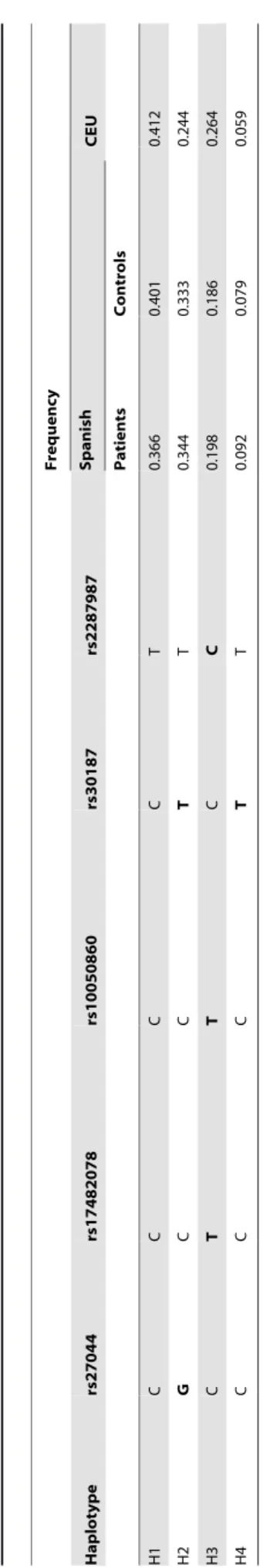
As it was previously described, linkage disequilibrium (LD) among these 5 SNPs was found in the Spanish population. Table 2 shows the four haplotypes (named from H1 to H4) with frequencies greater than 0.05 which were found in our patient and control cohorts. The rs27044G, a risk factor for AS, tags the haplotype H2, whereas the rs17482078T, which has been described to be a risk factor in BD, and also the rs100504860T and rs2287987C tag the haplotype H3. The three haplotypes containing mutations: H2, H3 and H4 were slightly more common among BD patients although the allelic model was not significant. Table 3 displays the conditional multivariate models which include the ERAP1 haplotypes in recessive models and the HLA-B risk factors (HLA-B*51 or B*57 yes/no). The OR values among individuals carrying the HLA-B risk factors were increased more than 2 fold in both, the H2H2 and H3H3 individuals, whereas this value remains the same for H1H1 subjects. Of note, individuals bearing simultaneously H4H4 and the HLA-B risk factors were all patients. In order to analyze the influence of the doses, individuals were grouped in four different categories. Homozygous, subjects with the same set of mutations in all their molecules (‘‘Ho’’, H2H2, H3H3, H4H4); double heterozygous, individuals in which all the molecules are mutated but each half has a different set of mutations (‘‘dHe’’, H2H3, H2H4 and H3H4); heterozygous, subject having half of molecules with and the other half without mutations (‘‘He’’, H1H2, H1H3 and H1H4); and finally, normal individuals, those having no molecules with these mutations (‘‘N’’, H1H1). Next, conditional multivariate models taking into account both, the condition HLA-B risk associated with BD (HLA-B*51 or
B*57 yes/no) and theERAP1mutation status (Ho, He, dHe and
N) were constructed. Table 4 displays the results of this analysis, the OR values among individuals carrying the HLA-B risk factors
Table
2.
ERAP1
haplotypes
obtained
by
the
combination
of
the
five
SNPs
included
in
this
study.
Frequency
Haplotype
rs27044
rs17482078
rs10050860
rs30187
rs2287987
Spanish
CEU
Patients
Controls
H1
C
C
C
C
T
0.366
0.401
0.412
H2
G
CC
T
T
0.344
0.333
0.244
H3
C
TT
C
C
0.198
0.186
0.264
H4
C
C
C
T
T
0.092
0.079
0.059
Only
those
haplotypes
having
frequencies
g
reater
than
0
.05
are
displayed.
Minor
alleles
are
in
bold.
doi:10.1371/journal.pone.
Table 3.Conditional logistic regression models of HLA-B and ofERAP1haplotypes.
HLA-B Risk H1H1 PatientsN = 343 Controls N = 450 P OR (95%CI)
+ + 28 13 ,1025 4.35 (2.19–8.64)
+ - 150 70 ,1025 4.33 (3.07–6.11)
- + 14 62 0.01 0.46 (0.25–0.84)
- - 151 305 1.0
HLA-B Risk H2H2
+ + 23 6 ,1025 8.54 (3.40–21.42)
+ - 155 77 ,1025 4.48 (3.20–6.28)
- + 20 44 .0.05
- - 145 323 1.0
HLA-B Risk H3H3
+ + 6 1 0.015 13.82 (1.65–115.72)
+ - 172 82 ,1025 4.83 (3.49–6.68)
- + 10 10 .0.05
- - 155 357 1.0
HLA-B Risk H4H4
+ + 3 0 .0.05
+ - 175 83 ,1025 4.73 (3.44–6.52)
- + 2 1 .0.05
- - 163 366 1.0
doi:10.1371/journal.pone.0102100.t003
Table 4.Conditional logistic regression models of HLA-B and three different conditions ofERAP1.
Factor B Ho Patients N = 343 Controls N = 450 P OR (95% CI)
+ + 32 7 ,1025 10.72 (4.62–24.91)
+ - 146 76 ,1025 4.51 (3.20–6.35)
- + 32 55 .0.05
- - 133 312 1.0
Factor B He
+ + 79 40 ,1025 5.03 (3.16–7.98)
+ - 99 43 ,1025 5.86 (3.75–5.14)
- + 88 171 .0.05
- - 77 196 1.0
Factor B dHe
+ + 39 23 ,1025 3.64 (2.09–6.34)
+ - 139 60 ,1025 4.98 (3.45–7.18)
- + 31 79 .0.05
- - 134 288 1.0
Factor B N
+ + 28 13 ,1025 4.35 (2.19–8.64)
+ - 150 70 ,1025 4.33 (3.07–6.11)
- + 14 62 0.01 0.46 (0.25–0.84)
- - 151 305 1.0
‘‘Ho’’ are individuals H2H2,H3H3 and H4H4; ‘‘He’’ are individuals H1H2,H1H3 and H1H4; ‘‘dHe’’ are individuals H2H3,H2H4 and H3H4; ‘‘N’’ are individuals H1H1. CI confidence interval.
were increased from 4.51 to 10.72 in the ‘‘Ho’’ positive individuals. Nevertheless, this effect was not observed in ‘‘He’’ nor in ‘‘dHe’’ subjects which have similar results as those obtained in the group ‘‘N’’.
Discussion
Our results are consistent with the previously described
association of ERAP1 with BD in the Turkish population [12],
because the effects detected in our population have the same direction as those described in the original paper, nevertheless, associations and interactions were not statistically significant in the Spanish population.
We decided to attempt the replication in our cohort of BD
patients because, additionally to the findings in GWAS,ERAP1is
an interesting candidate in immune-mediated diseases associated to HLA class I. In fact, the relationship of this gene with AS, which like BD is associated with HLA class I molecules, seems well established. Most of the studies investigating the influence of the
ERAP1 gene in susceptibility to AS include the rs27044 and
rs30187 and the authors report the minor alleles of these SNPs as risk factors for this pathology in Caucasian [18,26–30] as well as in Asian populations [31,32] and in meta-analysis studies [33,34]. Regarding the other 3 SNPs included in our study, rs17482078, rs100504860 and rs2287987, their minor alleles have been described as protective to AS in some studies but they have been non-associated in others [18,26–28,31]. Notably, these last 3 SNPs have MAFs lower than rs27044 and rs30187 in different populations and the difference is particularly marked in Asian populations (about 0.07 rs17482078, rs100504860 and rs2287987 vs. 0.50 for rs27044 and rs30187). The low frequency of these SNPs determines a low statistical power, especially in some Asian populations where BD is relatively common, which makes difficult the replication of this finding. In fact, the association between the rs17482078 and susceptibility to this disease which was described in the Turkish population in the original report was not replicated in the same study in the Japanese population. Additionally, the strength of the association could be different among cohorts. Thus, the OR values found in our cohort were lower than those detected in the original study, causing a reduction in statistical power,
which a priori was higher than 99% for all the SNPs included.
Nevertheless, our results were not discrepant with the original report because according to the recessive model found by Kirino et al [12], the frequencies of the homozygous for the minor alleles were higher among patients for all the SNPs and according to an epistatic interaction with the HLA, the OR values were higher among patients with the HLA-B risk factors, although the differences were not significant.
The rs27044 and the rs30187 are in LD with each other
(r2= 0.75, D9= 0.97) and the rs17482078, the rs100504860 and
the rs2287987 are also in LD with each other (r2and D9 values
greater than 0.90 in the European population). In spite of this strong LD most of the studies in AS include these 5
non-synonimous ERAP1 polymorphisms. Four major haplotypes
(named from H1 to H4 in Table 2) are obtained by the
combination of the variations of these five SNPs according to data from CEU and also from our population. The H2 and H3 correspond respectively with risk and protection haplotypes in AS [34,35], however, our data suggest that both haplotypes could be
risk factors in BD. Association ofERAP1with AS is restricted to
individuals bearing the B risk factor for this disease,
HLA-B27, [35,36] whereas in psoriasis the relationship betweenERAP1
and HLA-C remains unclear [19,37]. Althought our results require further confirmation, data displayed in Tables 3 and 4 support a model in which the presence of the same mutation in both chromosomes increased the susceptibility risk in individuals
bearing the HLA-B risk factors. ERAP1 is located in the ER
where it is one of the enzymes trimming peptides to reach the optimal size for class I MHC binding. A reduction of the trimming activity for the protective alleles for AS has been found in studies investigating individual changes associated with AS [38–40]. Very
recently, the influence of the natural haplotypes of ERAP1 in
trimming activity has been explored [41]. These authors suggested
that there is a range of trimming activity among theERAP1alleles
with some of them encoding molecules that overtrim and others that undertrim. Both conditions, over and under activity could be associated with diseases. The ‘‘Ho’’ status is a scenario in which all the ERAP1 molecules of one individual have the same mutations, producing a lower number of peptides with the appropriate size. Nevertheless, the ‘‘dHe’’ individuals have two different ERAP1 molecules. If one of them over trims whereas the other under trims, the compensation of the enzimatic activity could generate a larger number of units with the appropriate size.
In conclusion, data in the Spanish population were consistent with association between ERAP1 and BD as well as with an epistatic interaction between ERAP1 and HLA-B described in the Turkish population, although our results were not statistically significant.
Supporting Information
File S1 Supplementary Tables S1–S2. Table S1:Different models of association with Behc¸et disease for the single nucleotide
polymorphisms studied inERAP1.Table S2:Data of the SNPs
studied inERAP1according to a recessive model for the minor
alleles. (DOC)
Acknowledgments
The authors would like to thank the Asociacio´n Andaluza de Enferme-dades Autoinmunes (AADEA) and all patients and donors enrolled in the present study for their cooperation.
Author Contributions
Conceived and designed the experiments: MAMC JRGL MFGE. Performed the experiments: MCJ LOF. Analyzed the data: MAMC MFGE. Contributed reagents/materials/analysis tools: NOC RGL GE GGG MAGG ACBM RS PF MRC TC SC JSB JM. Wrote the paper: MAMC MFGE.
References
1. Mendes D, Correia M, Barbedo M, Vaio T, Mota M, et al. (2009) Behc¸et’s disease: a contemporary review. J Autoimmun 32:178–188.
2. Kone´-Paut I, Geisler I, Wechsler B, Ozen S, Ozdogan H, et al. (1999) Familial aggregation in Behc¸et’s disease: high frequency in siblings and parents of pediatric probands. J Pediatr 135:89–93.
3. Sakane T, Takeno M, Suzuki N, Inaba G (1999) Behc¸et’s disease. N Engl J Med 341:1284–1291.
4. Menthon de M, Lavalley MP, Maldini C, Guillevin L, Mahr A (2009) HLA-B51/B5 and the risk of Behc¸et’s disease: a systematic review and meta-analysis of case-control genetic association studies. Arthritis Rheum 61:1287–1296. 5. Yazici H, Fresko I, Yurdakul S (2007) Behc¸et’s syndrome: disease manifestations,
7. Meguro A, Inoko H, Ota M, Katsuyama Y, Oka A, et al. (2010) Genetics of Behc¸et disease inside and outside the MHC. Ann Rheum Dis 69:747–754. 8. Karasneh J, Gu¨l A, Ollier WE, Silman AJ, Worthington J (2005) Whole-genome
screening for susceptibility genes in multicase families with Behc¸et’s disease. Arthritis Rheum 52:1836–1842.
9. Fei Y, Webb R, Cobb BL, Direskeneli H, Saruhan-Direskeneli, et al. (2009) Identification of novel genetic susceptibility loci for Behc¸et’s disease using a genome-wide association study. Arthritis Res Ther 11:R66.
10. Remmers EF, Cosan F, Kirino Y, Ombrello MJ, Abaci N, et al. (2010) Genome-wide association study identifies variants in the MHC class I, IL10, and IL23R-IL12RB2 regions associated with Behc¸et’s disease. Nat Genet 42:698–702. 11. Mizuki N, Meguro A, Ota M, Ohno S, Shiota T, et al. (2010) Genome-wide
association studies identify IL23R-IL12RB2 and IL10 as Behc¸et’s disease susceptibility loci. Nat Genet 42:703–706.
12. Kirino Y, Bertsias G, Ishigatsubo Y, Mizuki N, Tugal-Tutkun I, et al. (2013) Genome-wide association analysis identifies new susceptibility loci for Behc¸et’s disease and epistasis between HLA-B*51 and ERAP1. Nat Genet 45:202–207. 13. Saric T, Chang SC, Hattori A, York IA, Markant S, et al. (2002) An IFN-gamma-induced aminopeptidase in the ER, ERAP1, trims precursors to MHC class I-presented peptides. Nat Immunol 3:1169–1176.
14. Serwold T, Gonzalez F, Kim J, Jacob R, Shastri N (2002) ERAAP customizes peptides for MHC class I molecules in the endoplasmic reticulum. Nature 419:480–483.
15. Cui X, Hawari F, Alsaaty S, Lawrence M, Combs CA, et al. (2002) Identification of ARTS-1 as a novel TNFR1-binding protein that promotes TNFR1 ectodomain shedding. J Clin Invest 110:515–526.
16. Cui X, Rouhani FN, Hawari F, Levine SJ (2003) Shedding of the type II IL-1 decoy receptor requires a multifunctional aminopeptidase, aminopeptidase regulator of TNF receptor type 1 shedding. J Immunol 171:6814–6819. 17. Cui X, Rouhani FN, Hawari F, Levine SJ (2003) An aminopeptidase, ARTS-1,
is required for interleukin-6 receptor shedding. Biol Chem 278:28677–28685. 18. Wellcome Trust Case Control Consortium; Australo-Anglo-American
Spondy-litis Consortium (TASC) (2007) Association scan of 14,500 nonsynonymous SNPs in four diseases identifies autoimmunity variants. Nat Genet 39:1329– 1337.
19. Genetic Analysis of Psoriasis Consortium & the Wellcome Trust Case Control Consortium 2 (2010) A genome-wide association study identifies new psoriasis susceptibility loci and an interaction between HLA-C and ERAP1. Nat Genet 42:985–990
20. Sun LD, Cheng H, Wang ZX, Zhang AP, Wang PG, et al. (2010) Association analyses identify six new psoriasis susceptibility loci in the Chinese population. Nat Genet 42:1005–1009.
21. Cargill M, Schrodi SJ, Chang M, Garcia VE, Brandon R, et al. (2007) A large-scale genetic association study confirms IL12B and leads to the identification of IL23R as psoriasis-risk genes. Am J Hum Genet 80:273–290.
22. Australo-Anglo-American Spondyloarthritis Consortium (TASC) (2010) Ge-nome-wide association study of ankylosing spondylitis identifies non-MHC susceptibility loci. Nat Genet 42:123–127.
23. Pimentel-Santos FM, Ligeiro D, Matos M, Moura˜o AF, Sousa E, et al. (2009) Association of IL23R and ERAP1 genes with ankylosing spondylitis in a Portuguese population. Clin Exp Rheumatol 27:800–806.
24. International Study group for Behcet’s disease (1990) Criteria for diagnosis of Behcet’s disease. Lancet 335:1078–1080.
25. Montes-Cano M, Conde-Jaldo´n M, Garcı´a-Lozano J, Ortiz-Ferna´ndez L, Ortego-Centeno N, et al. (2013) HLA and non-HLA genes in Behc¸et’s disease: A multicentric study in the Spanish population. Arthritis Res Ther 15:R145
26. Szczypiorska M, Sa´nchez A, Bartolome´ N, Arteta D, Sanz J, et al. (2011) ERAP1 polymorphisms and haplotypes are associated with ankylosing spondylitis susceptibility and functional severity in a Spanish population. Rheumatology (Oxford) 50:1969–1975.
27. Tsui FW, Haroon N, Reveille JD, Rahman P, Chiu B, et al. (2010) Association of an ERAP1 ERAP2 haplotype with familial ankylosing spondylitis. Ann Rheum Dis 69:733–736.
28. Harvey D, Pointon JJ, Evans DM, Karaderi T, Farrar C, et al. (2009) Investigating the genetic association between ERAP1 and ankylosing spondylitis. Hum Mol Genet 18:4204–4212.
29. Maksymowych WP, Inman RD, Gladman DD, Reeve JP, Pope A, et al. (2009) Association of a specific ERAP1/ARTS1 haplotype with disease susceptibility in ankylosing spondylitis. Arthritis Rheum 60:1317–1323.
30. Paza´r B, Sa´fra´ny E, Gergely P, Sza´nto´ S, Szekanecz Z, et al. (2010) Association of ARTS1 gene polymorphisms with ankylosing spondylitis in the Hungarian population: the rs27044 variant is associated with HLA-B*2705 subtype in Hungarian patients with ankylosing spondylitis. J Rheumatol 37:379–384. 31. Choi CB, Kim TH, Jun JB, Lee HS, Shim SC, et al. (2010) ARTS1
polymorphisms are associated with ankylosing spondylitis in Koreans. Ann Rheum Dis 69:582–584.
32. Davidson SI, Wu X, Liu Y, Wei M, Danoy PA, et al. (2009) Association of ERAP1, but not IL23R, with ankylosing spondylitis in a Han Chinese population. Arthritis Rheum 60:3263–3268.
33. Lee YH, Choi SJ, Ji JD, Song GG (2011) Associations between ERAP1 polymorphisms and ankylosing spondylitis susceptibility: a meta-analysis. Inflamm Res 60:999–1003.
34. Chen R, Yao L, Meng T, Xu W (2012) The association between seven ERAP1 polymorphisms and ankylosing spondylitis susceptibility: a meta-analysis involving 8,530 cases and 12,449 controls. Rheumatol Int 32:909–914. 35. Bettencourt BF, Rocha FL, Alves H, Amorim R, Caetano-Lopes J, et al. (2013)
Protective effect of an ERAP1 haplotype in ankylosing spondylitis: investigating non-MHC genes in HLA-B27-positive individuals. Rheumatology (Oxford) 52: 2168–2176.
36. Kadi A, Izac B, Said-Nahal R, Leboime A, Van Praet L, et al. (2013) Investigating the genetic association between ERAP1 and spondyloarthritis. Ann Rheum Dis 72:608–613.
37. Lysell J, Padyukov L, Kockum I, Nikamo P, Sta˚hle M (2013) Genetic association with ERAP1 in psoriasis is confined to disease onset after puberty and not dependent on HLA-C*06. J Invest Dermatol 133:411–417.
38. Evnouchidou I, Kamal RP, Seregin SS, Goto Y, Tsujimoto M, et al. (2011) Cutting Edge: Coding single nucleotide polymorphisms of endoplasmic reticulum aminopeptidase 1 can affect antigenic peptide generation in vitro by influencing basic enzymatic properties of the enzyme. J Immunol 186:1909– 1913.
39. Australo-Anglo-American Spondyloarthritis Consortium (TASC); Wellcome Trust Case Control Consortium 2 (WTCCC2) (2011) Interaction between ERAP1 and HLA-B27 in ankylosing spondylitis implicates peptide handling in the mechanism for HLA-B27 in disease susceptibility. Nat Genet 43:761–7. 40. Kochan G, Krojer T, Harvey D, Fischer R, Chen L, et al. (2011) Crystal
structures of the endoplasmic reticulum aminopeptidase-1 (ERAP1) reveal the molecular basis for N-terminal peptide trimming. Proc Natl Acad Sci U S A 108:7745–7750.