Cannabinoids Regulate Bcl-2 and Cyclin D2
Expression in Pancreatic
β
Cells
Jihye Kim1☯, Kyung Jin Lee2☯, Jung Seok Kim1, Jun Gi Rho1, Jung Jae Shin1, Woo
Keun Song3, Eun Kyung Lee4, Josephine M. Egan5*, Wook Kim1*
1Department of Molecular Science and Technology, Ajou University, Suwon, 16499, South Korea, 2Department of Convergence Medicine, Asan Institute for Life Sciences, University of Ulsan College of Medicine, Asan Medical Center, Seoul, 05505, Korea,3Department of Life Science, Bio Imaging and Cell Dynamics Research Center, Gwangju Institute of Science and Technology, Gwangju, 61005, South Korea, 4Department of Biochemistry, College of Medicine, The Catholic University of Korea, Seoul, 06591, South Korea,5Laboratory of Clinical Investigation, National Institute on Aging, National Institutes of Health, Baltimore, MD, 21224, United States of America
☯These authors contributed equally to this work.
*[email protected](WK);[email protected](JME)
Abstract
Recent reports have shown that cannabinoid 1 receptors (CB1Rs) are expressed in pancre-aticβcells, where they induce cell death and cell cycle arrest by directly inhibiting insulin receptor activation. Here, we report that CB1Rs regulate the expression of the anti-apopto-tic protein Bcl-2 and cell cycle regulator cyclin D2 in pancreaanti-apopto-ticβcells. Treatment of MIN6 andβTC6 cells with a synthetic CB1R agonist, WIN55,212–2, led to a decrease in the
expression of Bcl-2 and cyclin D2, in turn inducing cell cycle arrest in G0/G1 phase and cas-pase-3-dependent apoptosis. Additionally, genetic deletion and pharmacological blockade of CB1Rs after injury in mice led to increased levels of Bcl-2 and cyclin D2 in pancreaticβ
cells. These findings provide evidence for the involvement of Bcl-2 and cyclin D2 mediated by CB1Rs in the regulation ofβ-cell survival and growth, and will serve as a basis for devel-oping new therapeutic interventions to enhanceβ-cell function and growth in diabetes.
Introduction
The number of people diagnosed with diabetes worldwide has increased exponentially. How-ever, it is currently not possible to directly treat the cause of diabetes. Type 1 diabetes (T1D) results fromβ-cell destruction by an autoimmune reaction that leads to insulin deficiency, and type 2 diabetes (T2D) is caused by insulin resistance andβ-cell failure [1]. Therefore, insuffi-cient insulin secretion due toβ-cell loss is the common and major component in the pathogen-esis of T1D and T2D, and decreasedβ-cell survival and growth are the primary mechanisms forβ-cell loss [1]. Theβ-cell mass, which is governed by balancingβ-cell death and prolifera-tion, plays an essential role in maintaining optimal glucose homeostasis by determining the amount of insulin that is secreted into blood. Therefore, identifying the parameters that regu-lateβ-cell death and proliferation and understanding their molecular mechanisms are OPEN ACCESS
Citation:Kim J, Lee KJ, Kim JS, Rho JG, Shin JJ, Song WK, et al. (2016) Cannabinoids Regulate Bcl-2 and Cyclin D2 Expression in PancreaticβCells. PLoS ONE 11(3): e0150981. doi:10.1371/journal. pone.0150981
Editor:Dong-Gyu Jo, Sungkyunkwan University, REPUBLIC OF KOREA
Received:January 11, 2016
Accepted:February 22, 2016
Published:March 11, 2016
Copyright:This is an open access article, free of all copyright, and may be freely reproduced, distributed, transmitted, modified, built upon, or otherwise used by anyone for any lawful purpose. The work is made available under theCreative Commons CC0public domain dedication.
Data Availability Statement:All relevant data are within the paper and its Supporting Information files.
Funding:This work was supported by a Basic Science Research Program through the National Research Foundation of Korea (NRF) funded by the Ministry of Science, ICT & Future and the Ministry of Education (2015R1A2A1A15054227 and 2009-0093826 for WK and
especially important, and many molecules and signaling pathways have been identified. Among them, cyclin D2 and Bcl-2 are essential molecules in the regulation ofβ-cell growth and survival. Cyclin D2 is an essential regulator ofβ-cell expansion and stimulates cell cycle progression from G1 to S phase. Additionally, cyclin D2-deficient mice showed reducedβ-cell growth and glucose intolerance [2–4]. The anti-apoptotic protein Bcl-2 is an essential molecule in the regulation ofβ-cell death. An imbalance between pro-apoptotic and anti-apoptotic Bcl-2 family proteins causesβ-cell death via the mitochondrial pathway, and overexpression of Bcl-2 protectsβcells from cytokine- and lipotoxic stress-induced cell death [5–7].
Because insulin is a key hormone that regulates not only energy homeostasis but alsoβ-cell proliferation and death [8–10], many studies have focused on identifying factors that influence the insulin signaling pathway. Our recent studies have shown that the cannabinoid 1 receptor (CB1R), a G protein-coupled receptor that is activated by endogenous cannabinoids (ECs), is present in pancreaticβcells, in which its activation directly inhibits insulin receptor kinase activity by binding to the tyrosine kinase domain of the insulin receptor [11,12]. Activation of CB1Rs by ECs and synthetic cannabinoids induceβ-cell death and cell cycle arrest by inhibit-ing insulin receptor signalinhibit-ing via IRS2-AKT-BAD and IRS2-AKT-p27, respectively [11,12]. Additionally, it has been reported that CB1Rs induce cell cycle arrest and death by inhibiting the PI3K-AKT cascade in various types of cancer cells [13–15]. Furthermore, treatment of can-cer cells with the synthetic cannabinoid WIN55,212–2 led to the dose-dependent down-regula-tion of both cyclin D2 and Bcl-2 [15]. However, whether CB1Rs influenceβ-cell growth and survival by regulating the levels of cyclin D2 and Bcl-2 remains unclear. Here, we demonstrate that CB1R activation inducesβ-cell death and cell cycle arrest at G1 phase by decreasing Bcl-2 and cyclin D2 levels, respectively, bothin vitroandin vivo.
Materials and Methods
Materials and Reagents
AM251(N-(piperidin-1-yl)-5-(4-iodophenyl)-1-(2,4-dichlorophenyl)-4-methyl-1H-pyrazole-3-carboxamide),WIN55,212-2([(3R)-2,3-dihydro-5-methyl-3-(4 morpholinylmethyl)pyrrolo [1,2,3-de]-1,4-benzoxazin-6-yl]-1-naphthalenyl-methanone) and streptozotocin (STZ) (2-deoxy-2-[[(methylnitrosoamino)carbonyl] amino]-D-glucose) were purchased from Cayman Chemical. Rimonabant (biaryl pyrazole N-(piperidinyl)-5-(4-chlorophenyl)-1-(2,4-dichlorophenyl)-4-methyl-1H-pyrazole-3-carboxamide) was synthesized at Ajou Univer-sity. Antibodies against caspase-3, cyclin D2, and Bcl-2 were purchased from Cell Signaling Technology. An antibody againstβ-actin was purchased from Abcam. Antibody against Bax was purchased from Santa Cruz. Anti-mouse and anti-rabbit secondary horseradish peroxidase conjugate (HRP) was obtained from Bio-Rad Laboratories.
Cell Culture and Treatment
The mouse pancreaticβcell lines MIN6 andβTC6 were cultured in Dulbecco’s modified Eagle’s medium supplemented with 10% heat-inactivated fetal bovine serum and 1% antibiotic penicillin and streptomycin and were maintained under standard cell culture conditions at 37°C and 5% CO2 in a humid environment. Cells were treated with WIN55,212–2 at 0, 1, 2.5, and 5μM for 24 or 48 h in DMEM with 0.5% FBS with or without AM251.
Cell Viability Assay
Cell viability was determined by MTS (3-(4,5-dimethylthiazol-2-yl)-5-(3-carboxymethoxyphe-nyl)-2-(4-sulphenyl)-2H- tetrazolium, inner salt) assay (Cell Titer 96 AQueous One Solution
Cell Proliferation Assay; Promega). MIN6 andβTC6 cells were plated on 96-well plates and allowed to adhere to the plates overnight. Cells were treated with WIN-55,212–2 (0, 1, 2.5, 5μM) for 24 h, followed by incubation with MTS dye for 2 h. Absorbance was determined at
490 nm using iMark (Bio-Rad).
Western Blot Analysis
Whole cell lysates were prepared using modified radioimmune precipitation assay buffer (10 mM Tris-HCl, pH 7.4, 150 mM NaCl, 1% NonidetP-40, 1 mM EDTA, and 0.1% SDS), sepa-rated by electrophoresis in SDS-containing polyacrylamide gels, and transferred onto PVDF membranes (Millipore). Incubation with primary antibodies against caspase-3, cyclin D2, Bcl-2, Bax, andβ-actin was followed by incubations with the appropriate secondary antibodies con-jugated with HRP.
Cell Cycle Analysis by Flow Cytometry
Cells were plated at a density of 1x10⁶cells on 6-well culture dishes. After an overnight incuba-tion, cells were treated with WIN-55,212–2 (1.0, 2.5, 5.0μM) for 48 h, washed twice with cold
phosphate-buffer saline (PBS), detached with 0.25% trypsin-EDTA, and pelleted. The pellet was suspended in cold PBS, fixed in a final concentration of 70% ethanol for 1 h at 4°C, washed with cold PBS, and incubated with 100μg/ml RNase A for 15 min at room temperature. Nuclei
were stained with 50μg/ml propidium iodide (PI; Sigma-Aldrich) for 30 min at 4°C in the
dark. Samples were analyzed by flow cytometry using a fluorescence activated cell sorter (FACS). Results were analyzed using Mod-Fit LT software (Verity Software House, Topsham, ME) to determine cell cycle distribution.
Animal Experiments
All animal experiments were carried out in compliance with the protocol specifically approved for this study by the Ajou University Animal Care and Use Committee.CB1R−/−mice and their wild-type littermates were developed and backcrossed into a C57Bl/6J background, as previously described [16]. For regeneration experiments, low-dose (50 mg/kg) STZ was admin-istered by daily i.p. injection into 2-month-old CD1 (Fig A inS1 File) orCB1R−/−andCB1R +/+mice (n = 5 per group) for 5 days (Fig B inS1 File). DMSO or AM251 (10 mg/kg) was then
administered into CD1 mice by daily i.p injection without STZ. Three weeks after STZ with-drawal, mice were euthanized with a lethal dose of isoflurane and pancreata were collected for the metabolic and morphological analyses. DMSO or rimonabant (5 mg/kg) was administrated by daily intraperitoneal (i.p.) injection into 6-week-olddb/dbmice (n = 5 per group) for 4 weeks (Fig C inS1 File). Pancreata were rapidly dissected, fixed, and sectioned at a thickness of 7μm. After antigen unmasking, the slides were blocked with 5% bovine serum albumin (BSA)/
PBS and incubated at 4°C with a specific primary antibody, followed by secondary antibodies along with DAPI, in some cases, for nuclear staining. Slides were viewed with a Leica DMRBE microscope equipped with a 400X objective lens. Signal intensity was assessed using ImageJ software (http://rsb.info.nih.gov/ij/).
Statistical Analysis
Results and Discussion
Activation of CB1R with WIN55,212
–
2 leads to decreased
β
cell survival
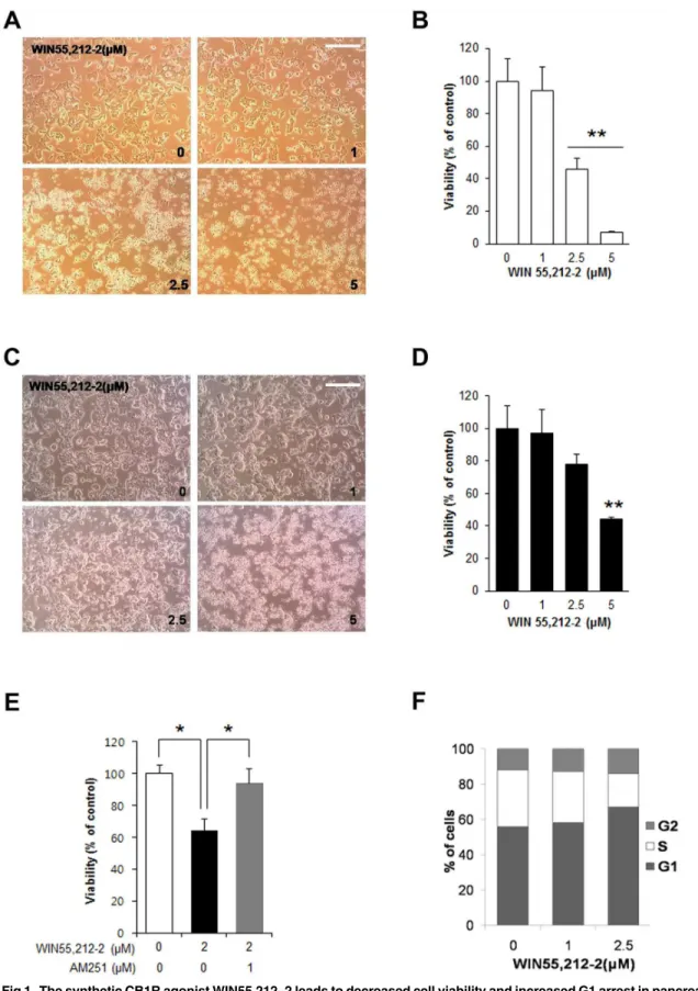
We first investigated the effects of the synthetic CB1R agonist WIN55,212–2 on pancreaticβ -cell viability. Treatment of mouse insulinoma MIN6 -cells with WIN55,212–2 caused morpho-logical changes characteristic of apoptosis, including cell rounding, in a dose-dependent man-ner (Fig 1A). Additionally, the viability of MIN6 cells dose-dependently decreased with WIN55,212–2 treatment (Fig 1B). Similar effects of WIN55,212–2 on morphological changes (Fig 1C) and viability (Fig 1D) were observed in another mouse insulinoma cell line (βTC6). The effects of WIN55,212–2 on viability were prevented by AM251, a selective CB1R antago-nist (Fig 1E), suggesting that these effects are mediated by CB1Rs. We next examined whether WIN55,212–2 regulates cell cycle progression in pancreaticβ-cell lines. To analyze cell cycle distribution, DNA content analysis was performed with PI staining and subsequent FACS anal-ysis after treatment with WIN55,212–2. As shown inFig 1F, treatment ofβTC6 cells with WIN55,212–2 led to an increase in the proportion of cells in G1 phase and a decrease in the proportion of cells in S phase. These results indicate that activation of CB1R by WIN55,212–2 induces cell cycle arrest at G1 phase in pancreaticβcells.
WIN55,212
–
2 decreases Bcl-2 and cyclin D2 expression in pancreatic
β
cells
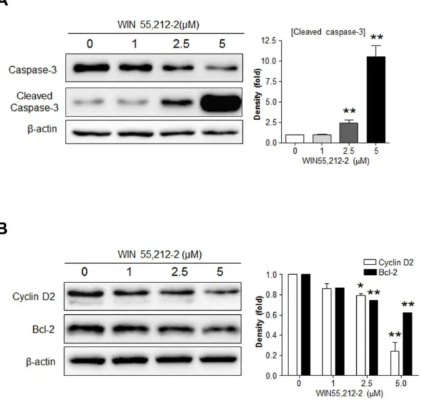
Because cyclin D2 and Bcl-2 are pivotal molecules in the regulation ofβ-cell growth and sur-vival [2–7], we next investigated the potential role of these molecules as a/the mediator of WIN55,212-2-controlledβ-cell growth and survival. Consistent with our previous results (Fig 1), WIN55,212–2 induced caspase-3 activation inβTC6 cells in a dose-dependent manner (Fig 2A). WIN55,212–2 caused a dose-dependent decrease in the levels of both cyclin D2 and Bcl-2 inβTC6 cells (Fig 2B). Similar effects of WIN55,212–2 on caspase-3 activation and cyclin D2 and Bcl-2 levels were also observed in MIN6 cells (data not shown). These results suggest that WIN55,212-2-induced cell cycle arrest and cell death might be mediated by decreasing cyclin D2 and Bcl-2 levels, respectively.
CB1R blockade in diabetic mouse models increases Bcl-2 and cyclin D2
levels in pancreatic
β
cells
Our previous report demonstrated that the pharmacological and genetic blockade of CB1Rs has beneficial effects onβ-cell growth and survival in mouse models of T1D [12]. Multiple injections of low-dose STZ cause insulin-dependent diabetes and produce a progressive increase in blood glucose levels due to selectiveβ-cell destruction, and over time, the remaining
βcells attempt to survive and proliferate [17,18]. Compared with control mice, genetic deletion and pharmacological blockade of CB1Rs in STZ-injected mice lead to decreased blood glucose levels and increasedβ-cell growth and survival resulting from decreased levels of cyclin-depen-dent kinase inhibitor p27 and active caspase-3 caused by enhanced insulin signaling via the IRS2-AKT pathway [12]. Similar effects were also observed in AM251-injected normal and db/dbmice [11]. Thus, using pancreata from those mice, we further examined whether the
Fig 1. The synthetic CB1R agonist WIN55,212–2 leads to decreased cell viability and increased G1 arrest in pancreaticβ-cell lines.(A)
Representative images of MIN6 cells exposed to WIN55,212–2. Scale bar, 50μm. (B) The relative cell viability of MIN6 cells was determined by MTS assay 24 h after treatment with WIN55,212–2. Scale bar, 50μm. (C) Representative images ofβTC6 cells exposed to WIN55,212–2. (D) The relative cell viability of
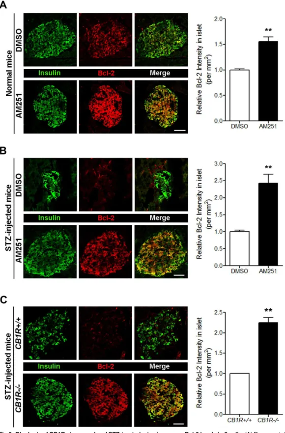
enhanced levels of Bcl-2 in pancreaticβcells compared with STZ-injectedCB1R+/+mice (Fig
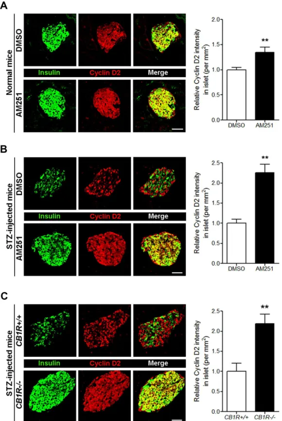
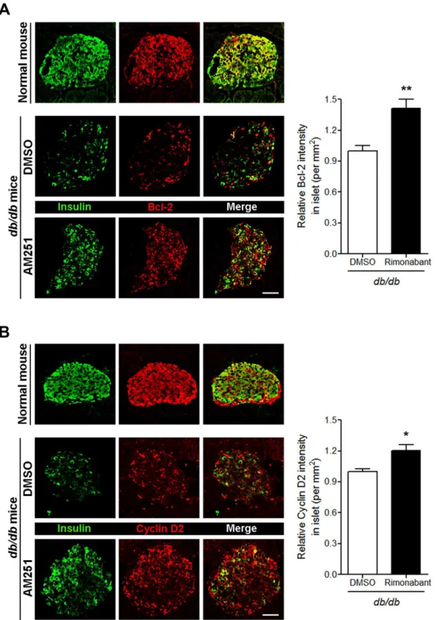
3C). Similar to Bcl-2, the levels of cyclin D2 in pancreaticβcells of normal mice were also sig-nificantly increased by AM251 (Fig 4A). Furthermore, CB1R blockade by AM251 in STZ-injected mice also significantly increased cyclin D2 levels in pancreaticβcells (Fig 4B), and a similar pattern was evident in pancreatic sections from STZ-injectedCB1R-/-mice (Fig 4C). The pharmacological blockade of CB1Rs in the T2D mouse model resulted in decreased blood glucose levels as well as increased intra-islet insulin content andβ-cell mass due to enhancedβ-cell proliferation [11]. Thus, we then investigated the expression levels of Bcl-2 and cyclin D2 in pancreaticβcells ofdb/dbmice. The Bcl-2 level was reduced in pancreatic
βcells ofdb/dbmice compared with normal mice (Fig 5A). Daily injection of another selective or without a CB1R inverse agonist AM251. MIN6 cells were exposed to WIN55,212–2 with or without AM251 for 24 h, and relative cell viability was
determined by MTS assay. (F) WIN55,212–2 treatment inβTC6 cells increases the proportion of cells in G0/G1 phase.βTC6 cells exposed to WIN55,212–2 for 24 h were analyzed using a fluorescence activated cell sorter (FACS). Data are shown as the mean±SEM from three independent experiments.*P< 0.05;**P<0.01.
doi:10.1371/journal.pone.0150981.g001
Fig 2. Effects of WIN55,212–2 treatment on the expression of Bcl-2 and cyclin D2 inβTC6 cells.βTC6 cells exposed to WIN55,212–2 for 24 h were subjected to western blot analysis for cleaved caspase-3 (A) or cyclin D2 and Bcl-2 (B). Relative densities for the indicated proteins are shown on the right. Data are shown as the mean±SEM from three independent experiments.*P<0.05;**P<0.01.
Fig 3. Blockade of CB1Rs in normal and STZ-treated mice increases Bcl-2 levels inβcells.(A) Representative images for insulin and Bcl-2 inβcells of DMSO- and AM251-injected normal mice. The relative signal intensity for Bcl-2 inβcells is shown on the right. (B) Representative images for insulin and Bcl-2 inβcells of DMSO- and AM251-injected mice after STZ treatment. The relative signal intensity for Bcl-2 inβcells is shown on the right. (C) Representative images for insulin and Bcl-2 inβcells ofCB1R+/+andCB1R-/-mice 4 weeks after STZ treatment. The relative signal intensity for Bcl-2 inβcells is shown on the right. Data are shown as the mean±SEM from n = 3–5 animals per group.*P<0.05;**P<0.01. Scale bar, 50μm.
Fig 4. Blockade of CB1Rs in normal and STZ-treated mice increases cyclin D2 levels inβcells.(A) Representative images for insulin and cyclin D2 inβ
cells of DMSO- and AM251-injected normal mice. The relative signal intensity for cyclin D2 inβcells is shown on the right. (B) Representative images for insulin and cyclin D2 inβcells of DMSO- and AM251-injected mice after STZ treatment. The relative signal intensity for cyclin D2 inβcells is shown on the right. (C) Representative images for insulin and cyclin D2 inβcells ofCB1R+/+andCB1R-/-mice 4 weeks after STZ treatment. The relative signal intensity for cyclin D2 inβcells is shown on the right. Data are shown as the mean±SEM from n = 3–5 animals per group.*P<0.05;**P<0.01. Scale bar, 50μm.
CB1R antagonist (rimonabant) for 4 weeks indb/dbmice led to significantly increased levels of Bcl-2 in pancreaticβcells compared with DMSO-treated mice (Fig 5A). A similar pattern for cyclin D2 was also observed in pancreaticβcells of rimonabant-injecteddb/dbmice (Fig 5B). These results suggest that the effects of CB1R blockade onβ-cell growth and survival are medi-ated, at least in part, by enhanced Bcl-2 and cyclin D2 levels.
CB1Rs play critical roles in regulation ofβ-cell mass and function by influencing insulin recep-tor signaling pathway [11,12,19]. Recent reports have shown that activation of CB1Rs by ECs and synthetic cannabinoids inhibited insulin receptor signaling, resulting in reducedβ-cell mass [11,12]. The serine/threonine kinase AKT is the primary mediator of insulin receptor signaling and many downstream targets of AKT have been identified that may underlie the ability of this cascade to regulateβ-mass. It has been well known that AKT directly phosphorylate several cru-cial downstream molecules that mediate cell survival and proliferation signals, such as pro-apo-ptotic protein BAD, forkhead transcription factor FoxO1, and cyclin-dependent kinase inhibitor p27, leading to the suppression of the cell death and growth arrest signals [20–24]. Consistently, our previous studies have shown that activation of CB1Rs inducesβ-cell death and cell cycle arrest by inhibiting AKT-mediated phosphorylation of BAD and p27, respectively [11,12]. Both cyclin D2 and Bcl-2 are also crucial downstream targets of AKT that play essential roles in regula-tion ofβ-cell mass [3–7,25,26]. Therefore, our data suggest that CB1Rs could regulate the expres-sion of cyclin D2 and Bcl-2 by influencing insulin receptor signaling via IRS2-AKT cascade.
Conclusions
In the present report, we provide evidence for a molecular mechanism by which CB1Rs regu-lateβ-cell growth and survival through influencing cyclin D2 and Bcl-2. We found that activa-tion of CB1R leads to a decrease in the expression of cyclin D2 and Bcl-2, in turn inducing cell cycle arrest in G0/G1 phase and caspase-3-dependent apoptosis bothin vitroandin vivo. Taken together, these findings suggest involvement of Bcl-2 and cyclin D2 in the regulation of
β-cell survival and growth by CB1Rs. Further studies examining the direct relationship between EC-inducedβ-cell death and Bcl-2 as well as EC-inducedβ-cell growth arrest and cyclin D2 are warranted, which would contribute to identify the exact mechanism by which CB1Rs regulate
β-cell growth and survival.
Supporting Information
S1 File. Experimental timeline of the study. (TIF)
Acknowledgments
We are deeply grateful to Dr. J. Pickel, NIMH Transgenic Core/NIH, for providing theCB1R -/-mice, and the animal facilities of NIA/NIH carried out the genotyping and husbandry.
Author Contributions
Conceived and designed the experiments: JK KJL JME WK. Performed the experiments: JK KJL JSK JGR JJS WK. Analyzed the data: JK KJL JSK JGR JJS WKS EKL JME WK. Contributed reagents/materials/analysis tools: WKS EKL. Wrote the paper: JK KJL JME WK.
insulin and cyclin D2 inβcells of normal and DMSO- or rimonabant-injecteddb/dbmice. The relative signal intensity for cyclin D2 inβcells is shown on the right. Data are shown as the mean±SEM from n = 5 animals per group.*P<0.05;**P<0.01. Scale bar, 50μm.
References
1. Cnop M, Welsh N, Jonas JC, Jörns A, Lenzen S, Eizirik DL. Mechanisms of pancreatic beta-cell death in type 1 and type 2 diabetes: many differences, few similarities. Diabetes. 2005; 54(Suppl 2):S97– S107. PMID:16306347
2. Georgia S, Bhushan A. Beta cell replication is the primary mechanism for maintaining postnatal beta cell mass. J Clin Invest 2004; 114(7):963–968. PMID:15467835
3. Kushner JA, Ciemerych MA, Sicinska E, Wartschow LM, Teta M, Long SY, et al. Cyclins D2 and D1 are essential for postnatal pancreatic beta-cell growth. Mol Cell Biol 2005; 25(9):3752–3762. PMID: 15831479
4. Georgia S, Hinault C, Kawamori D, Hu J, Meyer J, Kanji M, et al. Cyclin D2 is essential for the compen-satory beta-cell hyperplastic response to insulin resistance in rodents. Diabetes. 2010; 59(4):987–996. doi:10.2337/db09-0838PMID:20103709
5. Gurzov EN, Eizirik DL. Bcl-2 proteins in diabetes: mitochondrial pathways ofβ-cell death and dysfunc-tion. Trends Cell Biol. 2011; 21(7):424–431. doi:10.1016/j.tcb.2011.03.001PMID:21481590 6. Iwahashi H, Hanafusa T, Eguchi Y, Nakajima H, Miyagawa J, Itoh N, et al. Cytokine-induced apoptotic
cell death in a mouse pancreatic beta-cell line: inhibition by Bcl-2. Diabetologia. 1996; 39(5):530–536. PMID:8739912
7. Rabinovitch A, Suarez-Pinzon W, Strynadka K, Ju Q, Edelstein D, Brownlee M, et al. Transfection of human pancreatic islets with an anti-apoptotic gene (bcl-2) protects beta-cells from cytokine-induced destruction. Diabetes. 1999; 48(6):1223–1229. PMID:10342808
8. Kulkarni RN, Brüning JC, Winnay JN, Postic C, Magnuson MA, Kahn CR. Tissue-specific knockout of the insulin receptor in pancreatic beta cells creates an insulin secretory defect similar to that in type 2 diabetes. Cell. 1999; 96(3):329–339. PMID:10025399
9. Withers DJ, Gutierrez JS, Towery H, Burks DJ, Ren JM, Previs S, et al. Disruption of IRS-2 causes type 2 diabetes in mice. Nature. 1998; 391(6670):900–904. PMID:9495343
10. Kasuga M. Insulin resistance and pancreatic beta cell failure. J Clin Invest. 2006; 116(7):1756–1760. PMID:16823472
11. Kim W, Doyle ME, Liu Z, Lao Q, Shin YK, Carlson OD, et al. Cannabinoids inhibit insulin receptor sig-naling in pancreaticβ-cells. Diabetes. 2011; 60(4):1198–1209. doi:10.2337/db10-1550PMID: 21346174
12. Kim W, Lao Q, Shin YK, Carlson OD, Lee EK, Gorospe M, et al. Cannabinoids induce pancreaticβ-cell death by directly inhibiting insulin receptor activation. Sci Signal. 2012; 5(216):ra23. doi:10.1126/ scisignal.2002519PMID:22434934
13. Blázquez C, Carracedo A, Barrado L, Real PJ, Fernández-Luna JL, Velasco G et al. Cannabinoid receptors as novel targets for the treatment of melanoma. FASEB J. 2006; 20(14):2633–2635. PMID: 17065222
14. Sarfaraz S, Afaq F, Adhami VM, Mukhtar H. Cannabinoid receptor as a novel target for the treatment of prostate cancer. Cancer Res. 2005; 65(5):1635–1641. PMID:15753356
15. Sarfaraz S, Afaq F, Adhami VM, Malik A, Mukhtar H. Cannabinoid receptor agonist-induced apoptosis of human prostate cancer cells LNCaP proceeds through sustained activation of ERK1/2 leading to G1 cell cycle arrest. J Biol Chem. 2006; 281(51):39480–39491. PMID:17068343
16. Zimmer A, Zimmer AM, Hohmann AG, Herkenham M, Bonner TI. Increased mortality, hypoactivity, and hypoalgesia in cannabinoid CB1 receptor knockout mice. Proc Natl Acad Sci U S A. 1999; 96
(10):5780–5785. PMID:10318961
17. Krishnamurthy J, Ramsey MR, Ligon KL, Torrice C, Koh A, Bonner-Weir S, et al. p16INK4a induces an age-dependent decline in islet regenerative potential. Nature. 2006; 443(7110):453–457. PMID: 16957737
18. Like AA, Rossini AA. Streptozotocin-induced pancreatic insulitis: new model of diabetes mellitus. Sci-ence. 1976; 193(4251):415–417. PMID:180605
19. González-Mariscal I, Krzysik-Walker SM, Kim W, Rouse M, Egan JM. Blockade of cannabinoid 1 recep-tor improves GLP-1R mediated insulin secretion in mice. Mol Cell Endocrinol. 2015; 423:1–10. doi:10. 1016/j.mce.2015.12.015PMID:26724516
20. Datta SR, Dudek H, Tao X, Masters S, Fu H, Gotoh Y, et al. Akt phosphorylation of BAD couples sur-vival signals to the cell-intrinsic death machinery. Cell. 1997; 91(2):231–241. PMID:9346240 21. Zha J, Harada H, Yang E, Jockel J, Korsmeyer SJ. Serine phosphorylation of death agonist BAD in
22. Brunet A, Bonni A, Zigmond MJ, Lin MZ, Juo P, Hu LS, et al. Akt promotes cell survival by phosphorylat-ing and inhibitphosphorylat-ing a Forkhead transcription factor. Cell. 1999; 96(6):857–868. PMID:10102273 23. Medema RH, Kops GJ, Bos JL, Burgering BM. AFX-like Forkhead transcription factors mediate
cell-cycle regulation by Ras and PKB through p27kip1. Nature. 2000; 404(6779):782–787. PMID: 10783894
24. Shin I, Yakes FM, Rojo F, Shin NY, Bakin AV, Baselga J, et al. PKB/Akt mediates cell-cycle progression by phosphorylation of p27(Kip1) at threonine 157 and modulation of its cellular localization. Nat Med. 200; 8(10):1145–1152. PMID:12244301
25. Pugazhenthi S, Nesterova A, Sable C, Heidenreich KA, Boxer LM, Heasley LE, et al. Akt/protein kinase B up-regulates Bcl-2 expression through cAMP-response element-binding protein. J Biol Chem. 2000; 275(15):10761–10766. PMID:10753867