intraocular lens positioning in a transscleral fixation
technique
Purpose: To assess, through ultrasound biomicroscopy (UBM), the positioning of intraocular lens (IOL) haptics as related to the ciliary sulcus by using a scleral fixation technique, as well as to evaluate if two fixation points are sufficient to avoid tilting of the lenses.
Methods: Sixteen aphakic eyes underwent an IOL implantation by means of the same scleral fixation technique, performed by the same surgeon. One month after surgery, both the positioning of the IOL haptics and the distances between the IOLs and the cornea were assessed by UBM. Results were statistically studied.
Results: Of the 32 intraocular lens haptics fixed to the sclera, eight were placed inside the ciliary sulcus and 24 were placed outside the ciliary sulcus. There was no statistical difference in the distances measured between the intraocular lenses and the cornea for the haptics placed outside the ciliary sulcus as compared to the haptics placed inside the ciliary sulcus, suggesting that other factors, besides the distance to the limbus, may be relevant in the positioning of haptics in the ciliary sulcus. The same measurements performed at 3, 6, 9, and 12 hours, on the periphery of the lenses, were also similar, suggesting that 2-point fixation is sufficient to avoid tilting of the lenses in the eye.
Conclusions: Other factors (e.g. the ciliary sulcus opening angle), than the distance from the limbus where the sclera is transfixed, are important for the placement of the lenses in the ciliary sulcus. Two fixation points are sufficient to avoid tilting of the IOL in the eye.
Biomicroscopia ultra-sônica na avaliação da posição das lentes intra-oculares em
uma técnica de fixação escleral
Study sponsored by a CNPq fellowship.
(1) Department of Ophthalmology, Universidade Federal
de São Paulo, Escola Paulista de Medicina.
(2) Department of Ophthalmology, Universidade Federal
de São Paulo, Escola Paulista de Medicina.
(3) Department of Ophthalmology, Universidade Federal
de São Paulo, Escola Paulista de Medicina.
(4) Associate Professor, Department of Ophthalmology,
Universidade Federal de São Paulo, Escola Paulista de Medicina
The authors have no financial interest in the results of the study.
Address for correspondence: Raul C. Vianna Filho Rua Cel. José Monteiro, 287 - S. José dos Campos (SP) Brazil. CEP 12210-140. E-mail: raulvianna@uol.com.br Raul de Camargo Vianna Filho (1) Lincoln de Freitas (2)
Norma Allemann (3)
Ana Luísa Hofling de Lima (4)
SUMMARY
Keywords: Intraocular lens implantation; Pseudophakia; Aphakia; Ultrasonography.
INTRODUCTION
In cataract surgery, intraocular lens (IOL) implantation is a reality and in order to duly implant it in the posterior chamber, it has to be supported by the posterior capsule. If this support does not exist, several surgical techniques may be used, such as IOL implantation in the anterior chamber, lenses with iris support, iris fixation lenses and transsclerally fixated lenses 1, 2.
and if two fixation points were sufficient to avoid tilting of the optical part.
PATIENTS AND METHODS
This study was prospectively performed at the Cataract Sector of the Department of Ophthalmology, Universidade Federal de São Paulo - Escola Paulista da Medicina (UNIFE-SP-EPM). Sixteen eyes of previously aphakic patients were studied.
Inclusion criteria were: 1- at least 3 months between cataract extraction surgery, resulting in aphakia, and intrao-cular lens implantation by the transscleral fixation technique; 2- best corrected visual acuity on examination better than or equal to 20/80.
The ophthalmologic examination, which included refrac-tion and measurement of best corrected visual acuity, biomi-croscopy, fundoscopy under mydriasis and applanation tonometry, excluded: 1 - patients with corneal decompensa-tion, inactive chronic uveitis or active uveitis and anatomical congenital alterations; 2 - retina and optical nerve diseases which might increase surgical risk or be potentiated by the new surgery; 3 - uncontrolled glaucoma cases or glaucoma under clinical control but with a cup larger than 0.5; 4 -patients with a single eye.
This study followed the protocol liberated by the Ethics Committee of UNIFESP - EPM, including informed consent. All surgeries were performed by the same surgeon with experience in transscleral fixation. The used technique was transscleral fixation at the 3 and 9 hour positions with lifting of the scleral flaps. Prolene 10-0 thread was used for fixation with two straight needles (STC-6). The thread in the needle was passed from the exterior to the interior of the eye, at 0.75 mm from the surgical limbus, under the scleral flap at the 9 hour position, independent of the operated eye. Introduction of the thread into the sclera was made perpendicularly to the ocular wall so that the site of the scleral orifice corresponded to the site of the intraocular orifice regarding distance from the surgical limbus.
At the 3 hour position (independent of the operated eye), under the scleral flap and at 0.75 mm from the limbus a 13x4 (27) gauge needle was introduced, also perpendicularly to the ocular wall. The prolene thread needle was introduced in the 13x4 (27G) needle which served as guide for the emergence of the prolene needle at the 3 hour position.
A wide anterior vitrectomy was performed in all cases, first cleaning the anterior chamber, followed by the pupillary region and anterior vitreous.
A model SF-70H intraocular lens, produced by Mediphacos, specific for transsceral fixation, was used in all surgeries.
On the return visit after 1 month, ultrasound biomicros-copy was performed using a Humphrey model 840 Ultrasound Biomicroscope with a 50 MHz transducer. All examinations were performed by the same examiner, who was different
from the surgeon. On UBM examination, the following were evaluated:
1 - Positioning of the intraocular lens haptics at the 3 and 9 hour regions;
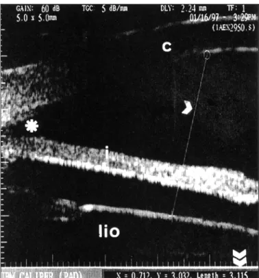
2 - Measurement of the distance between the posterior surface of the cornea and the anterior surface of the intraocular lens, at the 4 quadrants. This measurement was performed at 4 mm from the scleral spur (apex of the camera sinus), tracing a straight line perpendicularly to the surface of the intraocular lens at the 3, 6, 9 and 12 hour meridians (Fig. 1).
RESULTS
This study involved 16 patients, nine (56.25%) males and seven (43.75%) females, with seven (43.75%) right eye and nine (56.25%) left eye surgeries.
Mean age of the studied patients was 68.4 years, ranging from 56 to 79 years.
The observed interval between cataract surgery, leading to aphakia, and scleral fixation surgery for intraocular lens implantation ranged from three months to 38 years.
The results obtained through ultrasound biomicroscopy in the evaluation of positioning of intraocular lens haptics after scleral fixation are shown in Table 1. It is observed that of the 32 haptics (16 intraocular lenses), eight (25%) were placed in the scleral
Fig. 1 - Longitudinal ultrasound biomicroscopy examination (UBM with 50 MHz transducer, under immersion). Cornea (c), iris (i), intraocular lens (lio) and anterior chamber angle (*) echoes are observed. At this meridian, the straight line segment (arrow) indicates the distance from the posterior surface of the cornea to the anterior IOL surface (length =
sulcus (Fig. 2), and of the 24 (75%) outside the ciliary sulcus, 22 (91.6%) were posterior to the ciliary sulcus (Fig. 3) and two (8.3%) were anterior to the ciliary sulcus (Fig. 4). Statistical analysis showed that there was a difference between the positio-ning inside or outside the scleral sulcus and that, using this technique, it was easier to place the haptics outside than inside the ciliary sulcus.
On comparing the placement of the 3 hour haptics in relation to the ciliary sulcus, it was observed that, of the sixteen 3 hour haptics, two (12.5%) were placed inside and 14 (87.5%) outside the ciliary sulcus (all posterior to the ciliary sulcus), showing that at this position there was a greater tendency to place the haptic outside the ciliary sulcus (Table 1). Repeating the comparison for the 9 hour positioning of the haptics, there were six (37.5%) haptics placed inside and ten (52.5%) placed outside the sulcus (two anterior and 8 posterior to the ciliary
sulcus), showing that, in this position, there is no difference in the positioning of the haptics (Table 1).
Through ultrasound biomicroscopy also the distance
bet-Table 1. Results of the positioning of intraocular lens haptics in eyes submitted to secondary implantation with scleral fixation, according
to localization and orientation of fixation.
Haptics Inside the ciliary Outside the ciliary Total sulcus (%) sulcus (%) (%)
3 hours 2 (12.5%) 14 (87.5%) 16 (100.0%) 9 hours 6 (37.5%) 10 (52.5%) 16 (100.0%)
Total 8 24 32
Chi square test : X2 calc 8.00; X2 crit 3.84.
Chi square test for the 3 hour position: X2 calc 9.00; X2 crit 3.84.
Chi square test for the 9 hour position: X2 calc 1.00, X2 crit 3.84.
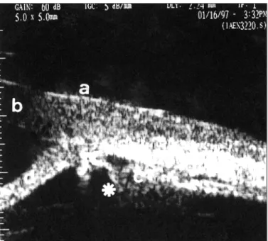
Fig. 2 - Longitudinal ultrasound biomicroscopy (UBM) examination showing, in profile, the echoes corresponding to sclerocorneal transition (a), cornea (b), ciliary body (c) and iris (d) and ciliary sulcus (*). The intraocular lens haptic (arrow) appears as a cylindrical structure
causing reverberation, located in the ciliary sulcus.
Fig. 3 - Longitudinal ultrasound biomicrospy (UBM) examination, showing, in profile, the echoes corresponding to sclerocorneal transition (a), cornea (b), ciliary body (c) and iris (d) and ciliary sulcus (*). The intraocular lens haptic (arrow) appears as a cylindrical structure
causing reverberation, located posterior to the ciliary sulcus.
Fig. 4 - Longitudinal ultrasound biomicroscopy (UBM) examination showing, in profile, the echoes corresponding to sclerocorneal transition (a), cornea (b), ciliary body (c) and iris (e) and ciliary sulcus (*). The intraocular lens haptic (arrow) appears as a cylindrical structure causing reverberation, located anterior to the ciliary sulcus, originating
ween the optical part of the intraocular lens and the cornea was measured at a point located at 4 mm from the limit of the corneal-sceral limbus, at the 3, 6, 9, and 12 hour positions. The obtained values are shown in Table 2. The mean of the distances measured for the 3 hour position was 3.77 mm; for the 6 hour position, 3.85 mm; for the 9 hour position, 3.74 mm; and for the 12 hour position, 3.63 mm. Conclusion of the analysis of the data by Friedman’s rank variance (with a calculated χ2 of6.08 and critical χ2 of 7.82) was that there is no difference between the means of the distances at the four positions (Table 2).
These same distances, when analyzed only for the 3 and 9 hour positions, and on comparing the distance of the lenses whose haptics were placed in the sulcus in the 3 hour position with the distance of the lenses whose haptics were placed, also, in the sulcus at the 9 hour position, did not show significant differences (Table 3), using Mann-Whitney’s test far a calculated U of 3.0 and a critical U of 0.
Repeating the calculations for the lenses whose haptics were implanted outside the ciliary sulcus at the 3 hour position, as compared with the lenses whose haptics were implanted outside the ciliary sulcus at the 9 hour position, also no difference was observed (Table 3) using Mann-Whitney’s test.
On comparison of the distances of the haptics implanted inside the sulcus with the distances of the haptics implanted outside the sulcus for the 3 hour position haptics, no signifi-cant difference was observed using Mann-Whitney’s test (Table 3).
Repetition of the analysis (Mann-Whitney test) only for the haptics implanted at the 9 hour position showed that there was no difference between the lens - cornea distances for the haptics
positioned inside or outside the ciliary sulcus (Table 3).
DISCUSSION
Pavlin et al.3 and Vianna Filho et al. 4 had already used ultrasound biomicroscopy examination to assess the positio-ning of haptics after scleral fixation, however, Pavlin et al.’s 3 study only showed that ultrasound biomicroscopy is a useful propaedeutic method for the localization of the haptics. Vianna Filho et al.4 used that method to study a standardized surgical technique, performed by several surgeons. In the present study, ultrasound biomicroscopy was used as propae-deutic method to assess a surgical technique of scleral intrao-cular lens fixation, performed by the same surgeon.
Ultrasound biomicroscopy detected that of the 32 implanted haptics (16 intraocular lenses), eight (25%) were placed inside and 24 (75%) outside the ciliary sulcus (Table 1), this being a significant difference when analyzed by the chi square test.
In order to study if there was a tendency to place the haptics inside the sulcus at the 3 and 9 hour positions, the placing of the haptics at those two positions was assessed (Table 1). The statistical chi square test showed a tendency to place the haptics outside the ciliary sulcus at the 3 hour position, while there was
Table 2. Values of the distance from the cornea (point located at 4 mm from the scleral spur) to the anterior surface of the intraocular lens for the 3, 6, 9, and 12 hour positions, in eyes submitted to scleral
fixation surgery
Distance (mm)
Case 3 hours 6 hours 9 hours 12 hours
1 3.50 3.74 3.70 3.16 2 4.26 4.20 4.24 4.25 3 4.05 4.07 4.20 4.02 4 3.46 3.59 3.54 3.68 5 3.81 3.67 3.55 3.77 6 3.98 3.95 3.93 3.62 7 3.77 3.45 3.65 3.76 8 3.11 3.58 2.89 2.47 9 3.43 3.73 3.49 3.12 10 4.07 3.89 3.95 4.22 11 3.78 3.92 3.74 3.69 12 3.68 4.37 3.72 3.53 13 3.60 3.69 4.05 3.89 14 3.99 4.04 3.91 3.81 15 4.22 3.86 3.90 3.82 16 3.70 3.91 3.45 3.31 Mean 3.77 3.85 3.74 3.63
Friedman’s rank variance analysis (3 x 6 x 9 x 12 hours) x2 calc =6.08; x2 crit = 782
Table 3. Distance between the intraocular lens and the cornea at the 3 and 9 hour positions of the lenses with haptics outside and inside the ciliary sulcus, in patients submitted to scleral fixation surgery
Distance (mm)
3 hour haptic 9 hour haptic Ciliary Outside the Ciliary Outside the sulcus ciliary sulcus sulcus ciliary sulcus
3.11 3.50 3.70 4.24 3.68 4.26 3.55 4.20
.- 4.05 3.93 3.54
- 3.46 3.65 2.89
- 3.81 3.72 3.49
- 3.98 3.45 3.95
- 3.77 - 3.74
- 3.43 - 4.05
- 4.07 - 3.91
- 3.78 - 3.90
- 3.60 -
-- 3.99 -
-- 4.22 -
-- 3.70 -
-Mean 3.39 3.83 3.66 3.79 Mann-Whitney test
Ciliary sulcus x outside the ciliary sulcus 3 hour haptic 9 hour haptic
U calc = 4.0 U calc= 19.0 U crit = 1.0 U crit = 11.0
3 hour haptic x 9 hour haptic
ciliary sulcus outside the ciliary sulcus U calc = 3.0 U calc = 69.5
no difference between hitting or not the ciliary sulcus at the 9 hour position, showing that another factor influenced this result.
If the site to transfix the sclera, in the utilized technique, was not correct, taking into account the anatomical studies by Duffey et al.5 who suggested a localization of the scleral sulcus at the 3 and 9 hour positions at 0.50 mm from the limbus, a greater number of haptics outside the ciliary sulcus than in this study should have been observed. A difference was observed only for the haptics at the 3 hour position. If, however, the localization of the ciliary sulcus was correct, more haptics would be inside than outside the sulcus; but, again, there is a difference between placing the haptics inside or outside the ciliary sulcus at the 3 hour but not at the 9 hour position.
The position of the intraocular lens was assessed through ultrasound biomicroscopy by means of measuring in milli-meters the distance between the posterior surface of the cornea and the anterior IOL surface, in 4 quadrants. It was not possible to show a difference between the measurements (Table 2), indicating that this method with two fixation points does not tend to cause tilting of the intraocular lens.
Using the above mentioned measuring method to compare distances between the intraocular lens and the cornea at the 3 and 9 hour positions with the haptics placed in the ciliary sulcus (Table 3), equivalence between the distances was shown.
The former measurements were repeated for haptics placed outside the ciliary sulcus at the 9 hour position (Table 3). Again there were no differences, meaning that the haptics positioned outside the ciliary sulcus both at one and the other side were placed at a practically equal distance from the limbus, pro-bably with the same inclination of the needles, indicating that the used technique was equivalent for all cases.
Comparing the measurements of the distances between the intraocular lenses and the cornea whose haptics were placed in the ciliary sulcus at the 3 hour position with those whose haptics were outside the ciliary sulcus, also no differences were observed (Table 3). The same occurs on comparing these measurements for haptics placed at the 9 hour position inside with those placed outside the ciliary sulcus (Table 3).
These data of formerly described measurements reinforce the idea that besides the chosen distance from the limbus for scleral fixation, other factors must be involved in the positioning of the haptics in the ciliary sulcus because, except for the two haptics in an anterior position, if the haptics placed outside the ciliary sulcus were in a posterior position (Table 1), they should be more distant from the cornea than those placed inside the ciliary sulcus. Since these measurements are equivalent, probably anatomical differences between indi-viduals, anatomical differences which occurred after cataract extraction or even anatomical differences during the scleral fixation surgery could be occurring.
Pavlin et al.6, studying eyes of normal individuals through ultrasound biomicroscopy, found alterations in angle opening of the ciliary body. Passing a line tangentially to the sclera at 500
µm posterior to the scleral spur and another through the ciliary body, always at the same environmental illumination, will determine what would be the angle opening of the ciliary body, which varied from 18 to 87 degrees in 11 studied individuals.
This variation obtained in Pavlin et al.’s 6 study may be one of the reasons leading to find haptics located in the sulcus, while others placed at the same distance from the cornea, probably with smaller ciliary body angle openings, are located posterior to the ciliary sulcus. However, this cannot be affirmed, but only suggested, since variations in angle opening of the ciliary body of these patients were not studied. Vajpayee et al. 7 mention in their study that, after performing anterior vitrectomy, they injected air behind the iris to “open” the ciliary body in order to then pass the fixation thread from the inside to the outside and, thus, to reach the ciliary sulcus.
Although not providing references about this ciliary body “opening” by injecting air, Vajpayee et al.’s 7 study already indicates that variations in angle opening of the ciliary body may occur after cataract extraction and even during scleral fixation surgery.
It is easy to imagine that after removal of the opacified lens there is loss of tension exerted by the zonules on the ciliary processes which could promote anatomical alterations with alterations in the angle opening of the ciliary body. Pavlin et al.3 assert that zonular tension loss after lens extraction leads to a contraction of the ciliary process with subsequent narro-wing of the ciliary sulcus.
CONCLUSIONS
Ultrasound biomicroscopy showed that there is a differen-ce regarding positioning of intraocular lens haptics inside or outside the ciliary sulcus, using the described surgical technique, for the 3 hour position.
The results of the study evidenced, through ultrasound biomicroscopy, that there was no difference between the distance of the intraocular lens and the cornea, measured at the 3, 6, 9, and 12 hour positions.
It can be concluded that the scleral fixation method with only two fixation points and intraocular lenses which are appropriate for this fixation technique did not elicit, in this study, tilting of the intraocular lenses.
Ultrasound biomicroscopy is a good propaedeutic method to assess the position of intraocular lens haptics by the scleral fixation technique, providing important data for the study of factors related to the surgical success and for comparison with other techniques of aphakia correction.
RESUMO
escleral, avaliando-se também se dois pontos de fixação são suficientes para que não haja inclinação da parte óptica.
Métodos: Dezesseis olhos afácicos foram submetidos a implante de LIO por uma mesma técnica de fixação escleral, realizados por um mesmo cirurgião. Um mês após a cirurgia, o posicionamento das alças das LIO foram avaliados pelo UBM, assim como distâncias entre as LIO e córnea. Os resultados foram submetidos a testes estatísticos.
Resultados: Das 32 alças fixadas à esclera, oito estavam localizadas no sulco ciliar e 24 fora deste. Não houve diferença estatística nas distâncias entre LIO e córnea para alças posicionadas no sulco cilar quando comparadas àquelas localizadas fora do sulco. Isto sugere que, além da distância ao limbo na qual se transfixa a esclera, outros fatores devem estar associados ao posicionamento da alça no sulco ciliar. As medidas LIO – córnea realizadas na periferia das LIO às 3, 6, 9, e 12 horas foram semelhantes, mostrando que dois pontos de fixação são suficientes para que a LIO não fique inclinada.
Conclusões: Outros fatores (por exemplo o ângulo de abertura do corpo ciliar), além da distância ao limbo na qual se transfixa a esclera, são importantes para o posicionamento das alças no sulco ciliar. Dois pontos de
fixação são suficientes para que a LIO não apresente inclinação dentro do olho.
Palavras-chave: Implante de lente intra-ocular; Pseudofacia; Afacia; Ultra-sonografia.
REFERENCES
1. Champion R, McDonnell PJ, Green WR. Intraocular lenses. Histopathologic characteristics of a large series of autopsy eyes. Surv Ophthalmol 1985;30:1-32. 2. Heidemann DG, Dunn SP. Visual results and complications of transsclerally sutured intraocular lenses in penetrating keratoplasty. Ophthalmic Surg 1990;21:609-14.
3. Pavlin CJ, Rootman D, Arshinoff S, Harasiewicz K, Foster FS. Determination of the haptic position of transsclerally fixated posterior chamber intraocular lenses by ultrasound biomicroscopy. J Cataract Refract Surg 1993;19:573-7. 4. Vianna Filho RC, Mori ES, Allemann N, Agmont W, Araújo Filho A. Avalia-ção do posicionamento das alças de lentes intra-oculares após fixaAvalia-ção trans-escleral através de biomicroscopia ultra-sônica. Arq Bras Oftal 1996;59:307-10.
5. Duffey RJ, Holland EJ, Agapitus PJ, Lindstrom RL. Anatomic study of transsclerally sutured intraocular lens implantation. Am J Ophthalmol 1989;108:300-9.
6. Pavlin CJ, Harasiewicz K, Stuart Foster F. Ultrasound biomicroscopy of anterior segment structures in normal and glaucomatous eyes. Am J Ophthalmol 1992;113:381-9.
7. Vajpayee RB, Angr SK, Sandramouli S, Rewari R. Direct scleral fixation of posterior chamber intraocular lenses using a special needle-holder. Ophthalmic Surg 1992;23:383-7.