*Corresponding author: Hojjatollah Nozad Charoudeh, Tel/Fax: +98 41 33342086, Email: [email protected] ©
2016 The Authors. This is an Open Access article distributed under the terms of the Creative Commons Attribution (CC BY), which permits Adv Pharm Bull, 2016, 6(2), 153-161
doi: 10.15171/apb.2016.022 http://apb.tbzmed.ac.ir
Advanced
Pharmaceutical
Bulletin
Key Immune Cell Cytokines Affects the Telomere Activity of Cord
Blood Cells
In vitro
Balal Brazvan1,2, Raheleh Farahzadi1, Seyede Momeneh Mohammadi1,2, Soheila Montazer Saheb1, Dariush Shanehbandi3, Laurent Schmied4, Jafar Soleimani Rad1, Masoud Darabi5, Hojjatollah Nozad Charoudeh1* 1
Stem Cell Research Center, Tabriz University of Medical Sciences, Tabriz, Iran. 2
Student Research Committee, Tabriz University of Medical Sciences, Tabriz, Iran. 3
Immunology Research Center, Tabriz University of Medical Sciences, Tabriz, Iran. 4
Immunotherapy Laboratories, Department of Biomedicine, University Hospital Basel, Basel, Switzerland.
5 Department of Clinical Biochemistry and Laboratory Medicine, Faculty of Medicine, Tabriz University of Medical Sciences, Tabriz,
Iran.
Introduction
Telomeres are nucleoprotein structures at the end of eukaryotic chromosomes. As it is proven, DNA hexamer (TTAGGG)n repeat have an important role in chromosomes stability.1,2 In differentiated cells, the number of DNA hexamer repeats decreases with each cell division.3-5
Telomerase is encoded by the telomerase reverse transcriptase (TERT) gene and it associated with a telomerase RNA component (TERC) as RNA template. This enzyme adds telomeric repeats to the chromosome ends and prevents telomere shortening in germ line cells and the majority of tumor cells.6,7 Therefore, telomere length and expression level of telomerase positively correlate with the immortality of cancer cells, germ-line cells and embryonic stem cells.8-11
In many previous studies has been demonstrated that telomere lengths change in lymphocytes with activation and differentiation of T and B cells. In parallel
telomerase is expressed in normal lymphocytes, and this expression is regulated in the time of development and activation. These findings indicate that the maintenance of telomere length may be important to regulate T- and B-cell responses.12,13 There is a strong correlation between telomere length and replicative capacity in all normal cells. This correlation extends in T cells to differences in telomere length and replicative capacity in subsets such as naive and memory CD4+ cells. Furthermore, telomerase expression is highly regulated during development and activation of both T and B cells.14
IL-2, IL-7 and IL-15 are critical cytokines, regulating in hematopoiesis and proliferation, self-renewal, differentiation and senescence of HSCs (Hematopoietic stem cells).15,16 Interleukin- 2 (IL-2) supports hematopoiesis, T cell proliferation (both CD4+ and CD8+) and cellular metabolism.17-20 As well as the Research Article
Article History:
Received: 11 March 2016 Revised: 14 March 2016 Accepted: 15 March 2016 ePublished: 30 June 2016
Keywords: Telomere Telomerase Interleukin Mononuclear cells CD34+ cells
Abstract
Purpose: Telomere is a nucleoprotein complex at the end of eukaryotic chromosomes and its length is regulated by telomerase. The number of DNA repeat sequence (TTAGGG)n is reduced with each cell division in differentiated cells. The aim of this study was to evaluate the effect of SCF (Stem Cell Factor), Flt3 (Fms- Like tyrosine kinase-3), Interleukin-2, 7 and 15 on telomere length and hTERT gene expression in mononuclear and umbilical cord blood stem cells (CD34+ cells) during development to lymphoid cells.
Methods: The mononuclear cells were isolated from umbilical cord blood by Ficoll-Paque density gradient. Then cells were cultured for 21 days in the presence of different cytokines. Telomere length and hTERT gene expression were evaluated in freshly isolated cells, 7, 14 and 21 days of culture by real-time PCR. The same condition had been done for CD34+ cells but telomere length and hTERT gene expression were measured at initial and day 21 of the experiment.
Results: Highest hTERT gene expression and maximum telomere length were measured at day14 of MNCs in the presence of IL-7 and IL-15. Also, there was a significant correlation between telomere length and telomerase gene expression in MNCs at 14 days in a combination of IL-7 and IL-15 (r = 0.998, p =0.04). In contrast, IL-2 showed no distinct effect on telomere length and hTERT gene expression in cells.
Brazvan et al.
expansion of activated T lymphocytes (T cells) and differentiation of B cells.21 In addition, previous studies revealed that in T-cells recently exposed to IL-2 apoptosis is more likely induced upon antigen-dependent T-cell receptor stimulation.22 IL-2 along with Interleukin-15 (IL-15) are growth factors for natural killer cells.23 It is shown that IL- 7 influences the expansion of B cells, promotes T cell differentiation and has an important role in natural killer cell development.24 Additionally, Interleukin-15 (IL-15) promotes expansion, proliferation and homeostasis of T cells.25
We have previously shown that B and T cell expansion increased in cord blood mononuclear cells using IL-2 and IL-7.26 In another study, we illustrated that IL-2 and IL-15 are important for the expansion of NKP46 positive cells (NK cells) in cord blood cells in vitro.27
Cord blood is a major source of CD34 positive cells used HSC-transplantation in leukemia treatment or cancer immunotherapy. So far, it remains unclear if cytokines can interfere with telomerase dynamic in umbilical cord blood cells.
The aim of this study was to evaluate the influence of IL-2, IL-7 and IL-15 on the telomere length and hTERT gene expression on mononuclear and CD34+ umbilical cord blood stem cells during development to the lymphoid cell.
Materials and Methods
Mononuclear cord blood isolation and CD34+ cells
enrichment
Umbilical cord blood sample were obtained from full-term normal deliveries in Tabriz Alzahra hospital, Iran. The collected cord blood was diluted 1:2 with phosphate buffered saline (PBS) plus 10% fetal bovine serum (FBS). Mononuclear cells (MNCs) were isolated by Ficoll-Hypaque (GE Healthcare, Piscatta, NJ, USA) gradient centrifugation at 400×g for 25 min at 4º C. Subsequently MNCs were collected and washed twice in PBS plus 5% FBS. The isolated MNCs were co-incubated with 100 µl of CD34+ micro beads (Miltenyi Biotec, Germany Cat no: 130100453) for 30 minutes. Thereafter resuspended cells were passed through one LS MACS column (Miltenyi Biotec, Germany). Thereafter enriched CD34+ cells were retrieved by flushing the column. For purity assessment of CD34+ cells, flow cytometry was performed by FACSCalibur (BD Bioscience) and the output data were processed with Flow software version X.0.7.
Culture condition
MNCs and isolated CD34+ cells were seeded in 96-well flat bottom cell culture plates at the density of 5×10⁵ cells per well. For culturing RPMI 1640 supplemented
with 20% (v/v) FBS and 1% (v/v)
penicillin/streptomycin was used. Different combinations of cytokines (Pepro Tech, London, UK) were added to the culture medium in final concentrations of 80 ng/ ml and cell cultures were maintained for 21 days. SCF and Flt3 were same in all groups. Cells were harvested and
analyzed at day 7, 14 and 21. All cytokines used in this study including SCF, FLT3, IL-2, IL-7 and IL-15 were purchased from Peprotech.
Flow cytometry
Monoclonal antibodies were CD3 (UCHT1; R&D) for T cells, CD20 (PE; clone 2H7; BD Biosciences) for B cells and Anti-NKp46-PE (BD,biosience) for NK cells. Flow cytometry was performed at 14 day of culture period time. Shortly, the cells were incubated with these antibodies in each group (for 20 min at 4ºC). We used of Propidium iodide (1.0 mg/mL; Invitrogen) to eliminate of dead cells. The flow cytometry analysis of the cell suspensions was performed on a BD FACSCalibur analyser (BD Biosciences). Between 10000 and 30000 events were collected and analysed by the flow cytometry software Perttu Terho (version: 2.5.1., Cyflogic, Finland).
RNA and DNA extraction
Total RNA was extracted from harvested MNCs and CD34+ cells at different time points using Thermo Scientific Gene JET RNA Purification Kit (K0731) according to the manufacturer's instruction. Extracted RNA was dissolved in nuclease-free water. For integrity assessment of extracted RNA we use of %1.5 agarose gel electrophoresis and for purity assessment we use of spectrophotometry. DNA was extracted using the pure link genomic DNA mini kit (Invitrogen, USA) according to the manufacturer's instruction. Briefly, the cells were collected and digested with Proteinase K (Invitrogen, USA) at 55 °C. Any residual RNA was removed by incubation with RNase A (Invitrogen, USA) for 2min. After digestion, 200 µl ethanol (96~100%) was added, mixed thoroughly by pulse vortexing for 30 sec, and transferred carefully to a DNA binding pure link spin column. Finally, impurities were removed by washing buffer. The genomic DNA was then eluted in the low salt elution buffer. For integrity assessment we use of %1 agarose gel electrophoresis and purity assessment was analysed by spectrophotometry.
c-DNA synthesis and Real-time PCR
Reverse transcription was carried out using the RevertAidTM first strand cDNA synthesis kit (K1622; Fermentas, Germany). Two microgram RNA was used for the first-strand cDNA synthesis in a total volume of 20 µL according to the manufacturer's guidelines. Briefly, the reaction tubes were put at 65°C for 10 min, 42°C for 60 min preceded with 70°C for 10 min and followed at 4°C for 5 min. For every reaction set, one RNA sample was prepared without RevertAidTM M-MuLV reverse transcriptase (RT reaction) to provide a negative control in the subsequent PCRs.
Effects of immune cell cytokines on telomere activity
mRNA-specific primers were designed using Oligo 7 software (v. 7.52, Molecular Biology Insights, Inc, USA). The sequences are listed in Table 1 and 2. Both β
-actin and hTERT amplification were done in triplicates
for each sample. β-actin was selected as an endogenous housekeeping gene. Forty-five thermal cycles were performed in the following order: 2 min at 94ºC, 40
cycles, 94ºC for 15 sec and 63ºC for 1 min. PCR data were analyzed using Rotor-Gene 6000 Software (version: 1.7) to determine CT values. Delta CT values were calculated in relation to β-actin CT values by the 2 -ΔΔCT method, in which ΔCt represents the difference
between the CT value of target genes and the CT value
of β-actin.
Table 1. Primers for quantitative Real-time RT-PCR
No. Gene Primer pair sequence (5'-3') Product length (bp)
NM_001193376.1 TERT CCGCCTGAGCTGTACTTTGT
CAGGTGAGCCACGAACTGT 234
NM_001101.3 β-actin AAACTGGAACGGTGAAGGTG
TATAGAGAAGTGGGGTGGCT 174
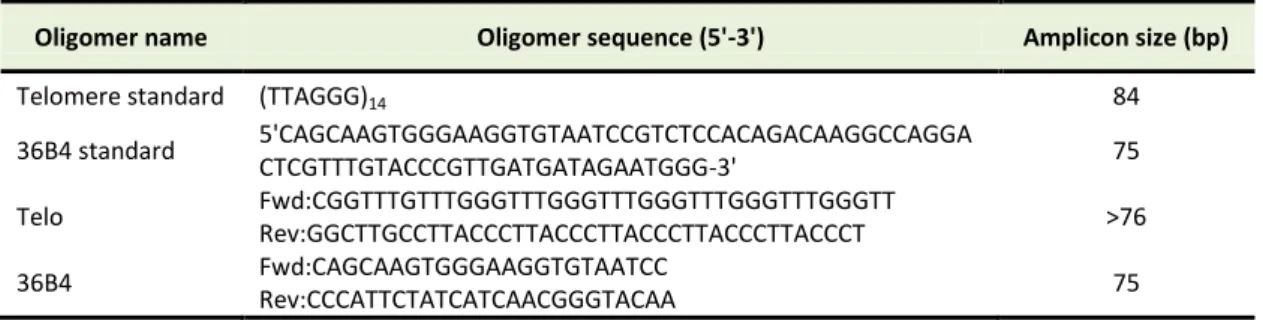
Table 2. Oligomers used for aTL assay
Oligomer name Oligomer sequence (5'-3') Amplicon size (bp)
Telomere standard (TTAGGG)14 84
36B4 standard 5'CAGCAAGTGGGAAGGTGTAATCCGTCTCCACAGACAAGGCCAGGA
CTCGTTTGTACCCGTTGATGATAGAATGGG-3' 75
Telo Fwd:CGGTTTGTTTGGGTTTGGGTTTGGGTTTGGGTTTGGGTT
Rev:GGCTTGCCTTACCCTTACCCTTACCCTTACCCTTACCCT >76
36B4 Fwd:CAGCAAGTGGGAAGGTGTAATCC
Rev:CCCATTCTATCATCAACGGGTACAA 75
Standard curves and associated calculations for aTL A standard curve was obtained from dilution series of known quantities of a synthesized 84 mer oligonucleotide (84 bp in length) containing only TTAGGG repeated 14 times. The number of repeats in each standard is calculated as previously described by
O’Callaghan.2
For generating a standard curve the serial dilutions of TEL STD A (10-1 [1.18 ×10⁸] through to 10-6 [1.18 × 10³] dilution) is performed. Plasmid DNA (pet 28a) was added to each standard to maintain a constant 20 ng of total DNA per reaction tube (Table 3).
Table 3. Amounts of calculation for aTL
Oligomer Molecular
weight (MW)
Weight of telomere standard and 36B4
(g)
Number copies of 36B4
Number molecules of oligomer in TEL STD A
Amount of telomere sequence in TEL STD A
(kbp)
SCG STD A
(TTAGGG)14 26667.2. 2.6667 × 10
4
/6.02 ×
1023= 0.44 × 10⁻¹⁹ -
60 × 10-12/0.44 × 10-19 =1.36×10⁹
1.36 × 109× 84=
1.18 × 10⁸ -
synthesized 36B4
oligomer standard 23268.1
2.32681 × 10⁴/6.02 × 1023= 0.38 × 10⁻¹⁹
200 × 10-12/0.44
× 10-19= 5.26×10⁹ - -
2.63 × 10⁹ For the single copy gene (SCG) standard curve, we
routinely used 36B4, which encodes the acidic ribosomal phosphoprotein P0. Although telomeric DNA sequence is consistent in mammals, the SCG will be different, thus, an SCG standard curve and amplicon must be generated for each target species. SCG amplification is crucial for the accuracy and reliability of the results generated in the aTL assay.
For generating a standard curve the serial dilutions of SCG STD a (10-1 through to 10-6 dilution) is performed (Figure 1). As same as telomere standard, plasmid DNA (pet 28a) is added to each standard to maintain a constant 20 ng of total DNA per reaction tube.
Statistical analysis
In this study data were studied by one-way ANOVA followed by the Tukey test. For graph we used of Prism software (GraphPad Software, Inc., San Diego, CA version; 6). Values were measured statistically significant at P < 0.05.
Results
Brazvan et al.
cytometry using CD 20 for B cells, CD3 for T cells and NKP46 for NK cells.
Figure 1. Standard curve used to calculate absolute telomere length.(A) Graph shows standard curve for calculating length of telomere sequence per reaction tube. X-axis represents number of cycle and Y-axis show the standards concentration.(B) Graph shows standard curve for calculating genome copies using 36B4 copy number. X-axis represents number of cycle and Y-axis show standard concentrations per each reaction.
As shown in Figure 2 the IL-2 are involved in expansion of T (94%), B (96%) and NK cells(38%). However IL-7 increased T cell expansion (92%) as well as B cells (97%), but not significantly increased NK cells. Our data were shown that IL-15 can increase the expansion of NK cells (28%), but it was not important for B cells. The Figure 2 shows that IL-2 and IL-15 can stimulate expansion of mononuclear cells toward T and NK cells and IL-7 was important for B cells.
Figure 2. Cord blood mononuclear cell derived T, B and NK cells in presence of IL-2, IL-7 and IL-15 at day 14 in vitro. Harvested cells evaluated by flow cytometry and measured the percentage of B, T and NK cells. In all groups SCF and FLt3 included. All data showed mean ± SD. Differences between groups are significant at *p <0.05.
The effect of Interleukin-7 on telomere length and hTERT gene expression in cord blood mononuclear cells
To investigate the effects of IL-7 on both telomere length and hTERT gene expression, quantitative real-time PCR was carried out in MNCs cultured with IL-7 for 21 days. We found that IL-7 could actively alter the dynamic of telomere length over a period of 21 days: The minimal telomere length was found in non-cultured cells (0.86 kb). The absolute telomere length was elongated in day14 (56.82 kb) in compared to day7 while it shortened (9.09 kb) in day 21 days in compared to day14 (Figure 3A). A simultaneous evaluation of mean fold of hTERT in day 7, 14 and 21 was 2.36, 3.94 and 0.97, respectively (Figure 3B). In parallel highest hTERT expression was seen at day 14 (3.94) and the significant difference was seen in days 14 and 21 (P<0.01).
Figure 3. comparison of telomere length and hTERT gene expression folds in MNCs after treatment with 80 ng/ml of IL-7.(
A) Telomere length (Kbp) in 7, 14 and 21 days of MNCs. (B) hTERT gene expression fold in MNCs in 7, 14 and 21 days. Differences between groups are significant at *p <0.05, **p <0.01, and ***p<0.001.
Association between telomere length and hTERT gene expression in cord blood mononuclear cells is an
outcome of CD34+ stem cells expression using
Interleukin-7 (IL-7)
Effects of immune cell cytokines on telomere activity
expression were measured innon-cultured and late time point of our experiment (day 21). Similar to MNCs, telomere length and hTERT gene expression were measured. The mean telomere length of CD34+ cells was recorded 0.12 and 0.129 kb at day 0 and 21 respectively. No significant differences were seen in telomere length at the beginning and day 21 (P>0.05) (Figure 4A). Accordingly, the mean hTERT gene expression fold was 0.14 in day 0 and reached 9.79 at day 21 while no significant differences were observed in gene expression levels of hTERT between day 0 and 21 (P= 0.06) (Figure 4B).
Figure 4. Relationship between of telomere length and hTERT gene expression folds in CD34+
cells in using 80 ng/µl of IL- 7. (A) Telomere length (Kbp) of CD34+ cells in 0 and 21 days. (B) hTERT gene expression fold of CD34+ in 0 and 21 days. Differences between groups are significant at *p <0.05, **p <0.01, and ***p<0.001.
Association between Immune cell key cytokines with Telomere length and hTERT gene expression in cord blood MNCs
To evaluate telomere length and hTERT gene expression, quantitative real-time PCR was performed at day 7, 14 and 21 for harvested cells.
Average telomere length of MNCs after 14 days of co-culture with IL- 2, IL- 7 and IL- 15 was 7.42 kb, 58.62 kb and 22.52 kb. Interestingly, the highest length of telomere was observed after co-culture with IL- 7 (Figure 5A). Also, the mean hTERT gene expressions measured in cultured cells with IL-2, IL-7 and IL-15 and were in order 1.35, 17.34, 3.94 and 48.54. The
significant differences were indicated for cultured with IL- 2 (P< 0.01), IL-7 (P<0.05) and IL-15 (P<0.001) (Figure 5B). The highest hTERT gene expression fold was seen in cultured with IL-15.
Figure 5. Comparisons of telomere length and hTERT gene expression fold in different groups at 14th day of MNCs (using 80ng/µl of each cytokines). (A) telomere length (Kbp) in SCF+FLT3, SCF+FLT3+IL-2, SCF+FLT3+IL-7 and SCF+FLT3+IL-15 groups at 14th
day of MNCs.(B) hTERT gene expression fold in SCF+FLT3, SCF+FLT3+IL-2, SCF+FLT3+IL-7 and SCF+FLT3+IL-15 groups at 14th
day of MNCs. Differences between groups are significant at *p <0.05, **p <0.01, and ***p<0.001.
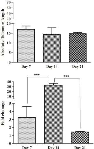
Correlation between hTERT gene expression and cellular turnover
Brazvan et al.
Telomere length at day 7, 14 and 21 accounted for 17.2 kb, 14.4 kb and 14.4 kb, respectively and there were no significant differences between groups (P>0.05) (Figure 6A). Mean hTERT gene expression was 3.26, 33.11 and 1.44 in days 7, 14 and 21 of culture respectively. Significant differences were seen at day 14 and 21 (P<0.001) (Figure 6B). Also there was correlation between telomere length and hTERT gene expression at 14 day (r = 0.998, p =0.04).
Figure 6. association between of telomere length and hTERT gene expression Fold in SCF+FLT3+IL-2+IL-7+IL-15 group of MNCs (using 80ng/µl of each cytokines). (A) Telomere length (Kbp) in 7, 14 and 21 days of MNCs. (B) hTERT gene expression fold in 7, 14 and 21 days of MNCs. Differences between groups are significant at *p <0.05, **p <0.01, and ***p<0.001.
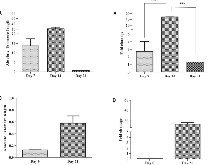
Both Telomere length and hTERT gene expression in MNCs and CD34 positive cells are high using Interleukin-15 (IL-15)
Both MNC and CD34+ cells (n= 4 ×10⁵) were co_cultured with IL-15 and telomere length and hTERT gene expression were measured at different time points. Telomere length was measured in MNCs by real-time PCR. Absolute telomere length was 13.68 kb at day 7, 22.52 and 0.7 kb at day 14 and 21, respectively. The highest telomere length was seen at day 14. There was no significant difference between groups (P>0.05) (Figure 7A). hTERT gene expression fold at day 7, 14 and 21
day was 2.73, 48.54 and 1.34, respectively. Results showed that there are significant differences in hTERT gene expression between day 14 and 21(P<0.001). The highest hTERT gene expression fold was observed at day 14 (Figure 7B).
Final telomere length of CD34+ accounted for 0.12 kb in non-cultured cells and 0.58 kb in cells cultured for 21 days. There was no significant difference between telomere length at noncultured cells and cells cultured for 21 days (P>0.05) (Figure 7C). hTERT gene Expression fold was 0.14 at noncultured and 0.11at day 21. There was no significant difference between hTERT expression in non-cultured and at day 21 in CD34+ cells (P>0.05) (Figure 7D).
Discussion
Natural killer cell, T and B lymphocyte development are controlled by immune cell cytokines (2, 7 and IL-15) which known as common cytokine gamma chain. Previous study demonstrated that there is a relationship between B cell development and IL-2 and IL-7.28 Our results showed that IL-2 and IL-7 affect the expansion of T and B cells more than NK cells at 14 day of culture. NK cell development is dependent to two member of common cytokine gamma chain on the other hand IL-2 and IL-15.29 Similar to previous study, NK cells expansion percent was higher in IL-2 and Il-15 group more than IL-7 group.
Effects of immune cell cytokines on telomere activity
experiment which may be due to the late expansion of T cell in response to IL-7. Yu Li et al described the potency of IL-15 to induce telomerase expression during activation of the Jak3 and PI3K/ AKT signaling pathways in T lymphocytes. Particularly in memory CD8+T cells.37 On the other hand, IL-15-induced telomerase dynamic is associated with the relative stable
telomere length in long-term cultured memory phenotype CD8+T cells.37 The induction of hTERT via cytokines is distinctly mediated by PI3K/Akt, or the NF-kB pathways. Among the possible mechanism suggested for NF-kB pathway, protein kinase C (PKC) has been shown to have a critical role in up-regulation of TERT expression.38
Figure 7. Comparison of telomere length and hTERT gene expression folds in MNCs and CD34+
in using 80 ng/ µl of IL-15. (A) Telomere length (Kbp) in MNCs in different days of culture period time. (B) hTERT gene expression fold of MNCs in different days of culture period time. (C) Telomere length (Kbp) of CD34+ cells in 0 and 21 days. (D) hTERT gene expression fold of CD34+ in 0 and 21 days. Differences between groups are significant at *p <0.05, **p <0.01, and ***p<0.001.
In CD34+ cells, telomere length and hTERT gene expression analysis showed that telomere length was higher at the 21th day, thereby indicating that prolonged exposure to IL-7 its effects. The CD34+ cord blood cells proliferated faster in response to cytokine stimulation and total cell proliferation was increased sevenfold compared to their counterpart bone marrow.39 Study of colony-forming dynamic by sorted cord blood and bone marrow CD34+ cells revealed that, although the number of CD34+ cells in cord blood is less than bone marrow, cord blood CD34+ cells have a significantly higher number of colony formation.36 This is possible that
telomere length and hTERT gene expression in MNCs is an outcome of CD34+ cells.
Brazvan et al.
mechanism. Another theory may be is when cytokines used with together telomere length not affected by hTERT expression level and remodeling in chromatin structure cause to this condition. In the other manner, in this condition telomere length not regulated by hTERT level.40,41
Conclusion
In conclusion, this study showed that telomere length and hTERT gene expression are affected by IL-7 ad IL-15. The longest telomere length and highest hTERT gene expression were observed at 14th day of culture when cells treated with Il-7 and Il-15. But IL-2 has no distinct effect on telomere length and hTERT gene expression.
Acknowledgments
The authors thank the Stem Cell Research Center, Tabriz University of Medical Sciences. This work was supported by a grant (5/104/618) from the Tabriz University of Medical Sciences. We further thank Mrs. Montazer saheb for her great help in this study.
Ethical Issues Not applicable.
Conflict of Interest
All author declared no conflict of interest.
References
1. Lu W, Zhang Y, Liu D, Songyang Z, Wan M. Telomeres-structure, function, and regulation. Exp
Cell Res 2013;319(2):133-41. doi:
10.1016/j.yexcr.2012.09.005
2. O’Callaghan NJ, Fenech M. A quantitative PCR method for measuring absolute telomere length. Biol
Proced Online 2011;13:3. doi:
10.1186/1480-9222-13-3
3. Arenas-Aranda DJ, Hernández-Caballero E, Salamanca-Gómez F. Cellular Senescence and Its Relation with Telomere. INTECH Open Access Publisher; 2012.
4. Oeseburg H, de Boer RA, van Gilst WH, van der Harst P. Telomere biology in healthy aging and disease.
Pflugers Arch 2010;459(2):259-68. doi:
10.1007/s00424-009-0728-1
5. Price LH, Kao HT, Burgers DE, Carpenter LL, Tyrka AR. Telomeres and early-life stress: an overview.
Biol Psychiatry 2013;73(1):15-23. doi:
10.1016/j.biopsych.2012.06.025
6. Donate LE, Blasco MA. Telomeres in cancer and ageing. Philos Trans R Soc Lond B Biol Sci 2011;366(1561):76-84. doi: 10.1098/rstb.2010.0291 7. Hao LY, Armanios M, Strong MA, Karim B, Feldser
DM, Huso D, et al. Short telomeres, even in the presence of telomerase, limit tissue renewal capacity.
Cell 2005;123(6):1121-31. doi:
10.1016/j.cell.2005.11.020
8. Blasco MA. Telomere length, stem cells and aging.
Nat Chem Biol 2007;3(10):640-9. doi:
10.1038/nchembio.2007.38
9. Flores I, Blasco MA. The role of telomeres and telomerase in stem cell aging. FEBS Lett
2010;584(17):3826-30. doi:
10.1016/j.febslet.2010.07.042
10. Hiyama E, Hiyama K. Telomere and telomerase in stem cells. Br J Cancer 2007;96(7):1020-4. doi: 10.1038/sj.bjc.6603671
11. Shay JW, Wright WE. Role of telomeres and telomerase in cancer. Semin Cancer Biol
2011;21(6):349-53. doi:
10.1016/j.semcancer.2011.10.001
12. Kimura M, Gazitt Y, Cao X, Zhao X, Lansdorp PM, Aviv A. Synchrony of telomere length among hematopoietic cells. Exp Hematol 2010;38(10):854-9. doi: 10.1016/j.exphem.2010.06.010
13. Lin J, Epel E, Cheon J, Kroenke C, Sinclair E, Bigos M, et al. Analyses and comparisons of telomerase activity and telomere length in human T and B cells: insights for epidemiology of telomere maintenance. J
Immunol Methods 2010;352(1-2):71-80. doi:
10.1016/j.jim.2009.09.012
14. Dioni L, Hoxha M, Nordio F, Bonzini M, Tarantini L, Albetti B, et al. Effects of short-term exposure to inhalable particulate matter on telomere length, telomerase expression, and telomerase methylation in steel workers. Environ Health Perspect 2011;119(5):622-7. doi: 10.1289/ehp.1002486 15. Copley MR, Beer PA, Eaves CJ. Hematopoietic stem
cell heterogeneity takes center stage. Cell Stem Cell 2012;10(6):690-7. doi: 10.1016/j.stem.2012.05.006 16. Zhang CC, Lodish HF. Cytokines regulating
hematopoietic stem cell function. Curr Opin Hematol 2008;15(4):307-11.
doi: 10.1097/MOH.0b013e3283007db5
17. Frauwirth KA, Thompson CB. Regulation of T
lymphocyte metabolism. J Immunol
2004;172(8):4661-5. doi: 10.4049/
jimmunol.172.8.4661
18. Gaffen SL, Liu KD. Overview of interleukin-2 function, production and clinical applications.
Cytokine 2004;28(3):109-23. doi:
10.1016/j.cyto.2004.06.010
19. Kandasamy K, Mohan SS, Raju R, Keerthikumar S, Kumar GS, Venugopal AK, et al. NetPath: a public resource of curated signal transduction pathways. Genome Biol 2010;11(1):R3. doi: 10.1186/gb-2010-11-1-r3
20. Rathmell JC, Vander Heiden MG, Harris MH, Frauwirth KA, Thompson CB. In the absence of extrinsic signals, nutrient utilization by lymphocytes is insufficient to maintain either cell size or viability. Mol Cell 2000;6(3):683-92. doi: 10.1016/S1097-2765(00)00066-6
Effects of immune cell cytokines on telomere activity
J Med 2011;365(22):2067-77. doi:
10.1056/NEJMoa1105143
22. Guo Q, Chen X, Du Y, Guo J, Su Y. Cyclic AMP-Responsive Element Modulator alpha Polymorphisms Are Potential Genetic Risks for Systemic Lupus Erythematosus. J Immunol Res 2015;2015:906086. doi: 10.1155/2015/906086
23. Suzuki S, Iwamoto M, Saito Y, Fuchimoto D, Sembon S, Suzuki M, et al. Il2rg gene-targeted severe combined immunodeficiency pigs. Cell Stem Cell 2012;10(6):753-8. doi: 10.1016/j.stem.2012.04.021 24. Janot-Sardet C, Assouline B, Cheynier R, Morre M,
Beq S. A validated assay to measure soluble IL-7 receptor shows minimal impact of IL-7 treatment. J
Immunol Methods 2010;353(1-2):115-23. doi:
10.1016/j.jim.2009.12.003
25. Anthony SM, Schluns KS. Emerging roles for IL-15 in the activation and function of T-cells during immune stimulation. Rese Rep Biol 2015;6:25-37. doi: 10.2147/RRB.S57685
26. Aliyari Z, Alami F, Mostafavi T, Taiefi Nasrabadi H, Soleimanirad J, Nozad Charoudeh H. The roles of IL-2, IL-7, and IL15 ligands in B cells development from cord blood mononuclear cells. Iran J Ped Hematol Oncol 2015;5(3):155-60.
27. Aliyari Z, Alemi F, Brazvan B, Tayefi Nasrabadi H, Nozad Charoudeh H. CD26+ Cord Blood Mononuclear Cells Significantly Produce B, T, and NK Cells. Iran J Immunol 2015;12(1):16-26. 28. Parrish YK, Baez I, Milford TA, Benitez A,
Galloway N, Rogerio JW, et al. IL-7 Dependence in human B lymphopoiesis increases during progression of ontogeny from cord blood to bone marrow. J
Immunol 2009;182(7):4255-66. doi:
10.4049/jimmunol.0800489
29. Rochman Y, Spolski R, Leonard WJ. New insights into the regulation of T cells by γc family cytokines.
Nat Rev Immunol 2009;9(7):480-90. doi:
10.1038/nri2580
30. Blackburn EH. Telomere states and cell fates. Nature 2000;408(6808):53-6. doi: 10.1038/35040500 31. Zhang B, Qian D, Ma HH, Jin R, Yang PX, Cai MY,
et al. Anthracyclines disrupt telomere maintenance by telomerase through inducing PinX1 ubiquitination and degradation. Oncogene 2012;31(1):1-12. doi: 10.1038/onc.2011.214
32. Cong Y, Shay JW. Actions of human telomerase beyond telomeres. Cell Res 2008;18(7):725-32. doi: 10.1038/cr.2008.74
33. Schuller CE, Jankowski K, Mackenzie KL. Telomere length of cord blood-derived CD34(+) progenitors predicts erythroid proliferative potential. Leukemia 2007;21(5):983-91. doi: 10.1038/sj.leu.2404631 34. Wallace DL, Berard M, Soares MV, Oldham J, Cook
JE, Akbar AN, et al. Prolonged exposure of naive CD8+ T cells to interleukin-7 or interleukin-15 stimulates proliferation without differentiation or loss of telomere length. Immunology 2006;119(2):243-53. doi: 10.1111/j.1365-2567.2006.02429.x
35. Martens UM, Brass V, Sedlacek L, Pantic M, Exner C, Guo Y, et al. Telomere maintenance in human B lymphocytes. Br J Haematol 2002;119(3):810-8. doi: 10.1046/j.1365-2141.2002.03910.x
36. Cao JN, Gollapudi S, Sharman EH, Jia Z, Gupta S. Age-related alterations of gene expression patterns in human CD8+ T cells. Aging Cell 2010;9(1):19-31. doi: 10.1111/j.1474-9726.2009.00534.x
37. Li Y, Zhi W, Wareski P, Weng NP. IL-15 activates telomerase and minimizes telomere loss and may preserve the replicative life span of memory CD8+ T cells in vitro. J Immunol 2005;174(7):4019-24. doi: 10.4049/jimmunol.174.7.4019
38. Barsov EV. Telomerase and primary T cells: biology and immortalization for adoptive immunotherapy.
Immunotherapy 2011;3(3):407-21. doi:
10.2217/imt.10.107
39. Khan S, Jutzy JMS, Aspe JR, McGregor DW, Neidigh JW, Wall NR. Survivin is released from cancer cells via exosomes. Apoptosis 2011;16(1):1-12. doi: 10.1007/s10495-010-0534-4
40. Atkinson SP, Hoare SF, Glasspool RM, Keith WN. Lack of telomerase gene expression in alternative lengthening of telomere cells is associated with chromatin remodeling of the hTR and hTERT gene promoters. Cancer Res 2005;65(17):7585-90. doi: 10.1158/0008-5472.CAN-05-1715