3 artigo 410
ORIGINAL ARTICLE
1 – Resident Physician at the Passo Fundo Orthopedics Hospital, Passo Fundo, RS, Brazil.
2 – Orthopedist and Traumatologist in the Hand Group, Passo Fundo Orthopedics Hospital, Passo Fundo, RS, Brazil. Work performed at the Passo Fundo Orthopedics Hospital, Passo Fundo, RS, Brazil.
Correspondence: Hospital Ortopédico de Passo Fundo/RS. Rua Sete de Setembro, 817 – 99010-121 – Passo Fundo, RS. E-mail: [email protected] Work received for publication: August 18, 2010; accepted for publication: March 22, 2011.
FIXATION OF FRACTURES OF THE DISTAL EXTREMITY OF THE RADIUS
USING THE MODIFIED KAPANDJI TECHNIQUE: EVALUATION
OF THE RADIOLOGICAL RESULTS
Antonio Piva Neto1, Fabio Colla Lhamby2
INTRODUCTION
In fractures of the distal extremity of the radius that present displacement and instability, some loss of the reduction initially obtained will occur unless the case is adequately managed. Percutaneous fixa-tion and plaster-cast immobilizafixa-tion are a simple and widely used method among trauma surgeons.
Today, several types of osteosynthesis devices are available on the market to aid surgeons in managing the different types of fracture encountered. However, Kirschner wires have their place in treating fractures of the distal extremity of the radius and still consti-tute one of the most common methods for fracture fixation, combined with plaster-cast immobilization.
ABSTRACT
Objective: To demonstrate a simple and efficacious option for treating fractures of the distal extremity of the radius using Kirschner wires. Methods: Between September 2008 and April 2009, 48 patients with fractures of the distal extremity of the radius, classified as A3 according to the AO classification, were treated surgically using a modification of the Kapandji technique. Results: Out of the 48 wrists operated, 42 (87.5%) presented postoperative measurements within the acceptable limits. We used the parameters of McQuenn and Caspers who considered that the radial angulation should be wider than 19° and the volar angulation should be narrower than -12°. All the postoperative volar inclination measurements
The authors declare that there was no conflict of interest in conducting this work
This article is available online in Portuguese and English at the websites: www.rbo.org.br and www.scielo.br/rbort
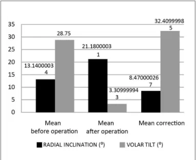
were narrower than -3°. The mean preoperative radial inclination was 13.14° and the mean postoperative value was 21.18°. The mean preoperative volar inclination was 28.75° and the mean postoperative value was 3.31°. The mean preoperative radial height was 5.25 mm and the mean postoperative value was 9.48 mm. Conclusion: The technique described here had excellent stability for treating fractures of the distal extremity of the radius classified as A3. It was easy to implement and minimally invasive, with minimal surgical complications, and it was inexpensive.
Keywords - Radius Fractures/surgery; Radius Fractures/ radiography;Bone Wires
The main disadvantages of fracture fixation using Kirschner fires are the need for postoperative immo-bilization, the need to remove the wires after fracture consolidation, the possibility of fracture displacement after percutaneous fixation and the possibility of tears in the extensor tendons.
pro-369 TECHNIQUE: EVALUATION OF THE RADIOLOGICAL RESULTS
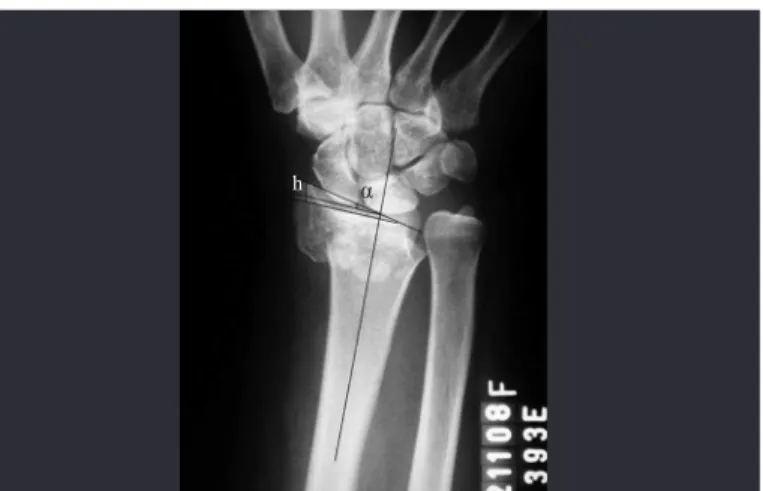
Figure 1 – Preoperative radiograph on wrist in posteroanterior (PA) view, showing an extra-articular fracture of the distal radius with metaphyseal comminution and diminished radial inclination. The radial inclination (α) and radial height (h) are demonstrated on the radiograph.
h α
vide support through sustaining the distal fragments and thus preventing displacement and shortening on these bone pieces (intrafocal fixation).
The surgical method described in the present stu-dy, with combined intrafocal and interfocal fixation with Kirschner wires, for fractures of the distal radius, had the aim of alleviating some of the complications associated with the intrafocal fixation method that was described by Adalbert I. Kapandji. Through
mo-difying Kapandji’s original technique, our objective
was to improve the stability of fracture fixation and demonstrate a simple and effective treatment option, with fewer complications.
MATERIALS AND METHODS
Between September 2008 and April 2009, a pros-pective analysis was conducted on 48 patients with fractures of the distal radius who were treated surgi-cally at Hospital São Vicente de Paulo and the City Hospital, in Passo Fundo, Rio Grande do Sul.
Measurements were made on radiographs produ-ced before the operation, immediately after the ope-ration and six months after the opeope-ration (at the time when the synthesis material was removed).
All the cases were evaluated by two traumatologists, of whom one was a hand surgeon, using the Arbeitsge-meinschaft für Osteosynthesefragen (AO) classification.
Only fractures classified as A3 were included in this study, which were the fractures capable of re-ceiving treatment through the technique presented, according to the proposed method.
The exclusion criteria were the following: frac-tures without the AO classification of A3, fracfrac-tures with volar deviation, “die punch” fractures, exposed fractures, bilateral fractures and multiple fractures. Multiple trauma patients were also excluded.
Fourteen patients were men and 34 were women.
The patients’ ages ranged from 20 to 89 years, with a
mean age of 55 years. The left wrist was affected in 27 patients and the right wrist in 21 patients.
All the fractures were evaluated before and after the operation in the posteroanterior (PA) and lateral radiographic views. The height and inclination an-gle of the radius were measured on PA radiographs (Figure 1), and the volar tilt was measured on lateral radiographs (Figure 2). The presence of joint steps and ulnar variance were also assessed.
Figure 2 – Preoperative radiograph on wrist in lateral view, showing a fracture of the distal radius with metaphyseal dorsal comminution and dorsal deviation. The volar tilt (β) is demonstrated on the radiograph.
We used the postoperative radiographic parameters established by McQueen and Caspers for evaluating the results. These authors considered that the results would be adequate if the radial angle was greater than or equal to 19º (normal: 24°) and if the volar angle was less than -12° (normal: between 4° and 12°).
SURGICAL TECHNIQUE
the reduction maneuver, the locations for inserting the Kirschner wires were marked out.
Application of traction and ulnar flexion and de-viation converted the dorsal angle to neutral and reco-vered the height of the radius through ligamentotaxis, thereby aiding in defining the locations for inserting the Kirschner wires.
After achieving an acceptable reduction, the first intrafocal Kirschner wire was placed at the lateral margin of the radius, by means of a small incision between the first and second compartments of the extensor tendons, one centimeter proximally to the fracture line. This Kirschner wire, which was used as a lever, as described by Kapandji, recovered and maintained the height of the distal radius, thus achie-ving correct alignment of the distal metaphysis over the diaphysis of the radius, in PA view.
With the aim of reduction of the dorsal deviation, another Kirschner wire was used as a level in the focus of the fracture. This second intrafocal wire is placed on the dorsum of the radius, between the third and fourth compartments of the extensor tendon. A small incision was made distally to the focus of the fracture in order to avoid skin traction and tension. Care was required, to ensure that the entire zone of dorsal comminution was situated distally to the wire.
Following this, another three static (transosseous) Kirschner wires were inserted. Through a small radial incision, the first of these wires was positioned at the apex of the radial styloid process and was introduced as far as the opposite cortical bone, thereby comple-menting the fixation of the radial styloid. The other two Kirschner wires were placed on the dorsum of the radius. The path of the wires started on the dorsal rim of the joint, making use of the denser structure of the subchondral bone in order to achieve better support for the assembly. The reference points used for introducing the wires were the intervals between the extensor compartments that have been described for arthroscopic portals. The first Kirschner wire was positioned in portal 3-4 of the wrist, which is located between the third and fourth compartments of the
ex-tensor tendon, distally to Lister’s tubercle. This wire
was placed proximally, going in the volar direction, to go through the opposite cortical bone. The next static Kirschner wire was used to complement the dynamic fixation of the ulnar fragment of the fracture and was
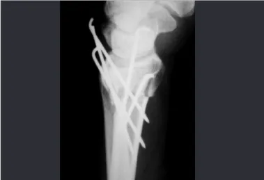
introduced into the portal 6R, which is located in the sixth compartment of the extensor tendon, and was directed proximally and radially. All the Kirschner wi-res were positioned subcutaneously and the incisions were sutured (Figures 3 and 4).
Figure 3 – Radiograph on wrist showing the immediate postoperative result in posteroanterior view, after fixation of the fracture of the distal radius using Kirschner wires, with correction of the radial angulation and restoration of the radial height.
Figure 4 – Radiograph on wrist showing the immediate postoperative result in lateral view, with correction of the dorsal deviation of the distal radius.
The wrist was immobilized using a brace from the axilla to the palm for one week, to control rotation and minimize skin irritation caused by the Kirschner wires. After this period, the splint was removed and, after producing a control radiograph, it was replaced by a plaster cast from the axilla to the palm.
371
The plaster-cast glove and the percutaneous wires were removed in the sixth week of immobilization, as an outpatient procedure under local anesthesia. The final radiograph was produced at that time and phy-siotherapy was then started.
RESULTS
The radiographic parameters before the operation and six weeks afterwards (Figures 5 and 6) were measured and compared (Table 1). It needs to be emphasized that all the patients achieved measure-ments in the immediate postoperative period that were considered adequate.
Out of the 48 wrists that were treated surgically, 42 (87.5%) presented postoperative measurements within the limits that were considered adequate accor-ding to McQueen and Caspers. All the postoperative
measurements on the volar tilt were less than -3° (range: -2° to 12°), with a mean correction of the de-formity of dorsal deviation of 32.41°. The six wrists that did not achieve the postoperative targets presen-ted smaller gains in radial inclination, although all of them presented values that were close to acceptable (16°, 18°, 17°, 15°, 18° and 17°). The ulnar variance was neutral in all these six wrists. In just one wrist, there was a loss of 3° in radial inclination, which went from 23° on the preoperative radiograph to 20° in the postoperative measurement.
The mean preoperative radial inclination was 13.14° (range: 4° to 26°) and the mean postoperative measurement after removal of the Kirschner wires was 21.18° (range: 15° to 28°), with a mean correc-tion of 8.47°. The mean preoperative volar tilt was 28.75° (range: 5° to 48° of dorsal deviation). The volar tilt measurement after removal of the Kirschner wires was 3.31° (range: 5° of dorsal tilt to 12° of volar tilt), with a mean correction of 32.41° (Figure 7).
Figure 5 – Radiograph on wrist in posteroanterior view after removal of the Kirschner wires, showing the final result from treatment of the fracture of the distal radius.
Figure 6 – Radiograph on wrist in lateral view after removal of the synthesis material, showing correction of the volar angulation of the distal radius.
Table 1 – Change in radiographic values from before to after the operation.
Before operation After operation Mean correction
Radial
inclination (0) 4 to 26 15 to 28 8.47
Volar tilt (0) -5 to -48 12 to -5 32.41
Radial height
(mm) 0 to 10 7 to -15 4.2
Figure 7 – Mean values before and after the operation and mean correction of radial inclination and volar tilt.
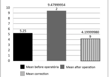
There was a mean correction of radial height of 4.2 mm (range: 1 to 9 mm), with a preoperative mean of 5.25 mm (range: 0 to 10 mm) and a postoperative mean of 9.48 mm (range: 7 to 15 mm) (Figure 8).
the need for postoperative plaster-cast immobiliza-tion. One of the criticisms of percutaneous fixation is that it does not provide stability, i.e. it is difficult to maintain the postoperative reduction(6-8). The fracture
tends to collapse. Geatting and Bisshop(9) used the
Ka-pandji technique in elderly individuals and observed that the deformity recurred(10-12).
In the present study, we observed that losses of the initial reduction occurred in patients with marked dorsal comminution in which the support provided by the dorsal cortical bone was lost. Some loss of the initial reduction was also expected in elderly pa-tients due to the osteopenia and comminution that are commonly present in such patients(13). Our
ob-jective through modifying Kapandji’s technique was
to improve the stability of fixation by bringing in transosseous wires, with the aim of diminishing the displacements and thus the complications coming from treating this type of fracture.
In only two (4.1%) of the 48 cases presented were there losses of the initial reduction. In one of the wrists, the loss was in the volar angle, which was initially a volar tilt of 2° and became a dorsal tilt of 5°. This patient presented a high degree of osteopenia and major comminution in the dorsal cortical bone of the distal radius, which demanded special care in relation to this type of fracture. In another 13 wrists (27%) with significant dorsal comminution, no losses of the reduction in volar tilt were observed. In another case of loss of the initial reduction, there was a loss of radial inclination from 23° to 20°, although this value was still within the desired range.
Several authors(14) attempted to implement early
mobilization and obtained unsatisfactory results, en-ding up with a variety of complications such as in-tense pain, reflex sympathetic dystrophy, loss of the initial reduction and torn tendons, among others. In the cases that we analyzed, there were no cases of ex-tensor tendon tears or of nerve injuries. Five patients (10.4%) presented skin lesions adjacent to the Kirsch-ner wires. There was one case of superficial infection in a patient who presented skin lesions. There were no cases of deep or bone infection.
It is known that there is a significant correlation between maintaining good radiological reduction un-til fracture consolidation is achieved and presenting good function. Moreover, wrist function depends on
Figure 8 – Mean values for radial height before and after the operation and mean correction.
It was observed in five cases that there was some loss of the initial reduction seen on radiographs pro-duced immediately after the operation, when these were compared with radiographs produced six weeks after the operation. In none of these cases was there any volar displacement of the distal fragment of the fracture. In one case, there was some loss of the ini-tial postoperative volar angle, which went from 2° of volar tilt to 5° of dorsal tilt.
There were no cases of torn extensor tendons or nerve injuries. Five patients (10.4%) presented skin lesions adjacent to the Kirschner wires. There was one case of superficial infection in one of the patients who presented skin lesions. There were no cases of deep or bone infection.
DISCUSSION
Adalbert I. Kapandji described a technique for fi-xation of fractures of the distal extremity of the radius using intrafocal Kirschner wires(1-3). This technique
was initially recommended for young patients. Becau-se of its success, its uBecau-se has spread and the indications have increased(4,5).
373
the degree of destructuring caused by loss of the nor-mal palmar angulation of the distal radial, which is difficult to restore and maintain through the methods of closed reduction and immobilization(15). In this
re-gard, the surgeon’s main aim is to achieve an anato -mical reduction that is maintained until consolidation,
independent of the patient’s age(16).
The method demonstrated here achieved this ob-jective. Out of the 48 wrists operated, six did not at-tain parameters that would be considered ideal, but the valued achieved were very close to the ideal. None of these wrists attained the desired radial inclination measurement, but were very close to the ideal value. The distal radioulnar joints were congruent and the ulnar variance measurements were neutral in all of them, thus indicating that the problems of mobility and ulnocarpal impaction were avoided.
Another of the possible complications from
Kapandji’s technique is that anterior displacement of
the distal fragment of the fracture may occur through exacerbated reduction(5,17-19). Associated use of the
technique of intrafocal fixation with transosseous wi-res attempts to improve the stability of the fixation, thereby avoiding this type of problem. In one wrist in this series (2%), this problem occurred. This case was analyzed, and it was perceived that the displace-ment was already present on the radiograph produced
immediately after the operation. Thus, we concluded that because of exacerbated reduction force (leverage on the dorsal intrafocal wire), volar displacement of the distal portion of the radius occurred. This could be corrected before performing the transosseous fixation.
Regarding the Kirschner wires used in this tech-nique, it is known that there is no significant diffe-rence in the final stability provided by wires of 2.0 or 1.6 mm in diameter. For this reason, we chose to use wires of 1.6 mm, which are more mallea-ble and therefore less aggressive towards the bone fragments(20,21).
From the results obtained, it was demonstrated that this variation in the technique described originally by Dr. Adalbert I. Kapandji is viable. It is a minimally invasive procedure that is simple to perform and fast. It can be done by trauma surgeons and produces ac-ceptable radiological results. The complications en-countered with the original method can be avoided.
CONCLUSION
We have presented an alternative surgical techni-que for treating wrist fractures. This operation is easy to carry out and minimally invasive, with minimal surgical complications. It is a low-cost method and produces reliable bone stability.
REFERENCES
1. Kapandji A. Focal pinning of radial distal fractures. Cahier Cirurg. 1987; 63:25 8.
2. Cooney WP 3rd, Linscheid RL, Dobyns JH. External pin fixation for unstable Colles’ fractures. J Bone Joint Surg Am. 1979;61(6A):840-5.
3. Stoffelen DV, Broos PL. Closed reduction versus Kapandji-pinning for extra-articular distal radial fractures. J Hand Surg Br. 1999;24(1):89-91.
4. Board T, Kocialkowski A, Andrew G. Does Kapandji wiring help in older pa-tients? A retrospective comparative review of displaced intra-articular distal radial fractures in patients over 55 years. Injury. 1999;30(10):663-9.
5. Hollevoet N, Verdonk R. Anterior fracture displacement in Colles’ fractures after Kapandji wiring in women over 59 years. Int Orthop. 2007;31(3):397-402.
6. Rosati M, Bertagnini S, Digrandi G, Sala C. Percutaneous pinning for fractures of the distal radius. Acta Orthop Belg. 2006;72(2):138-46.
7. Barton T, Chambers C, Lane E, Bannister G. Do Kirschner wires maintain reduction of displaced Colles’ fractures? Injury. 2005;36(12):1431-4.
8. Glickel SZ, Catalano LW, Raia FJ, Barron OA, Grabow R, Chia B. Long-term outcomes of closed reduction and percutaneous pinning for the treatment of distal radius fractures. J Hand Surg Am. 2008;33(10):1700-5.
9. Greatting MD, Bishop AT. Intrafocal (Kapandji) pinning of unstable fractures of the distal radius. Orthop Clin North Am. 1993;24(2):301-7.
10. Haas J L, Caffiniere de la J Y. Fixation of distal radial fractures: intramedullary pinning versus external fixation. In: Saffar P, Cooney WP, editors. Fractures of the distal radius. London: Martin Dunitz; 1995; p. 229-39.
11. Oskam J, Kingma J, Bart J, Klasen HJ. K-wire fixation for redislocated Colles’ fractures. Malunion in 8/21 cases. Acta Orthop Scand. 1997;68(3):259-61. 12. Snow M. Kelly M. Jeyam M. Fahmy N. Functional versus position: a randomized
controled trial of interfocal Kirschner wiring of unstable distal radial fractures. Eur J Trauma Emerg Surg. 2007;33(1):81-6.
13. Board T, Kocialkowski A, Andrew G. Does Kapandji wiring help in older pa-tients? A retrospective comparative review of displaced intra-articular distal radial fractures in patients over 55 years. Injury. 1999;30(10):663-9.
14. Lenoble E, Dumontier C, Goutallier D, Apoil A. Fracture of the distal radius. A prospective comparison between trans-styloid and Kapandji fixations. J Bone Joint Surg Br. 1995;77(4):562-7.
15. McQueen MM, Hajducka C, Court-Brown CM. Redisplaced unstable fractures of the distal radius: a prospective randomised comparison of four methods of treatment. J Bone Joint Surg Br. 1996;78(3):404-9.
16. Kumar S, Penematsa S, Sadri M, Deshmukh SC. Can radiological results be surrogate markers of functional outcome in distal radial extra-articular fractures? Int Orthop. 2008;32(4):505-9.
17. Lenoble E, Dumontier C, Goutallier D, Apoil A. Fracture of the distal radius. A prospective comparison between trans-styloid and Kapandji fixations. J Bone Joint Surg Br. 1995;77(4):562-7.
18. Nonnenmacher J, Kempf I. [Role of intrafocal pinning in the treatment of wrist fractures]. Int Orthop. 1988;12(2):155-62.
19. Peyroux LM, Dunaud JL, Caron M, Ben Slamia I, Kharrat M. The Ka-pandji technique and its evolution in the treatment of fractures of the distal end of the radius. Report on a series of 159 cases. Ann Chir Main. 1987;6(2):109-22.
20. Naidu SH, Capo JT, Moulton M, Ciccone W 2nd, Radin A. Percutaneous pinning of distal radius fractures: a biomechanical study. J Hand Surg Am. 1997;22(2):252-7.
21. Mittelmeier W, Braun C, Schäfer R. The Kapandji technique for fixation of distal radius fractures--a biomechanical comparison of primary stability. Arch Orthop Trauma Surg. 2001;121(3):135-8.