Peripheral Blood Mononuclear Cells from Chronic
Chagas’ Disease Cardiomyopathy Patients in Response
to
T. cruzi
Ribosomal P Proteins
Silvia A. Longhi1,2, Augusto Atienza3, Graciela Perez Prados4, Alcinette Buying5, Virginia Balouz6, Carlos A. Buscaglia6, Radleigh Santos7, Laura M. Tasso1, Ricardo Bonato3, Pablo Chiale3,
Clemencia Pinilla5., Valeria A. Judkowski5., Karina A. Go´mez1,2 *
1Instituto de Investigaciones en Ingenierı´a Gene´tica y Biologı´a Molecular (INGEBI) - Consejo Nacional de Investigaciones Cientı´ficas y Tecnolo´gicas (CONICET), Buenos Aires, Argentina,2Facultad de Farmacia y Bioquı´mica, Universidad de Buenos Aires, Buenos Aires, Argentina,3Hospital General de Agudos J.M. Ramos Mejı´a, Buenos Aires, Argentina,4Hospital General de Agudos J. A. Ferna´ndez, Buenos Aires, Argentina,5Torrey Pines Institute for Molecular Studies, San Diego, California, United States of America,6Instituto de Investigaciones Biotecnolo´gicas ‘‘Dr. Rodolfo Ugalde’’, Universidad Nacional de San Martı´n (UNSAM) - Consejo Nacional de Investigaciones Cientı´ficas y Te´cnicas (CONICET), Campus UNSAM, San Martı´n, Buenos Aires, Argentina,7Torrey Pines Institute for Molecular Studies, Port St. Lucie, Florida, United States of America
Abstract
Background:Trypanosoma cruziribosomal P proteins, P2band P0, induce high levels of antibodies in patients with chronic Chagas’ disease Cardiomyopathy (CCC). It is well known that these antibodies alter the beating rate of cardiomyocytes and provoke apoptosis by their interaction withb1-adrenergic and M2-muscarinic cardiac receptors. Based on these findings, we decided to study the cellular immune response to these proteins in CCC patients compared to non-infected individuals.
Methodology/Principal findings: We evaluated proliferation, presence of surface activation markers and cytokine production in peripheral blood mononuclear cells (PBMC) stimulated with P2b, the C-terminal portion of P0 (CP0) proteins andT. cruzilysate from CCC patients predominantly infected with TcVI lineage. PBMC from CCC patients cultured with P2b or CP0 proteins, failed to proliferate and express CD25 and HLA-DR on T cell populations. However, multiplex cytokine assays showed that these antigens triggered higher secretion of IL-10, TNF-aand GM-CSF by PBMC as well as both CD4+
and CD8+T cells subsets of CCC subjects. UponT. cruzilysate stimulation, PBMC from CCC patients not only proliferated but also became activated within the context of Th1 response. Interestingly,T. cruzilysate was also able to induce the secretion of GM-CSF by CD4+or CD8+T cells.
Conclusions/Significance:Our results showed that although the lack of PBMC proliferation in CCC patients in response to ribosomal P proteins, the detection of IL-10, TNF-a and GM-CSF suggests that specific T cells could have both immunoregulatory and pro-inflammatory potential, which might modulate the immune response in Chagas’ disease. Furthermore, it was possible to demonstrate for the first time that GM-CSF was produced by PBMC of CCC patients in response not only to recombinant ribosomal P proteins but also to parasite lysate, suggesting the value of this cytokine to evaluate T cells responses inT. cruziinfection.
Citation:Longhi SA, Atienza A, Perez Prados G, Buying A, Balouz V, et al. (2014) Cytokine Production but Lack of Proliferation in Peripheral Blood Mononuclear Cells from Chronic Chagas’ Disease Cardiomyopathy Patients in Response toT. cruziRibosomal P Proteins. PLoS Negl Trop Dis 8(6): e2906. doi:10.1371/journal. pntd.0002906
Editor:Mauricio Martins Rodrigues, Federal University of Sa˜o Paulo, Brazil ReceivedJanuary 29, 2014;AcceptedApril 15, 2014;PublishedJune 5, 2014
Copyright:ß2014 Longhi et al. This is an open-access article distributed under the terms of the Creative Commons Attribution License, which permits unrestricted use, distribution, and reproduction in any medium, provided the original author and source are credited.
Funding:This work was financially supported by grants from the Consejo Nacional de Investigaciones Cientı´ficas y Tecnolo´gicas (CONICET) and Universidad de Buenos Aires to KAG and the Agencia Nacional de Promocio´n Cientı´fica y Tecnolo´gica (ANPCyT) and Fundacio´n Bunge y Born to CAB and KAG. The funders had no role in study design, data collection and analysis, decision to publish, or preparation of the manuscript.
Competing Interests:The authors have declared that no competing interests exit. * E-mail: drkagomez@gmail.com
.These authors contributed equally to this work.
Introduction
Trypanosoma cruzi, the etiological agent of Chagas’ disease, affects approximately 8–10 million people, and its infection is one of the major human health problems in Central and South America, being extended now to Europe (especially Spain and Portugal), the
digestive alterations or both manifestations (cardiac plus digestive) [5]. Chronic Chagas’ disease Cardiomyopathy (CCC), the most frequent and severe consequence of the chronic infection by T. cruzi, is manifested predominately as an arrhythmogenic cardio-myopathy [6–9].
Up to now, the mechanisms of the pathophysiology of Chagas’ disease are not completely elucidated and two main hypotheses have been proposed. The first one is based on the inflammatory reaction elicited by the parasite leading to tissue damage, while the second argues for an autoreactive process resulting from an impaired immune response associated with molecular mimicry [10–13]. However, it is currently accepted that both mechanisms are not mutually exclusive and that Chagas’ disease is the result of both, parasite persistence in the chronic phase and the presence of autoantibodies/self-reactive T cells to host molecules [14,15]. As supporting evidence for the autoimmune hypothesis, previous work in our laboratory demonstrated the presence of circulating antibodies against ribosomal P proteins ofT. cruzi(anti-P Abs) with agonist-like properties on cardiac receptors in patients with CCC [16–24]. Those Abs predominantly recognized the C-terminal end of P2b (peptide R13, EEEDDDMGFGLFD) or P0 proteins (peptide P015, EEEDDDDDFGMGALF), which bear structural similarity to the acidic motif, AESDE, located on the second extracellular loop of the cardiac receptor [19,20,22]. Several studies including patients with CCC as well as experiments performed in mice immunized with recombinant P2b or P0 protein demonstrated a correlation between the presence of anti-P Abs and cardiac disorders [21,22]. These findings were confirmed by the generation of anti-R13 monoclonal Ab, mAb 17.2, which not only induce a dose-dependent increase on the beating frequency of rat cardiomyocytes in culture that is abolished by bisoprolol, a specificb1-adrenergic receptor antagonist [25], but also provoke apoptosis in the murine cardiac cell line HL-1 by its long-lasting b1-AR stimulatory activity [24]. The humoral immune response against ribosomal P proteins has been largely studied in patients with CCC; however, little is known about their recognition by T cells.
Most studies concerning the T cell immune response in Chagas’ disease, have been performed using freshly isolated peripheral
blood mononuclear cells (PBMC) but stimulated with epimastigote (the replicative form found in the midgut of insect vectors) or trypomastigote (the infective form found in the bloodstream and other human extracellular fluids) lysate [26–29]. Few investiga-tions have been focused on the reactivity of T cells against purified antigens of the parasite [30–40]. To date, studies performed with recombinant parasite proteins, such as the cytoplasmatic repetitive antigen (CRA), B13, trans-sialidase, and paraflagellar rod proteins on PBMC and cruzipain on T cells lines revealed that patients with CCC produced significant amount of IFN-cupon stimula-tion, which is in line with the typical pattern of inflammatory response described forT. cruzilysate [34–40]. However, Lorenaet al.also reported that the flagellar repetitive antigen (FRA) induced proliferation of PBMC by thymidine incorporation, but no difference was observed in IFN-cand TNF-asecretion between patients with CCC and non-infected individuals [37]. The aim of this study was to analyze the cellular immune response developed in patients with CCC against T. cruzi ribosomal P proteins, knowing the existence of a cross-reactive component at the humoral level. The specificity of the response was analyzed by proliferation and cytokine production using multiplex technology because it allows to quantify a large spectrum of cytokines in the same cell culture supernatant. Results showed that T. cruzi ribosomal P proteins, specifically P2band the C-terminal portion of P0 (CP0, 110 aa), did not induce the proliferation of PBMCs from CCC in a different manner than non-infected individuals. However, these antigens were able to induce the secretion of IL-10, TNF-a and GM-CSF by PBMC as well as both CD4+ and CD8+ T cells in patients with CCC. Surprisingly, ribosomal P proteins did not stimulate but actually reduced the secretion of IFN-cin cardiac patients. Furthermore, our results demonstrate for the first time that GM-CSF is produced in response not only to parasite lysate but also to ribosomal P proteins. These findings suggest that GM-CSF production could be included in the future to evaluate whole parasite and parasite protein specific T cell responses in Chagas’ disease.
Methods
Ethics statement
The research protocols followed the tenets of the Declaration of Helsinki and were approved by the Medical Ethics Committee of Ramos Mejı´a and Ferna´ndez Hospitals. All enrolled patients gave written informed consent, according to the guidelines of the Ethical Committee of the Hospitals, before blood collection and after the nature of the study was explained.
Study population
Patient selection was conducted at the Cardiovascular Division of the Ramos Mejı´a and Ferna´ndez Hospitals, Buenos Aires, Argentina. Positive serology for Chagas’ disease was determined by two or more tests (indirect immunofluorescence, enzyme-linked immunosorbent assay [ELISA], indirect hemagglutination, or complement fixation). Patients who had at least two of three tests were considered positive for Chagas’ disease. Patients underwent a complete clinical and cardiologic examination that included medical history, physical examination, electrocardiogram (ECG) at rest, laboratory and chest X-ray analysis, and echo doppler cardiography evolution. The exclusion criteria included the presence of systemic arterial hypertension, diabetes mellitus, thyroid dysfunction, renal insufficiency, chronic obstructive pulmonary disease, hydroelectrolytic disorders, alcoholism, history suggesting coronary artery obstruction and rheumatic disease, and the impossibility of undergoing the examinations. The study Author Summary
Chronic Chagas’ disease Cardiomyopathy (CCC) is the most frequent and severe consequence of the chronic infection by protozoan parasiteT. cruzi. Patients with CCC develop high levels of antibodies against ribosomal P proteins ofT. cruzi, called P2band P0. These antibodies can cross-react with, and stimulate, theb1-adrenergic and M2 muscarinic cardiac receptors, inducing a functional and pathological response in cardiomyocytes. In this study, we focused on the cellular immune response developed by CCC patients in response to T. cruzi ribosomal P proteins. Peripheral blood mononuclear cells (PBMC) from CCC patients stimulated with both proteins neither proliferated nor induced the expression of activation markers on CD4+and
CD8+ T cells. However, these cells responded by the
population consisted of 27 patients who completed the screening protocol and were diagnosed with Chronic Chagas’ disease Cardiomyopathy.
Twenty non-infected individuals (NI), within the same age range (30–70 years old) and showing negative serological tests for Chagas’ disease, were included as control group.
TSSA recombinant proteins
Due to its predominant clonal proliferation, theT. cruzispecies is composed by multiple strains showing extensive genetic diversity, which were recently grouped into 6 evolutionary lineages or discrete typing units (DTUs) known as TcI to TcVI [41].
Gluthatione S-transferase (GST)-fusion proteins bearing the entire TSSA from Sylvio X-10/1 strain (henceforth TSSA Sy, representative of TSSA isoforms from DTU TcI parasites) and CL Brener strain (henceforth TSSA CL, representative of TSSA isoforms from DTUs TcII/TcV/TcVI parasites) were expressed in Escherichia coli BL21 strain and purified as described [42]. Briefly, supernatants of bacterial cultures transformed with the indicated construct were induced for 3 h at 28uC with 0.250 mM isopropyl—b-D-thiogalactopyranoside, purified by glutathione-Sepharose chromatography and extensively dialyzed against PBS. The purity and integrity of GST-TSSA samples was assessed with silver-stained SDS-PAGE gels [42].
T. cruzilineage identification by immunophenotyping Enzyme-linked immunosorbent assay (ELISA). Polystyrene microplates (Nunc Maxisorp, Roskilde, Denmark) were coated overnight at 4uC with 1mg of either GST-TSSA Sy or GST-TSSA CL protein in 100mL of carbonate buffer. Additional wells were coated with recombinant GST expressed and purified as stated above to detect sera background reactivity. Plates were washed with TBS and then blocked with TBS containing 4% non-fat dry milk (TBS-M) for 1 h at 37uC. After washing, 100mL of each human serum (dilution 1/100 in TBS-M) was loaded onto plates and incubated for 1 h at 37uC. After washings with TBS, plates were incubated with 100ml of HRP-conjugated goat anti-human Ig (dilution 1/10,000 in TBS-M) (Sigma, St Louis, MO, USA). Enzyme activity was revealed with TMB (Sigma, St Louis, MO, USA) and Optical Density (OD) was read at 450 nm with an Automated Plate Reader (Molecular Devices, CA, USA). All samples were tested in duplicate, in two independent experiments. Sera from 4 non-infected individuals were also included on the plate to determine the baseline level. Serum samples showing OD values below NI baseline value+3 standard deviation (SD) were considered negative, while those rendering OD values between NI baseline value+ 3 SD and NI baseline+5 SD were considered non-conclusive.
Dot blot assays. A 1.5ml drop with 1mg of each GST-fusion protein was applied to a nitrocellulose filter (GE HealthCare, Uppsala, Sweden), allowed to dry at room temperature, blocked with TBS supplemented with 5% non-fat dry milk and incubated for 2 h with serum samples diluted 1/200. Washes were performed four times with TBS 0.2% Tween 20. Anti-human Ig Abs conjugated to HRP were diluted 1/5,000, incubated for 1 h in TBS 5% non-fat milk and revealed using either West-Pico or West-Fempto (both from Pierce, Rockford, USA) chemilumines-cent substrates.
Antigens
Whole antigenic lysate fromT. cruziepimastigotes was prepared as described previously [43]. Briefly, fresh epimastigotes (CL Brener, DTU Tc VI) cultured in a liquid medium (liver infusion tryptose), were collected by centrifugation and washed three times
with PBS. After centrifugation at 500xg during 5 min, the parasites were resuspended in lysis buffer (PBS, EDTA 1 mM, b-mercaptoethanol 5 mM, 0.1% SDS and protease inhibitors cocktail) and submitted to three cycles of freezing-thawing. The parasite lysate was diluted with PBS at 1 mg/ml, filter sterilized on 0.2mm-pore-size membranes, assayed for protein concentration, aliquoted, and stored at280uC until use.
TheT. cruzi recombinant proteins selected for this study were P2b-His and CP0-His; this last one corresponds to the C-terminal portion of P0 (110 aa). The ribosomal P proteins were obtained and purified by means as His6-tag as described [44]. The purity
and specificity of the recombinant proteins were analyzed by SDS-PAGE gels and Western-blot with a pool of chagasic and non-infected sera. Protein concentration was determined by Bradford (BioRad, Hercules, CA, USA), using BSA (Sigma, St Louis, MO, USA) as standard protein.
Peptides were prepared by solid-phase method of Merrifield as described by Mu¨lleret al. with a semi-automatic multi-synthesizer NPS 4000 (Neosystem, Strasbourg, France) [45]. Their purity was assessed by High Performance Liquid Chromatography (HPLC) and identified by mass spectrometry. Peptide R13 (EEEDDDMGFGLFD) was derived from the 13 C-terminal amino acids of P2b, P015 (EEEDDDDDFGMGALF) from 15 C-terminal region of P0 protein, and peptide H13 (EESDDDMGFGLFD) was derived from the corresponding region of mammalian ribosomal P proteins [46]. For ELISA, these peptides were coupled at a molar ratio of 1:30 to BSA (Sigma, St Louis, MO, USA) with 0.05% glutaraldehyde as previously described [45]. The products were assessed by analytical HPLC and amino acid analysis was used to calculate the peptide–BSA molar ratio.
Enzyme-linked immunosorbent assay (ELISA)
Microwell plates (Nunc Maxisorp) were coated overnight at 4uC with 50 ng protein/well ofT. cruzilysate, 2mg/well of recombi-nant proteins P2b-His and CP0-His or 2mM of synthetic peptide in 50mL of 0.05 M carbonate buffer pH = 9.6. Plates were washed with PBS containing 0.1% Tween-20 (PBST) and then blocked with PBST containing 2.5% non-fat dry milk (PMT) for 1 h at 37uC. After washing, 50mL of each diluted human serum (dilution 1/200 in PMT) was loaded onto plates and incubated for 1 h at 37uC. Following washing, plates were incubated with 50ml of peroxidase-conjugated goat anti-human IgG (dilution 1/3,000 in PMT) (Sigma, St Louis, MO, USA). Enzyme activity was revealed with TMB and, OD was read at 415 nm with an Automated Plate Reader (Molecular Devices, CA, USA). All samples were tested in duplicate. Sera from 8 non-infected individuals were also included on the plate to determine the baseline level, as the OD mean value
+3 SD. Antibody level is expressed as Reactivity index which was determined as the OD mean value of each serum sample/baseline value.
Cell preparation and proliferation assay
Peripheral blood mononuclear cells (PBMC) were isolated from heparinized blood by Ficoll-Hypaque density gradient centrifuga-tion (GE HealthCare, Uppsala, Sweden), washed once and resuspended in RPMI-1640 medium containing 100 U/ml penicillin, 100 mg/ml streptomycin, 2 mM L-glutamine and 5% of AB Rh-positive heat-inactivated normal human serum (Sigma, St Louis, MO, USA). Cell suspensions (200ml) were cultured as triplicates in the presence or absence of different stimuli for 4 or 6 days at a density of 2.56105 cells/well in 96-well sterile plates
6 days), peptides R13, P015 and H13 (at a final concentration of 5mg/ml for 6 days) while PHA (Phitohemaglutinin, Sigma, at a final concentration of 5mg/ml for 4 days) was used as positive control. All concentrations were determined by performing titration experiments. After the incubation period, cultures were exposed to 1 mCi/well of 3H-thymidine (3H-TdR, specific activity, 2 Ci/mmol, Amersham, Arlington Heights, IL) for 6 h and then harvested on glass fiber filters. The incorporated radioactivity was determined by liquid scintillation counting. All cultures were performed in triplicate. Results are expressed as Stimulation Index, calculated as the mean cpm of stimulated cultures/mean cpm of non-stimulated (culture medium only) cultures.
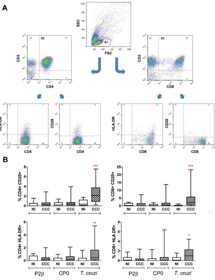
Phenotypic analysis of PBMC
2.56106cells were cultured in 24-well plates in 1 ml cultures
for 6 days with either medium alone, orT. cruzilysate, P2b-His, CP0-His (at a final concentration of 10mg/ml). After centrifu-gation, cells were washed, resuspended in ice-cold PBS, stained for 30 min at 4uC with the following fluorescent-labeled monoclonal antibodies: allophycocyanin (APC) conjugated anti-CD3 + phycoerythrin-cyano dye Cy5 (PE-Cy5) conjugated anti-CD4 + phycoerythrin (PE) conjugated anti-HLA-DR +
fluoresceinisothiocyanate (FITC) conjugated anti-CD25, or APC anti-CD3+PE-Cy5 anti-CD8+PE anti-HLA-DR+FITC anti-CD25. Cells were then fixed with 4% formaldehyde in PBS and kept at 4uC until analyzed by flow cytometry. In all cases, 10,000 to 15,000 events in the lymphocyte gate were acquired using a FACSAria flow cytometer (Becton Dickinson). Phenotypic analyses were carried out with FlowJo flow cytometric analysis software (TreeStar), selecting the small lymphocyte population. PBMC stained with FITC, PE-, APC- and PE-Cy5- labeled Ig control Abs were included in all experiments for background fluorescence. All Abs were purchased from BD Biosciences (San Diego, CA, USA).
Isolation of CD3+CD4+and CD3+CD8+T cells from
whole PBMC samples
CD8+T cells were isolated from PBMC by positive selection using EasySep CD8 Selection Kit (StemCell Technologies, Inc., Vancouver, Canada), while CD4+ T cells were separated from CD3+CD8neg T cells by negative selection (EasySep CD3 Selection Kit, StemCell Technologies). The purity of both populations was assessed by flow cytometry using specific conjugated mAb (see ‘‘Phenotypic analysis of PBMC’’) and, it was shown to be higher than 90% for both T cells subsets.
Cytokine determination by multiplex technology IL-2, IL-4, IL-10, IL-13, IL-17, IFN-c, GM-CSF and TNF-a were measured in the supernatants of whole PBMC cultures stimulated in the presence or absence of the indicated antigens and collected on days 1, 2 and 6 after stimulation. In addition, the same cytokines were quantified in cultures of isolated CD4+
or CD8+T cells (56105cells) co-cultured with irradiated CD3neg T cells (ratio 1:1) in the presence or absence of antigen after 6 days of stimulation. Cytokines were measured by using MILLI-PLEX MAP Human Cytokine/Chemokine Kit (for 8 cytokines) following the manufacturer’s directions (Millipore, St Charles, MO) and Luminex instrument and Beadlyte software were used for analysis. All samples were tested in duplicate. Results are expressed in ng/ml or Fold increase (FI) which was determined as [(cytokine in stimulated culture) - (cytokine in NS culture)]/
(cytokine in NS culture), where NS denotes non-stimulated cultured PBMC.
Data analysis
Statistical analysis was performed with GraphPad Prism statistical software (GraphPad Software). The nonparametric Mann-Whitney U test was used to generatePvalues comparing the median experimental values between groups each of the multiple sets of experimental data. Within each experiment, overall statistical significance of each result at both 10% and 5% significance was determined using Holm-Bonferroni Correc-tion. Differences were considered statistically significant at P, 0.05.
Results
Characteristics of patients with chronic Chagas’ disease Cardiomyopathy
Patients included in this study were all born in endemic areas from Argentina and Bolivia, and at the time of the enrollment they have been living in Buenos Aires (where no vectorial transmission occurs) for more than ten years, in average. The mean age was 54.2610.1, and 57% were female.
All T. cruzi-infected subjects were in the chronic phase of Chagas’ disease, involving only cardiac alterations. According to the New York Heart Association (NYHA) functional classification system, patients were classified as Class I, II, III/IV. Patients with no functional limitations but with some electrocardiographic alterations were classified as Class 0 [5].
Blood samples yielded negative results for currently used PCR protocols targeting parasite DNA [47], which is frequently the case in chronic chagasic patients due to low parasitemia. Taking this into account, we analyzed the profile of the humoral anti-TSSA (trypomastigote small surface antigen) response in our study patients as an indirect means of identifying the genotype of the infecting strain(s) [48,49]. To carry out this analysis, we evaluated the reactivity of serum samples against either TSSA Sy (the TSSA isoform from DTU TcI) or TSSA CL (the TSSA isoform from DTUs TcII/V/VI) in conventional ELISA and dot-blot (see Text S1 for details and Figure S1).
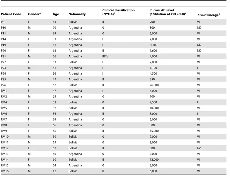
The main characteristics of the study population are summa-rized in Table 1.
Humoral immune response against ribosomal P proteins To characterize the humoral response in the subject population included in this study, the antibody reactivity against T. cruzi lysate, ribosomal P proteins, P2band CP0, together with their C-terminal peptides R13 and P015 was determined in sera of CCC patients and non-infected individuals by ELISA. The reactivity against peptide H13, which corresponds to the C-terminal region (residues 102–115) of the human ribosomal P protein was also measured.
The majority of CCC patients (24/27) showed reactivity (Reactivity index.1.7) to ribosomal P2bprotein and its peptide R13. The level of anti-CP0 antibodies was also elevated in the chagasic patients (17/27) compared to non-infected individuals, but the overall reactivity was lower than that observed for P2b protein (Figure 1). On the other hand, only marginal differences were determined in the median of the Reactivity Index for the anti-P015-antibodies in cardiac patients in comparison with non-infected subjects. No difference was observed against peptide H13 (human P ribosomal protein derived) between both groups of individuals (Figure 1). Together, these results showed that CCC patients mount a significant antibody response to ribosomal proteins as well as to peptides R13 and to a lower level to P015 in comparison to non-infected subjects.
Patients with chronic Chagas’ disease Cardiomyopathy and non-infected individuals showed similar proliferative profiles in response to ribosomal P proteins
In order to investigate the cellular response to ribosomal P proteins, PBMC from CCC patients and non-infected individuals were tested for their proliferative capacity in response to different T. cruziantigens. To determine the optimal protein and peptide concentration yielding the most consistent results, the prolifera-tive response was initially assayed in PBMC cultures from 4 cardiac patients non-included in this study. The results showed that 10mg/ml ofT. cruzilysate or ribosomal P proteins and 5mg/ ml of the peptides were optimal to trigger proliferative responses, and so these concentrations were used in the studies presented here.
Table 1.Characteristics of patients with chronic Chagas’ disease Cardiomyopathy.
Patient Code Gendera Age Nationality
Clinical classification (NYHA)b
T. cruziAb level
(1/dilution at OD = 1.0)c T.cruzilineaged
P8 F 63 Bolivia II 200 VI
P10 M 70 Argentina 0 300 ND
P11 M 34 Argentina 0 2,000 VI
P14 F 53 Argentina I 2,000 VI
P19 F 52 Argentina I ,200 ND
P20 F 63 Argentina II 1,600 ND
P21 M 56 Argentina III/IV 4,000 VI
P22 F 53 Bolivia I 2,000 VI
P23 M 42 Argentina I 1,100 I
P24 F 56 Argentina I 4,500 VI
P25 M 47 Argentina II 850 VI
P26 F 62 Bolivia II 26,000 VI
RM1 F 47 Argentina I 4,000 VI
RM2 M 65 Argentina 0 100 VI
RM4 F 52 Bolivia 0 9,500 I
RM5 F 37 Bolivia II 10,000 VI
RM6 F 56 Argentina II 8,000 I
RM7 F 54 Argentina 0 5,000 VI
RM8 F 66 Argentina 0 300 VI
RM9 F 46 Bolivia II 15,000 VI
RM10 M 50 Bolivia 0 7,000 VI
RM11 M 59 Bolivia 0 8,000 VI
RM12 F 67 Bolivia II 500 I-VI
RM13 M 40 Argentina 0 3,000 VI
RM14 F 60 Bolivia II 12,000 VI
RM15 M 64 Argentina 0 2,000 VI
RM16 M 42 Bolivia 0 6,000 VI
a
F: female, M: male.
bPatient’s heart failure was classified according to New York Heart Association (NYHA) Functional Classification. Class I: Patients with cardiac disease with slight
functional alterations but resulting in no limitation of ordinary physical activity; however, elevated activity causes symptoms, such as fatigue, palpitation, or dyspnea (shortness of breath); Class II: Patients with cardiac disease resulting in slight limitation of physical activity; comfortable at rest, but ordinary physical activity results in symptoms; Class III: Patients with cardiac disease resulting in marked limitation of physical activity; comfortable at rest, but less than ordinary activity causes symptoms; Class IV: Patients with cardiac disease resulting in marked limitation of physical activity, unable to carry out any physical activity without discomfort; symptoms of cardiac insufficiency at rest.
Patients with cardiac disease without any functional alteration were classified as Class 0.
cThe level of antibodies directed toT. cruzilysate was determined by in-house ELISA as described upon Methods.
dT cruzilineage was analyzed by TSSA recognition as described upon Methods. Results are shown in Figure S1. ND: not detected.
As shown in Figure 2, the majority of PBMC from CCC patients proliferated upon stimulation with T. cruzi lysate (Stimulation index median: 4.45) compared to PBMC from non-infected individuals (Stimulation index median: 1.07; P,0.001). On the contrary, the stimulation index of PBMC from cardiac patients and control subjects in response to ribosomal P proteins (Figure 2) as well as to peptides R13, P015 and H13 was not significantly different (data not shown). PBMC from all subjects proliferated in response to PHA and the responses were not significantly different between the cardiac and non-infected individuals (data not shown).
To characterize the phenotype of the cells after the stimulation with the different stimuli, cells were stained with different T cell markers and analyzed by flow cytometry. The forward vs side scatter dot plots revealed that the frequency of lymphocyte population in non-stimulated cultures was significantly lower in cardiac patients compared with non-infected individuals (48613% vs 62610%, respectively; P,0.001). However, the CD3+CD4+
:CD3+CD8+ ratio was approximately 2:1 in both groups. Interestingly, results showed that CCC patients present higher
subsets of CD25 and HLA-DR positive cells on both CD3+CD4+
and CD3+CD8+populations uponT. cruzistimulation (Figure 3). However, the expression of these markers was similar in T cells from cardiac patients and non-infected individuals when cells were stimulated with ribosomal P proteins (Figure 3).
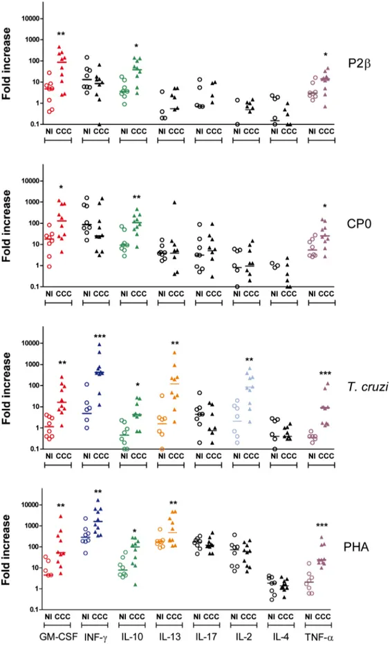
Cytokine response to ribosomal P proteins
Given the lack of proliferative response to ribosomal P proteins in the CCC patients, T cell activation was studied by analyzing cytokine secretion. Thus, PBMCs from 10 cardiac patients with different disease severity, and 8 non-infected donors were stimulated with P2band CP0 proteins andT. cruzilysate as well as PHA as positive control. Supernatants after 1, 2 and 6 days post-stimulation were collected and multiplex analysis was performed to evaluate the levels of GM-CSF, IFN-c, 10, IL-13, IL-17, IL-2, IL-4 and TNF-a. Despite the fact that cytokine responses have been studied by others afterT. cruzistimulation in patients with Chagas’ disease [50,51], reports have used different assays and stimulation/culture conditions making the direct comparison of all the cytokines difficult to achieve. In this study,
Figure 1. Humoral response against ribosomal P proteins and their C-terminal peptides.The presence of antibodies directed against P2b and CP0 proteins as well as peptides R13, P015 and H13 in the sera of 27 patients with chronic Chagas’ disease Cardiomyopathy patients (CCC) and 20 non-infected individuals (NI) was determined by ELISA as described under Methods. Results are expressed as Reactivity index, calculated as: (Optical Density mean value obtained of each serum sample/baseline value). Each symbol represents data from a single subject. Statistical analysis was performed using the Mann-Whitney U Test,***P,0.001, *P,0.05. The line for each of the scatters represents the median.
doi:10.1371/journal.pntd.0002906.g001
Figure 2. Parasite lysate but not ribosomal proteins trigger PBMC proliferative responses.PBMC isolated from chronic Chagas’ disease Cardiomyopathy patients (CCC; n = 27) and non-infected individuals (NI; n = 20) were seeded at 2.56105cells/well and stimulated withT. cruzilysate, P2bor CP0 proteins (10mg/ml) or medium alone for 6 days. Cell proliferation was determined by3H-thymidine incorporation. Results are expressed as Stimulation index, calculated as: (mean cpm of stimulated cultures/mean cpm of non-stimulated cultures (medium only)). Each symbol represents data from a single subject. Statistical analysis was performed using the Mann-Whitney U Test, ***P,0.001.
we aimed to simultaneously evaluate the kinetic responses of multiple cytokines in the same culture well. Figure 4 shows the maximum fold increase detected for each cytokine and in each subject among day 1, 2 and 6 determinations. The fold increase was determined by the difference between cytokine production (in pg/ml) in stimulated wells and the cytokine production in stimulated control wells divided the cytokine production in non-stimulated control wells. The actual fold increase for each of the days and the background production in pg/ml of each of the cytokines in non-stimulated wells are shown in Figures S2 to S5 and S6, respectively.
Upon stimulation with ribosomal P proteins, GM-CSF, IL-10 and TNF-a were secreted at higher levels in cardiac patients compared with non-infected individuals (Figure 4 and Figure S2 and S3). However, both proteins induced similar levels of IFN-c production in PBMC from cardiac patients and non-infected subjects (Figure 4). Furthermore, the fold increase of IFN-c production in response to both proteins was lower and statistically significant in the cardiac group after only the first days post-stimulation (Figure S2 and S3). The level of IL-2, IL-4, IL-13 and IL-17 secreted after stimulation with the ribosomal P proteins was very low or null at any of the 3 time points analyzed and, it was found to be similar between CCC patients and non-infected individuals (Figure 4 and Figure S2 and S3).
A larger number of cytokines were produced in response toT. cruzilysate or the universal stimulus PHA than in response to the individual ribosomal P proteins (Figure 4). Indeed, PBMC from cardiac patients in response to T. cruzi lysate also secreted statistically significant and higher levels of IFN-c, IL-2 and IL-13 compared with non-infected individuals. IFN-cand IL-13 were also increased in CCC patientsvsnon-infected individuals when PHA was used for stimulation. These results indicate that although the cells were capable of producing IFN-cand IL-13 in response to whole parasite or PHA, their production was not detected when the ribosomal P proteins were used as stimulus. The kinetic cytokine profile forT. cruzilysate and PHA is shown in Figure S4 and S5.
Profiles of cytokine production by CD4+and CD8+T cell
subsets derived from CCC patients
The results presented above revealed a cytokine signature expression upon stimulation with ribosomal P proteins andT. cruzi lysate in whole PBMC. To better understand the specific contribution of the T cells to this profile, CD3+CD4+ and CD3+CD8+ T cell subsets from three cardiac patients were enriched from PBMC and stimulated with the antigens in the presence of autologous antigen-presenting cells. Samples from patients RM11, RM12 and RM14 were chosen since they were among those that showed clear cytokines response after ribosomal P proteins stimulation.
As shown in Figure 5, GM-CSF was overall produced by both, CD4+ and CD8+ subsets by the 3 patients in response to the proteins andT. cruzi lysate. In general, IFN-cwas produced at very low levels by CD4+ and CD8+ T cells in all patients in response to the proteins, but enough to be different from the non-stimulated wells in the case of CD4+T cells (Figure 5). IL-10 was found to be secreted most frequently by both T cell subsets. IL-13 was not produced by CD8+ T cells in any of the 3 patients analyzed and in response to all the stimuli tested. However, IL-13
was produced by CD4+T cells in response toT. cruzilysate and/ or the proteins in the 2 of the 3 patients (RM11 and RM14). TNF-a was produced by both, CD4+ and CD8+ T cells and its production was higher in response to the proteins than toT. cruzi in 2 of the 3 patients. IL-2, IL-4 and IL-17 were not detected in response to any of the stimuli (data not shown).
Discussion
Since it has been widely demonstrated the relevance of antibodies directed to ribosomal P proteins in the pathophysiology of Chagas’ disease [21,23,24], this study aimed to further understand the cellular immune response raised against these proteins in CCC patients. Our results showed that PBMC did not proliferate uponin vitro stimulation with P2b and CP0 proteins. Additionally, the lack of proliferation in response to the proteins was associated with the absence of the expression of activation markers CD25 and HLA-DR on CD4+ and CD8+ T cell populations. These findings were also protein-specific, since T. cruzi lysate provoked an augmentation of both markers on the surface of T cells in agreement with data published by others [50,51]. Interestingly, the percentage of both T cell subtypes, CD3+CD4+ and CD3+CD8+ in PBMC were similar in cardiac patients and non-infected individuals independently of the stimulus. These results suggest that the lack of proliferative response was not due to an overall decrease on the size of the T cell population, nor to a shutdown of the proliferative capacity in these patients since the same cells responded toT. cruzilysate and a T cell specific universal mitogen such as PHA. However, it was possible to speculate that T cells specific to these proteins have been deleted by negative selection due to the similarity to the host specificities. In this regards, the analysis of the T cell response by cytokine release discarded this possibility since indeed, several cytokines were expressed in response to ribosomalT. cruziproteins. The use of multiplex technology allowed us to simultaneously analyze 8 cytokines, namely, IL-2, IL-4, IL-10, IL-13, IL-17, IFN-c, TNF-aand GM-CSF, corresponding to well-described CD4+
and CD8+ associated cytokines. In particular, GM-CSF was included because not only its production has been associated to antigen mediated activation of T cells by us and others but also, the threshold of antigen requirement for its production is lower than for other cytokines as TNF-a, IL-4 or IFN-c[52–54].
Our results showed that PBMC from CCC patients secreted high levels of GM-CSF, IL-10 and TNF-ain response to P2band CP0 proteins. Interestingly, the secretion of IFN-cat day 1 and 2 post-stimulation with ribosomal P proteins was similar or lower in cardiac patientsvs non-infected individuals. Moreover, our data demonstrated that patients with CCC developed a different cytokine profile in response toT. cruziand PHA stimulation than non-infected subjects.
Even though the secretion of GM-CSF, IL-10 and TNF-a in response to the proteins was significantly higher in cardiacvs non-infected individuals (P,0.05, nonparametric Mann-Whitney U Test), thesePvalues nonetheless did not stay significant at the 5% level when a multiple comparison (all 32 cytokine/stimulus pairings) was performed by using Holm-Bonferroni correction. In contrast, thePvalues for these cytokines in response toT. cruzi lysate did reach statistical significance at the 5% level. This difference could be explained by the fact that the frequency of percentage of CD25+or HLA-DR+cells in CD3+CD4+(R2 gate) or CD3+CD8+(R3 gate) lymphocytes. Horizontal lines represent the median and percentiles 25–75th, vertical lines represent percentiles 5–95th. Statistical analysis was performed using the Mann-Whitney U Test, ***P,0.001, **P,
0.01, *P,0.05.
single specific parasite protein T cells within the bulk population is lower than the frequency developed in response to wholeT. cruzi lysate and therefore it leads to lower cytokine secretion levels. However, it is important to remark that were the P values distributed at random amongst the proteins data, there would be
only a 8 3 16 6
~0:7%chance of the three exact same cytokines
(GM-CSF, IL-10 and TNF-a) being secreted in response to both proteins, demonstrating that the difference observed between cardiac patients and non-infected individuals was not a mere coincidence.
Following with T. cruzi lysate response, we observed that all studied cytokines were elevated and significantly different in the supernatants of cultured PBMC from cardiac patients with exception of IL-4 and IL-17. Upon PHA stimulation, PBMC from cardiac patients secreted higher amount of GM-CSF, IFN-c, IL-10, IL-13, and TNF-a; similar production was observed for IL-2, IL-4 and IL-17 between both groups of individuals. In addition, and independently of the stimulus, our results also showed that these cytokines were secreted by both T cells populations, except for IL-13 which was predominantly produced by CD4+T cells. Despite this finding, it is well-known that non-T cells, such as monocytes or B cells, also participate in the secretion of these cytokines. Indeed, Gomes et al. [28], by intracellular cytokine staining, reported that the majority of the IL-10-producing cells are monocytes (CD14+ cells) in asymptomatic patients, and the same group recently demonstrated that CD19+B cells is another important source of this cytokine in cardiac patients [55].
Furthermore, the spontaneous release of cytokines in non-stimulated PBMC, which provides information about the basal level of cytokine productionin vivo, showed a lower level for IFN-c, IL-10, IL-13, and TNF-ain CCC patients (Figure S6). It should be mentioned that Dutra et al.demonstrated that the expression of IFN-c, IL-10, IL-13 mRNAs was increased in PBMC from chagasic patients [33]. However, this discrepancy could depend either on the use ofex vivoPBMC or on the methodology used to determine cytokine expression. Our data, together with those reported by Giraldoet al.[56], may suggest thatT. cruzipersistence provokes a general dysfunction in peripheral T cell response.
The high levels of pro-inflammatory cytokines, like IFN-cand TNF-a, together with undetectable IL-4 production in response to PHA andT. cruzistimulation suggest that there is a shift towards polarized Th1-type of cytokine response in CCC patients. Although IL-10 was first described related to Th2 cells, now is known that is produced by all T cells, including Th1 and a regulatory T cell subsets, called Tr1 cells or IL-10-producing cells [57]. Recent studies with an experimental murine model revealed not only the protective role of IL-10 against fatal myocarditis, but also demonstrated that this cytokine was produced by both CD4+
and CD8+ subsets of IFN-c+IL-10+ double-producing T cells [58]. Similar data were obtained in studies by Belkaidet al.[59], where the main source of IL-10 in dermis and draining nodes of mice infected withLeishmania majoris a subset of CD4+T cells that produce both IL-10 and IFN-c.
Studies performed with others recombinant parasite proteins demonstrated that the majority of chagasic patients develop a strong humoral and cellular immune response with a tendency to
the typical pattern of inflammatory response described forT. cruzi lysate [34–40]. On the contrary, the cytokines released upon ribosomal P proteins stimulation made difficult to set a specific Th cells responsible for their secretion. This mixed cytokine profile which could be involved in balancing heart tissue damage and parasite persistence during chronic disease, strengthens in part the fact that B cells, through antibodies directed against P2band CP0 and not T cells, would have the major role in the development of cardiac symptoms by their interaction withb1-adrenergic and M2 muscarinic receptors.
Interestingly, GM-CSF was secreted at high levels by PBMC from CCC patients whenT. cruzi lysate, and both ribosomal P proteins were used as stimulus. To our knowledge, this is the first time that GM-CSF is used to evaluate theT. cruzispecific response of stimulated PBMC from cardiac patients. Instead, GM-CSF has been associated to a decrease in the rate of infection of both non-activated and IFN-cactivated macrophages infected withT. cruzi [60]. Moreover, Olivares Fontt et al.reported that the adminis-tration of exogenous recombinant GM-CSF improved the deficient immune response of chronically infected mice or, if neutralized by Ab anti-GM-CSF, it aggravated infection increas-ing parasitemia and host mortality in T. cruziinfected BALB/c mice [61]. In the aforementioned report, the role of GM-CSF was studied by correlating the outcome of infection with the titer of GM-CSF in plasma levels [61]. Even though it was not defined which cells were involved in GM-CSF secretion, it was speculated that lymphocytes could be in part contributing to the low but sustained amount of GM-CSF levels in infected mice. In our experiments, CD4+and CD8+T cells contributed almost equally with the secretion of this cytokine, independently of the stimulus, but it is not possible to discard that other cells as part of the PBMC pool also produced this cytokine.
While many questions remain regarding the pathogenesis of Chagas’ disease, this study represents one of the most comprehensive about the cytokine profile in response to T. cruzi and two recombinant proteins, like P2band CP0. The results show that a pool of PBMC in CCC patients has specificity forT cruziproteins and that this specificity is revealed by a Th1-cytokine dominant milieu, combined with regulatory cytokines like IL-10 and IL-13. This observation reinforces the idea that a delicate cytokine equilibrium prevails during the chronic phase of the disease. Interestingly, another cytokines, namely GM-CSF, were found significantly increased in cardiac compared to non-infected individ-uals, tempting us to suggest that this cytokine may be further applied for studying antigen responses at different stages of the disease.
Finally, due to the limited number of patients infected with TcI (3/27) compared with TcVI (20/27), it was not possible to determine a correlation between the intensity of humoral and cellular immune response and the T. cruzi lineage detected by TSSA reactivity. Further studies in that sense would provide valuable information on the role and contribution of genetic variability of T. cruzi to the immune response developed in humans.
As a whole, our findings also demonstrate that not all parasite proteins provoke a strong T cell activation combined with a pattern of cytokines similar to those described toT. cruzilysate or by infection with trypomastigote in CCC patients. In addition, as it was recently reported in the context of B cell-T cell recognition for other molecule-specificities [62,63], it is possible to hypothesize FI out of the 3 day-determinations for each subject and for each cytokine are shown. In color are denoted the cytokines for which the FI in the CCC patients were statistically significantly higher than in non-infected individuals. Each symbol represents data from a single subject. Statistical analysis was performed using the Mann-Whitney U Test, ***P,0.001, **P,0.01, *P,0.05.
Figure 5. Cytokine production profile by CD4+and CD8+T cells derived from chagasic patients.CD4+and CD8+T cells from three chronic Chagas’ disease Cardiomyopathy patients, called RM11, RM12 and RM14, were isolated as described under Methods. Enriched CD4+or CD8+ T cells were co-cultured with the autologous CD3(neg)fraction as antigen presenting cells and the indicated stimulus. Supernatants were collected on day 6 and cytokines quantified by multiplex technology. For comparison, results from whole PBMC from each of the patients are represented in the left panel. Symbol (+) indicates a fold induction between 3 to 5 and (++) a fold induction.5. The fold induction was calculated as: [(cytokine in stimulated culture) - (cytokine in NS culture)]/(cytokine in NS culture), where NS denotes non-stimulated cells.
that B cells specific for ribosomal P proteins could obtain help from T cells exhibiting different antigen reactivity. However, the interacting elements for T cell help recognition and activation may be the same for P2b and CP0, since a positive correlation was observed between the cytokines secreted by each of them (Figure S7). Currently, we are in the process of analyzing the immunoprevalence of recognition of these ribosomal P proteins and novel specificities involved in the immune response toT. cruzi infection in a large number of infected subjects at different stages of the disease.
Supporting Information
Figure S1 T. cruzi immunophenotyping of chagasic
patients using TSSA proteins. The presence of antibodies directed against GST, GST-TSSA Sy or GST-TSSA CL proteins was determined by ELISA in chronic Chagas’ disease Cardiomyopathy patients (CCC) and non-infected individuals (NI) as described under Methods. Results are expressed as means of duplicates. Negative samples were those rendering OD values below NI baseline value 63 SD (dark grey zone). Non-conclusive samples were those rendering OD values between NI baseline value 63 SD and NI baseline 65 SD (light grey zone). Samples indicated to the right were re-assayed by dot-blot revealed using West-Fempto except for patient P26, which was revealed using West-Pico chemiluminescent substrate.
(TIF)
Figure S2 Cytokine kinetics in PBMC stimulated with P2b protein. PBMC from patients with chronic Chagas’ disease Cardiomyopathy patients (CCC; n = 10) and non-infected individuals (NI; n = 8) were cultured in the presence of the indicated stimulus. Supernatants were collected on day 1, 2 and 6 and cytokines quantified by multiplex technology. The Fold increase was calculated as: [(cytokine in stimulated culture) - (cytokine in NS culture)]/(cytokine in NS culture), where NS denotes non-stimulated cultured PBMCs. Each symbol represents data from a single subject. The data were analyzed by using the Mann-Whitney U Test, ***P,0.001, **P,0.01, *P,0.05.
(TIF)
Figure S3 Cytokine kinetics in PBMC stimulated with CP0 protein. PBMC from patients with chronic Chagas’ disease Cardiomyopathy patients (CCC; n = 10) and non-infected individuals (NI; n = 8) were cultured in the presence of the indicated stimulus. Supernatants were collected on day 1, 2 and 6 and cytokines quantified by multiplex technology. The Fold increase was calculated as: [(cytokine in stimulated culture) - (cytokine in NS culture)]/(cytokine in NS culture), where NS denotes non-stimulated cultured PBMCs. Each symbol represents data from a single subject. The data were analyzed by using the Mann-Whitney U Test, ***P,0.001, **P,0.01, *P,0.05.
(TIF)
Figure S4 Cytokine kinetics in PBMC stimulated with
T. cruzi lysate. PBMC from patients with chronic Chagas’ disease Cardiomyopathy patients (CCC; n = 10) and non-infected individuals (NI; n = 8) were cultured in the presence of the indicated stimulus. Supernatants were collected on day 1, 2 and 6
and cytokines quantified by multiplex technology. The Fold increase was calculated as: [(cytokine in stimulated culture) -(cytokine in NS culture)]/-(cytokine in NS culture), where NS denotes non-stimulated cultured PBMCs. Each symbol represents data from a single subject. The data were analyzed by using the Mann-Whitney U Test, ***P,0.001, **P,0.01, *P,0.05. (TIF)
Figure S5 Cytokine kinetics in PBMC stimulated with PHA. PBMC from patients with chronic Chagas’ disease Cardiomyopathy patients (CCC; n = 10) and non-infected individuals (NI; n = 8) were cultured in the presence of the indicated stimulus. Supernatants were collected on day 1, 2 and 6 and cytokines quantified by multiplex technology. The Fold increase was calculated as: [(cytokine in stimulated culture) - (cytokine in NS culture)]/(cytokine in NS culture), where NS denotes non-stimulated cultured PBMCs. Each symbol represents data from a single subject. The data were analyzed by using the Mann-Whitney U Test, ***P,0.001, **P,0.01, *P,0.05.
(TIF)
Figure S6 Basal cytokine levels.PBMC from patients with chronic Chagas’ disease Cardiomyopathy patients (CCC; n = 10) and non-infected individuals (NI; n = 8) were cultured in media without any stimulation. Supernatants were collected on days 1, 2 and 6 and cytokines were quantified by multiplex technology. Each symbol represents data from a single subject. Statistical analysis was performed by using the Mann-Whitney U Test, ***P,0.001, **P,0.01, *P,0.05.
(TIF)
Figure S7 Correlation between cytokine releases by PBMC from CCC patients upon stimulation with P2b
and CP0 proteins. Results were expressed as the sum of maximum fold increase (FI) for GM-CSF, IL-10 and TNF-a, determined as indicated in Figure 2. Spearman’s correlation was performed using GraphPad Prism and data is shown (r: 0.988,P, 0.0001).
(TIF)
Text S1 T. cruzilineage identification by
immunophe-notyping.
(DOCX)
Acknowledgments
We thank all the patients and non-infected individuals who participated in this study. We are also very grateful to the staffs of the ‘‘Hospital General de Agudos J.M. Ramos Mejı´a’’ and Hospital General de Agudos Juan Ferna´ndez’’ and Violeta Chiauzzi for technical assistance. We are indebted to Carolina Cura and Alejandro Schijman who performed the PCR experiments forT. cruziparasite detection. We would like to dedicate this work to the memory of Dr. Mariano J. Levin.
Author Contributions
References
1. Moncayo A, Silveira AC (2009) Current epidemiological trends for Chagas disease in Latin America and future challenges in epidemiology, surveillance and health policy. Mem Inst Oswaldo Cruz 104 Suppl 1: 17–30.
2. Coura JR, Vin˜as PA (2010) Chagas disease: a new worldwide challenge. Nature 465: 56–57.
3. World Health Organization. Chagas disease: control and elimination. UNDP/ World Bank/WHO. 2010. Available: http://apps.who.int/gb/ebwha/pdf_ files/WHA63/A63_17-en.pdf. (Accessed 2011 Nov 9)
4. Junqueira C, Caetano B, Bartholomeu DC, Melo MB, Ropert C, et al. (2010). The endless race betweenTrypanosoma cruziand host immunity: lessons for and beyond Chagas disease. Expert Rev Mol Med 12:e29.
5. Rassi A Jr, Rassi A, Marin-Neto JA (2010) Chagas disease. Lancet, 375: 1388– 1402.
6. Coura JC, Borges-Pereira (2010) Chagas disease: 100 years after its discovery. A systemic review. Acta Tropica 115: 5–13.
7. Chiale PA, Ferrari I (2001) Autoantibodies in Chagas’ cardiomyopathy and arrhythmias. Autoimmunity 34: 205–210.
8. Elizari MV, Chiale PA (1993) Cardiac arrhythmias in Chagas’ heart disease. J CardiovascElectrophysiol 4: 596–608.
9. Zacks MA, Wen JJ, Vyatkina G, Bhatia V, Garg N (2005). An overview of chagasic cardiomyopathy: pathogenic importance of oxidative stress. An Acad Bras Cienc 77: 695–715.
10. Brandariz S, Schijman A, Vigliano C, Arteman P, Viotti R, et al. (1995) Detection of parasite DNA in Chagas’ heart disease.Lancet 346: 1370–1371. 11. Nagajyothi F, Machado FS, Burleigh BA, Jelicks LA, Scherer PE, et al. (2012)
Mechanism ofTrypanosoma cruzipersistence in Chagas disease. Cell Microbiol 14: 634–643.
12. Kierszenbaum F (1999) Chagas’ disease and the autoimmunity hypothesis. Clin Microbiol Rev12: 210–223.
13. Bonney KM, Engman DM (2008) Chagas Heart Disease Pathogenesis: One Mechanism or Many?. Clin Mol Med 8: 510–518.
14. Cunha-Neto E, Teixeira PC, Nogueira LG, Kalil J (2011) Autoimmunity. Adv Parasitol 76: 129–152
15. Teixeira ARL, Hecht MM, Guimaro MC, Sousa AO, Nitz N (2011) Pathogenesis of Chagas’ Disease: Parasite Persistence and Autoimmunity. Clin Microbio Rev 24: 592–630.
16. Mesri EA, Levitus G, Hontebeyrie-Joskowicz M, Dighiero G, Van Regenmortel MHV, et al. (1990) MajorTrypanosoma cruziantigenic determinant in Chagas’ heart disease shares homology with the systemic lupus erythematosus ribosomal P protein epitope. J Clin Microbiol 28: 1219–1224.
17. Rosenbaum M B, Chiale PA, Schejtman D, Levin M, Elizari MV (1994) Antibodies to beta-adrenergic receptors disclosing agonist-like properties in idiopathic dilated cardiomyopathy and Chagas’ heart disease. J. Cardiovasc Electrophysiol 5: 367–375.
18. Aznar C, Lopez-Bergami P, Brandariz S, Mariette C, Liegeard P, et al. (1995) Prevalence of anti-R-13 antibodies in humanTrypanosoma cruziinfection. FEMS Immunol Med Microbiol 12: 231–238.
19. Ferrari I, Levin MJ, Wallukat G, Elies R, Lebesgue D, et al. (1995) Molecular mimicry between the immunodominant ribosomal protein P0 ofTrypanosoma cruziand a functional epitope on the human beta1-adrenergic receptor. J Exp Med 182: 59–65.
20. Lopez-Bergami P, Scaglione J, Levin MJ (2001) Antibodies against the carboxyl-terminal end of theTrypanosoma cruzi ribosomal P proteins are pathogenic. FASEB J 15: 2602–2612.
21. Mahler E, Hoebeke J, Levin M (2004) Structural and functional complexity of the humoral response against theTrypanosoma cruziribosomal P2bprotein in patients with chronic Chagas’ heart disease. Clin Exp Immunol 136: 527–534. 22. Lopez Bergami P, Gomez KA, Levy GV, Grippo V, Baldi A, et al. (2005) The b1 adrenergic effects of antibodies against the C-terminal end of the ribosomal P2bprotein of Trypanosoma cruzi associate with a specific pattern ofepitope recognition. Clin Exp Immunol 142: 140–147.
23. Smulski C, Labovsky V, Levy G, Hontebeyrie M, Hoebeke J, et al. (2006) Structural basis of the cross-reaction between an antibody to theTrypanosoma cruziribosomal P2bprotein and the humanb1 adrenergic receptor. FASEB J 20: 1396–1406.
24. Levy GV, Tasso LM, Longhi SA, Rivello HG, Kyto¨ V, et al. (2011) Antibodies against theTrypanosoma cruziribosomal P proteins induce apoptosis in HL-1 cardiac cells. Int J Parasitol 41: 635–644.
25. Mahler E, Sepulveda P, Jeannequin O, Liegeard P, Gounon P, et al. (2001) A monoclonal antibody against the immunodominant epitope of the ribosomal P2beta protein ofTrypanosoma cruziinteracts with the human b1-adrenergic receptor. Eur J Immunol 31: 2210–2216.
26. Morato MJ, Brener Z, Canc¸ado JR, Nunes RM, Chiari E, et al. (1986) Cellular immune responses of chagasic patients to antigens derived from different Trypanosoma cruzistrains and clones. Am J Trop Med Hyg 35: 505–511. 27. Cuna WR, Encina JLR, Cuna CR (2000) Interferon-cor IL-10 production is
induced by relatedTrypanosoma cruziantigens. J Parasitol 86: 295–299. 28. Gomes JAS, Bahia-Oliveira LMG, Rocha MOC, Martins-Filho OA, Gazzinelli
G, et al. (2003) Evidence that Development of Severe Cardiomyopathy in Human Chagas’ Disease Is Due to a Th1-Specific Immune Response. Inf Immun 71: 1185–1193
29. de Barros-Mazon S, Guariento ME, da Silva CA, Coffman RL, Abrahamsohn IA (2004) Differential regulation of lymphoproliferative responses toTrypanosoma cruziantigen in patients with the cardiac or indeterminate form of Chagas disease. Clin Immunol 111: 137–145.
30. Scharfstein J, Gomes JA, Correa-Oliveira R (2009) Back to the future in Chagas disease: from animal models to patient cohort studies, progress in immuno-pathogenesis research. Mem Inst Oswaldo Cruz 104 Suppl 1: 187–198 31. Fiuza JA, Fujiwara RT, Gomes JA, Rocha MO, Chaves AT, et al. (2009) Profile
of central and effector memory T cells in the progression of chronic human chagas disease. Plos Negl Trop Dis 3: e512.
32. Dutra WO, Silva Menezes CA, Villani FNA, da Costa GC, da Silveira ABM, et al. (2009) Cellular and genetic mechanisms involved in the generation of protective and pathogenic immune responses in human Chagas disease. Mem Inst Oswaldo Cruz,104 Suppl. I: 208–218
33. Dutra WO, Gollob KJ, Pinto-Dias JC, Gazzinelli G, Correa-Oliveira R, et al. (1997) Cytokine mRNA profile of peripheral blood mononuclear cells isolated from individuals withTrypanosoma cruzichronic infection. Scand J Immunol 45: 74–80.
34. Abel LC, Rizzo LV, Ianni B, Albuquerque F, Bacal F, et al. (2001) Chronic Chagas’ disease cardiomyopathy patients display an increased IFN-gamma response toTrypanosoma cruziinfection. J Autoimmun 17: 99–107.
35. Ribeirao M, Pereira-Chioccola VL, Nia L, Augusto Fragata FA, Schenkman S, et al. (2000) Chagasic patients develop a type 1 immune response toTrypanosoma cruzitrans-sialidase. Parasite Immunol 22: 49–55.
36. Michailowsky V, Luhrs K, Rocha MOC, Fouts D, Gazzinelli RT, et al. (2003) Humoral and cellular immune responses toTrypanosoma cruzi-derived parafla-gellar rod proteins in patients with Chagas’ disease. Infect Immun 71: 3165– 3171.
37. Lorena VMB, Vercosa AFA, Machado RCA, Moitinho-Silva L, Cavalcanti MGA, et al. (2008) Cellular Immune Response From Chagasic Patients to CRAor FRA Recombinant Antigens ofTrypanosoma cruzi. J Clin Lab Anal 22: 91–98.
38. de Melo AS, de Lorena VM, de Moura Braz SC, Docena C, de Miranda Gomes Y (2012) IL-10 and IFN-cgene expression in chronic Chagas disease patients after in vitro stimulation with recombinant antigens of Trypanosoma cruzi. Cytokine 58: 207–212.
39. Mora´n-Utrera Y, Lo´pez-Monteon A, Rosales-Encina JL, Mendez-Bolaina E, Ramos-Ligonio A (2012) Trypanosoma cruziSSP4 Amastigote Protein Induces Expression of Immunoregulatory and Immunosuppressive Molecules in Peripheral Blood Mononuclear Cells. J Trop Med 2012: 829139.
40. Arnholdt AC, Piuvezam MR, Russo DM, Lima AP, Pedrosa RC, et al. (1993) Analysis and partial epitope mapping of human T cell responses toTrypanosoma cruzicysteinyl proteinase. J Immunol 151: 3171–179.
41. Zingales B, Andrade SG, Briones MR, Campbell DA, Chiari E, el al. (2009) A new consensus for Trypanosoma cruzi intraspecific nomenclature: second revision meeting recommends TcI to TcVI. Mem Inst Oswaldo Cruz 104: 1051–1054.
42. De Marchi CR, Di Noia JM, Frasch AC, Amato Neto V, Almeida IC, et al. (2011) Evaluation of a recombinant Trypanosoma cruzi mucin-like antigen for serodiagnosis of Chagas’ disease. Clin Vaccine Immunol 18: 1850–1855. 43. Gomez EB, Medina G, Ballesta JP, Levin MJ, Tellez-In˜on MT (2001) Acidic
ribosomal P proteins are phosphorylated inTrypanosoma cruzi. Int J Parasitol 31: 1032–1039.
44. Juri Ayub M, Levin MJ, Aguilar CF (2001) Overexpression and refolding of the hydrophobic ribosomal P0 protein fromTrypanosoma cruzi: a component of the P1/P2/P0 complex. Protein Expr Purif 22: 225–233.
45. Muller S, Couppez M, Briand JP, Gordon J, Sautiere P, et al. (1985) Antigenic structure of histone H2B. Biochem Biophys Acta 827: 235–246.
46. Elkon K, Bonfa E, Llovet R, Danho W, Weissbach H, et al. (1988) Properties of the ribosomal P2 protein autoantigen are similar to those of foreign protein antigens. Proc Natl Acad Sci USA 85: 5186–5189.
47. Burgos JM, Diez M, Vigliano C, Bisio M, Risso M, et al. (2010) Molecular identification of Trypanosoma cruzidiscrete typing units in end-stage chronic Chagas heart disease and reactivation after heart transplantation. Clin Infect Dis 51: 485–495.
48. Di Noia JM, Buscaglia CA, De Marchi CR, Almeida IC, Frasch AC (2002) A Trypanosoma cruzi small surface molecule provides the first immunological evidence that Chagas’ disease is due to a single parasite lineage. J Exp Med 195: 401–413.
49. Ca´nepa GE, Degese MS, Budu A, Garcia CR, Buscaglia CA (2012) Involvement of TSSA (trypomastigote small surface antigen) inTrypanosoma cruziinvasion of mammalian cells. Biochem J 444: 211–218.
50. Dutra WO, Martins-Filho OA, Canc¸ado JR, Pinto-Dias JC, Brener Z, et al. (1994) Activated T and B lymphocytes in peripheral blood of patients with Chagas disease. Int Immunol 6: 499–506.
51. Dutra WO, Martins-Filho OA, Canc¸ado JR, Pinto-Dias JC, Brener Z, et al. (1996) Chagasic patients lack CD28 expression on many of their circulating T lympho cytes. Scand J Immunol 43: 88–93.
53. Topalian SL, Rivoltini L, Mancini M, Markus NR, Robbins PF, et al. (1994) Human CD4+T cells specifically recognize a shared melanoma-associated antigen encoded by the tyrosinase gene. Proc Natl Acad Sci USA 91: 9461– 9465.
54. Shi Y, Liu CH, Roberts AI, Das J, Xu G, et al. (2006) Granulocyte-macrophage colony-stimulating factor (GM-CSF) and T-cell responses: what we do and don’t know. Cell Res 16: 126–133.
55. Fares RCG, Correa-Oliveira R, De Arau´jo FF, Keesen TSL, Chaves AT, et al. (2013) Identification of phenotypic markers of B cells from patients with Chagas disease. Parasite Immunol 35: 214–223
56. Giraldo NA, Bolan˜os NI, Cuellar A, Roa N, Cucunuba´ Z, et al. (2013) T Lymphocytes from Chagasic Patients Are Activated but Lack Proliferative Capacity and Down-Regulate CD28 and CD3f. PLoS Negl Trop Dis 7: e2038 57. Zhu J, Paul WE (2010) Heterogeneity and plasticity of T helper cells. Cell Res
20: 4–12.
58. Roffeˆ E, Rothfuchs AG, Santiago HC, Marino APMP, Ribeiro-Gomes FL, et al. (2012) IL-10 Limits Parasite Burden and Protects against Fatal Myocarditis in a Mouse Model ofTrypanosoma cruziInfection. J Immunol 188: 649–660.
59. Belkaid Y, Hoffmann KF, Mendez S, Kamhawi S, Udey MC, et al. (2001) The Role of Interleukin (IL)-10 in the Persistence ofLeishmania majorin the Skin after Healing and the Therapeutic Potential of Anti–IL-10 Receptor Antibody for Sterile Cure. J Exp Med 194: 1497–1506.
60. Olivares Fontt E, Vray B (1995) Relationship between granulocyte macrophage-colony stimulating factor, tumour necrosis factor-alpha andTrypanosoma cruzi infection of murine macrophages. Parasite Immunol 17: 135–141.
61. Olivares Fontt E, Heirman C, Thielemans K, Vray B (1996) Granulocyte-macrophage colony-stimulating factor: involvement in control ofTrypanosoma cruziinfection in mice. Infect Immun 64: 3429–3434.
62. Schultena V, Greenbauma JA, Hauserb M, McKinneya DM, Sidney J, et al. (2013) Previously undescribed grass pollen antigens are the major inducers of T helper 2 cytokine-producing T cells in allergic individuals. Proc Natl Acad Sci USA 110: 3459–3464.
63. Chen C, Dow C, Wang P, Sidney J, Read A, et al. (2011) Identification of CD4+