• Nenhum resultado encontrado

The lipidome, genotoxicity, hematotoxicity and antioxidant properties of andiroba oil from the Brazilian Amazon

N/A
N/A
Protected

Academic year: 2019

Share "The lipidome, genotoxicity, hematotoxicity and antioxidant properties of andiroba oil from the Brazilian Amazon"

Copied!
9
0
0

Texto

(1)

The lipidome, genotoxicity, hematotoxicity and antioxidant properties of

andiroba oil from the Brazilian Amazon

Susana Suely Rodrigues Milhomem-Paixão

1

, Maria Luiza Fascineli

2

, Mariana Matos Roll

3

, João Paulo

Figueiró Longo

2

, Ricardo Bentes Azevedo

2

, Julio Cesar Pieczarka

1

, Hugo Leonardo Crisóstomo Salgado

4

,

Alberdan Silva Santos

4

and Cesar Koppe Grisolia

3

1

Laboratório de Citogenética, Instituto de Ciências Biológicas, Universidade Federal do Pará (UFPA),

Belém, PA, Brazil.

2

Laboratório de Nanobiotecnologia, Departamento de Genética e Morfologia, Instituto de Ciências

Biológicas, Universidade de Brasília (UnB), Brasília, DF, Brazil.

3

Laboratório de Genética Toxicológica, Departamento de Genética e Morfologia, Instituto de Ciências

Biológicas, Universidade de Brasília (UnB), Brasília, DF, Brazil.

4

Laboratórios de Investigação Sistemática em Biotecnologia e Biodiversidade Molecular (LabISisBio),

Instituto de Ciências Exatas e Naturais, Universidade Federal do Pará (UFPA), Belém, PA, Brazil.

Abstract

Andirobeira is an Amazonian tree, the seeds of which produce a commercially valuable oil that is used in folk medi-cine and in the cosmetic industry. Andiroba oil contains components with anti-inflammatory, cicatrizing and in-sect-repellant actions. However, virtually nothing is known of the safety of this oil for humans. The aim of this work was therefore to investigate the hematotoxicity, genotoxicity and mutagenicity of andiroba oil using the comet and micronucleus assays, and to assess its antioxidant properties and lipidome as a means of addressing safety issues. For the experiments, andiroba oil was administered by gavage for 14 consecutive days in nulliparous female Swiss mice randomly distributed in four groups: negative control and three doses of oil (500, 1000 and 2000 mg/kg/day). These doses were chosen based on recommendations of the OECD guideline no. 474 (1997). GC/MS was used to investigate the free fatty acid, cholesterol and triterpene content of andiroba oil in a lipidomic analysis. No clinical or behavioral alterations were observed throughout the period of treatment, and exposure to andiroba oil at the doses and conditions used here did not result in hematotoxic, genotoxic or mutagenic effects. Testsin vitro showed that oil sample 3 from southwestern of Brazilian Amazon had a high antioxidant capacity that may protect biological systems from oxidative stress, although this activity remains to be demonstratedin vivo.

Keywords: acute toxicity, antioxidant activity,Carapa guianensis,genotoxicity, lipidomics. Received: April 13, 2015; Accepted: August 22, 2015.

Introduction

Andiroba oil is extracted from the seeds of the andi-roba tree, represented by two species,Carapa guianensis andCarapa procera, of the family Meliaceae that occurs in the Amazon (Fischet al., 1995). This oil has various uses in folk medicine, such as a cicatrizing and anti-inflammatory agent and as an insect repellent (Bauch and Dunisch, 2000; Ferrazet al., 2002; Shanleyet al., 2005); in recent years, andiroba oil has also been used intensively in the cosmetic industry (Ferreiraet al., 2010).

Previous studies have reported acaricidal (Farias et al., 2007, 2009), larvicidal (Silvaet al., 2004), insect repel-lent (Miotet al., 2004, 2011) and antiplasmodial (Miranda Júnior CRN, 2010, MSc dissertation, Universidade Federal do Pará, Belém, PA, Brazil) activities for andiroba oil. However, a general lack of knowledge regarding the toxic-ity and/or proof of pharmacological activities of natural products employed as medicines and their indiscriminate use may put Amazon populations at risk of adverse reac-tions (Frescura VDA, 2012, MSc dissertation, Universi-dade Federal de Santa Maria, Santa Maria, RS, Brazil).

Andiroba oil is rich in essential fatty acids, including oleic, palmitic, stearic and linoleic acids (Martinborough, 2002; Rain-Tree, 2014). Linoleic acid reduces cholesterol levels and blood pressure and is beneficial in the prevention of cancer. Andiroba oil also contains meliacines, a group of

DOI: http://dx.doi.org/10.1590/1678-4685-GMB-2015-0098

Send correspondence to C. K. Grisolia. Laboratório de Genética Toxicológica, Departamento de Genética e Morfologia, Instituto de Ciências Biológicas, Universidade de Brasília (UnB), Brasília, 70910-900, DF, Brazil. E-mail: grisolia@unb.br.

(2)

substances that confer a bitter taste to the oil and have antimalarial and antiparasitic activities; there are also limo-noids with anti-inflammatory, insect repellent and anti-tu-moral properties (Martinborough, 2002; Rain-Tree, 2014). Stearic, palmitic, oleic and linoleic fatty acids can cause apoptosis in different cell linesin vitro, depending on the concentration tested (Muet al., 2001; Luet al., 2003; Cury-Boaventuraet al., 2004). There has been much dis-cussion about the possible mechanisms involved in the in-duction of apoptosis by these acids and the physiological effects that they produce (Curiet al., 2002). One of the best characterized mechanisms in apoptosis involves damage to cellular DNA that may occur via various routes (Batista LFZ, 2008, PhD thesis, Universidade de São Paulo, São Paulo, SP, Brazil). This damage can be eliminated by the endogenous DNA repair system of cells, but if the damage persists it may induce genotoxicity, mutagenesis or even apoptosis (Wonget al., 2005; Asare et al., 2011; Batista LFZ, 2008, PhD thesis, Universidade de São Paulo, São Paulo, SP, Brazil).

Knowledge about the genotoxic potential of natural compounds and semi-synthetic and synthetic chemicals is

essential for regulatory agencies so that they can establish the risk to humans (Ribeiroet al., 2003). To date, few stud-ies have examined the toxicity of andiroba oil and there are no reports on the possible genotoxic or mutagenic effects of this oil. The aim of this work was therefore to investigate the possible toxicological, mutagenic and genotoxic effects of andiroba oil in Swiss mice and to examine the antioxi-dant properties of this oilin vitro. The data obtained con-tribute to our understanding of the safety of this oil and its saponified fraction.

Material and Methods

Plant material

Seeds from andiroba trees were collected from 21 sites in the state of Pará in the Brazilian Amazon under li-cense no. MMA/ICMBIO/SISBIO-33336-1 issued by the Brazilian Environmental Agency. The sample identifica-tions, mesoregions, municipalities, geographical coordi-nates and collection periods for these seeds are described in Table 1. The exsicates were deposited in the herbarium of the Brazilian Agricultural Research Company

Table 1- Samples, localities and periods of collection of andiroba seeds and the concentration of oil causing a 50% reduction in the DPPH radical (EC50).

Samples Mesoregion Municipality Geographical coordinates Collection

period EC50(mL/mL)

SE1 Southeast Nova Ipixuna S 04º 48’ 30.1’’ W 049º 21’ 42.8’’ 16-20/4/12 86.62 SE2 Jacundá S 04º 27’ 03.0’’ W 049º 06’ 59.5’’ 16-20/4/12 151.0

SE3 São Miguel do Guamá S 01º 35’ 28.5’’ W 047º 34’ 39.5’’ 3-7/5/2012 126.7

NE1 Northeast Abaetetuba S 01º 43’ 04’’ W 048º 52’ 58’’ 3-7/5/2012 2305 NE2 Acará S 01º 58’ 25.9’’ W 048º 18’ 49.5’’ 3-7/5/2012 302.0

NE3 Cametá S 02º 14’ 40.0’’ W 049º 29’ 45.0’’ 3-7/5/2012 204.3

LAM1 Lower Amazon Santarém S 02º 24’ 52’’ W 054º 42’ 36.0’’ 25-29/3/2012 43.56 LAM2 Oriximiná S 01º 30’ 55.3’’ W 055º 46’ 52.0’’ 25-29/3/2012 150.7

LAM3 Óbidos S 01º 39’ 21.5’’ W 055º 37’ 15.9’’ 25-29/3/2012 45.20 MAR1 Marajó Curralinho S 01º 45’ 59.1’’ W 049º 49’ 45.2’’ 12/4; 23/5 and

24/7/2012

468.9

MAR2 Salvaterra S 00º 48’ 02.0’’ W 048º 32’ 01.0’’ 12/4; 23/5 and 24/7/2012 90.23 MAR3 Breves S 01º 38’ 17.9’’ W 050º 27’ 99.5’’ 12/4; 23/5 and 24/7/2012 1894

MTR1 Metropolitan Santa Isabel do Pará S 01º 21’ 12.4’’ W 048º 08’ 37.3’’ 29/3-1/4/2012 326.3 MTR2 Castanhal S 01º 17’ 49.5’’ W 047º 55’ 19.7’’ 29/3-1/4/2012 559.3

MTR3 Bujarú S 01º 35’ 46.3’’ W 047º 57’ 32.9’’ 29/3-1/4/2012 252.6

SW1 Southwest Porto de Moz S 01º 45’ 00’’ W 052º 14’ 15.0’’ 25-29/3/2012 425.4 SW2 Medicilândia S 03º 23’ 59.0’’ W 052º 53’ 36.8’’ 12/3-24/4/2012 1181

SW3 Uruará S 03º 58’ 31.7’’ W 053º 37’ 32.1’’ 12/3-24/4/2012 59.95 SW4 Itaituba S 04º 16’ 34.0’’ W 055º 59’ 01.0’’ 29/3-5/4/2012 150.0 SW5 Jacareacanga S 06º 13’ 20.0’’ W 057º 45’ 10.0’’ 29/3-5/4/2012 100.4 SW6 Aveiro S 03º 50’ 30.0’’ W 055º 28’ 32.7’’ 29/3-5/4/2012 106.7

IANA* 8913

AMAZON* 204.8

(3)

(EMBRAPA) under accession number 191736. The seeds were identified by Dr. Regina Celia Viana Martins da Silva, curator of the IAN Embrapa Amazônia Oriental herbarium.

Collection of andiroba oil

Andiroba oil fromC. guianensisfrom each site was processed and characterized separately in the Laboratory for Systematic Investigation in Biotechnology and Fine Chemistry (LabISisBio) at the Federal University of Pará. After collection, the fruits were stored at 12 °C and trans-ported to the lab where the oil was extracted either with an artisanal extraction process (Shanley et al., 2005) or in naturausing organic solvent (Sousa Filho JVC, 2007, MSc dissertation, Universidade Federal do Pará, Belém, PA, Brazil). After artisanal extraction, the resulting mass was pressed and squeezed to reduce the oil content to < 5%, as described elsewhere (Gunstone and Padley, 1997). After extraction in organic solvent, the solvent was removed by evaporation to obtain oil without hexane that could be frac-tionated and separated. The derivatization technique to characterize the intact oil (sample SW3) and saponified and unsaponified compounds used in this study was essentially that described by Souzaet al.(2014), with some modifica-tions in the temperature programming and column change.

Antioxidant assay

To choose the sample that would be used in this study, the antioxidant capacity of the samples was evaluated by the 2,2-diphenyl-1-picryl-hydrazyl (DPPH) method. The protocol followed was that described by Brand-Williamset al. (1995) with adaptations described by Razali et al. (2008) and Atmaniet al.(2009) in which 900mL of DPPH

solution (0.06 mM) was mixed with 100mL of oil diluted in

methanol (12.5, 25, 50, 100 to 200mL/mL) followed by

in-cubation for 20 min. The absorbance was read in a Spectramax M2 spectrophotometer (Molecular Devices, Sunnyvale, CA, USA) at 515 nm. The results were ex-pressed as the percentage inhibition of the DPPH radical that was calculated using the absorbances measured above in conjunction with the following formula:

% inhibition=[A - ( )]´

A 0 A1 - AS

0 100

whereA0is the absorbance of DPPH,A1is the absorbance of DPPH + diluted oils andAsis the absorbance of diluted oils. The percentages of inhibition were used to calculate the EC50(effective concentration or minimum

concentra-tion required to reduce the DPPH radical by 50%) by non-linear regression. Based on the results of the DPPH as-say, the sample with the greatest antioxidant potential (sample SW3) and that was available in sufficient quantity was selected for thein vivotests.

Experimental design

For thein vivoassays, 90-day-old non-pregnant null-iparous female Swiss mice (Mus musculus) purchased from the Multidisciplinay Center for Biological Investigation (CEMIB) at the State University of Campinas (UNI-CAMP), Campinas, SP, Brazil, were used. The mice were maintained in the animal house of the Department of Ge-netics and Morphology at the University of Brasília under the following conditions: 23 ± 2 °C, relative humidity

30-70%, and a 12 h light/dark cycle, with 10-15 changes of air/h. The mice had access to drinking water and commer-cial rodent chowad libitum. The animal protocols were ap-proved by the Committee for Ethics in Animal Use (CEUA) of the Institute of Biological Sciences at the Uni-versity of Brasília (protocol no. 127331/2013).

The mice were randomly allocated to one of four ex-perimental groups (n = 6 mice/group): negative control and three doses of oil (500, 1000 and 2000 mg/kg/day). The negative control group received corn oil (Salada®, Bunge)

and the treated mice received a solution of andiroba oil and corn oil by gavage through a gastric tube. The treatments were done for 14 consecutive days and the dose limit corre-sponded to that recommended in the Organization for Eco-nomic Co-operation and Development (OECD) guidelines for the evaluation of genotoxicity by the micronucleus test (Macgregoret al., 1987) and of chromosomal aberrations in bone marrow (Prestonet al., 1987). Throughout the ex-perimental period, the mice were weighed on days 0, 3, 6, 9, 12 and 15, and food consumption was monitored on days 3, 6, 9, 12 and 15. All of the mice were examined daily throughout the treatment period to check for possible clini-cal alterations/symptoms.

On the 15thday the mice were sedated with a mixture

of ketamine chlorohydrate (10%) and xylazine (2%) and, after confirmation of narcosis, blood samples were ob-tained. Blood was collected by cardiac puncture and stored in plastic microtubes with 10% EDTA for hematological evaluation and preparation of slides for the comet assay. During autopsy, the mice were inspected for macroscopic alterations and the liver, kidneys and spleen were collected and weighed. For the micronucleus and nuclear abnormal-ity tests, slides were prepared using femur bone marrow.

Comet assay

The protocol for this assay was based on the alkaline comet assay (pH > 13) described by Singhet al. (1988), with some modifications. The slides were analyzed in a Zeiss-Axioskop 2 fluorescence microscope fitted with a 510-560 nm filter, a 590 nm blocking filter and a magnifi-cation of 400x. Ethidium bromide (20mg/mL) was used for

(4)

these data were then used to calculate the damage index (DI). The DI was defined as the product obtained by multi-plying the number of comets from each class with the digit denominating the class (0, 1, 2, 3 and 4), the formula being taken from Jaloszynskiet al.(1997).

Micronucleus test

The micronucleus test was done using mouse femur bone marrow, as described by Schmid (1975), with modifi-cations. The stained slides were examined with an Olympus BH2 light microscope at a magnification of 1000x. For each mouse, 4000 erythrocytes were counted, of which 2000 were polychromatic (PCE – polychromatic erythro-cytes) and 2000 were normochromatic (NCE – normo-chromatic erythrocytes). When the cell count reached 2000, the values for the two cell populations were recorded in or-der to assess the cytotoxicity and calculate the PCE/NCE ratio. The lower this ratio, the greater the cytotoxicity in-volving a significant reduction in PCE. In addition to this ratio, the appearance of the micronucleated polychromatic cells was also recorded.

Hematological analysis

For hematological analysis, 370mL of blood

contain-ing 10% disodium EDTA as anticoagulant was analyzed in an automatic veterinary hematocytometer (Sysmex pocH 100iV DiffTM) calibrated for mice.

Gas chromatography-mass spectrometry (GC-MS) analysis and compound identification

GC-MS analysis was done with a Thermo GC/MS system equipped with a quadrupole mass selective detector operated at 70eV in electron impact (EI) mode. The TRACE-1300 gas chromatograph (GC) was equipped with a RTX-5TG- RESTECK column (30 m x 0.25 mm di x 0.25

mm film thickness and the oven was operated at 40-300 °C

in increments of 10 °C/min with He as the carrier gas (flow: 1 mL/min). The injector was set at 220 °C and 1mL of the

sample was injected into the GC via an AI/AS-1310 auto-sampler. The mass spectrometer (MS) was operated in scan mode (start after 3 min, mass range from 40-800 a.m.u. at 1 scan/s). The transfer line and ion source were both operated at 280 °C. The compounds were identified by comparing their mass spectra with those of the NIST-11MS lipid li-brary database.

Statistical analysis

The EC50(effective concentration capable of

inhibit-ing 50% of the free radical) was calculated by non-linear re-gression from the percentage inhibition of the free radical in the DPPH assay for the 21 samples of andiroba oil. The quantitative data were evaluated by parametric or non-parametric statistical methods based on the normal distri-bution of the data. Parametric data were compared by anal-ysis of variance (ANOVA) followed by Dunnett’s test,

whereas non-parametric data were analyzed with the Wil-coxon text followed by the Kruskall-Wallis test. All data comparisons and statistical analyses were done using the softwares Instat 3.02 and Prism 5.0 (GraphPad Inc., La Jolla, CA, USA), with a value of p£0.05 indicating

signif-icance.

Results

All of the oil samples showed antioxidant activity to-wards the DPPH radical, as shown in Table 1, which sum-marizes the EC50data. Supplementary Figures S1-S7 show

that there was a correlation between the percentage of DPPH inhibition and the oil concentration needed to cause a 50% reduction of DPPH. Samples with high and low anti-oxidant properties showed regional variations,e.g., north-eastern Pará State, Marajó island and the metropolitan region of Belém, as well as variations within each region. Two commercial samples of andiroba oil (Amazon Ervas and Iana®D’amazônia) were also tested for their

antioxi-dant potential. The Iana sample, diluted in mineral oil as stated on the manufacturer’s label, had the lowest antioxi-dant capacity of all the samples tested, with an EC50of 8913

mL/mL, compared to 204.8mL/mL for the Amazon Ervas

sample, which was similar to the value for the Cametá sam-ple from northeastern Brazil.

Based on these results and the availability of each oil, the sample SW3 was chosen for biological assays. The lipid profiles were determined using the derivatization tech-nique. Saponified compounds represented > 97% of the sample and non-saponified compounds consisted of ste-roids and triterpenoids derivatives (Table 2). More than 97% of the fatty acids were represented by oleic, palmitic, stearic, linoleic and arachidonic acids. However, the deri-vatization technique was also capable of detecting traces of other acids in the same sample. These findings agreed with those reported for other analyses of andiroba oil (Martin-borough, 2002).

To evaluate the possible toxicity of SW3in vivo, the mice were observed daily for any alterations during treat-ment, but no clinical or behavioral alterations were ob-served and there were no changes in body weight. No macroscopic alterations were seen during autopsy nor were there any alterations in the absolute or relative weight of the kidneys, liver or spleen (Table 3).

(5)

Table 2- Profile of fatty acids, steroids and triterpenes present in two samples (SW3-1 and SW3-2) of andiroba oil.

Component Peak retention time (min) Retention index Oil profile SW3-1 SW3-2

Lipids 1 (%) Lipids 2 (%)

Fatty acids 15.52 1899 Miristic acid 0.07±0.03 0.13±0.02 17.54 2027 Palmitic acid 37.95±0.63 32.21±0.94

17.86 2048 Palmitoleic acid 0.07±0.12 0.81±0.07

20.49 2234 Oleic acid 55.23±0.43 41.92±0.52

21.37 2301 Linoleic acid 4.21±0.34 7.63±0.36

21.58 2318 Stearic acid 0.03±0.06 14.53±0.75

22.93 2428 Arachidic acid 1.07±0.19 2.12±0.39

25.52 2664 Behenic acid 0.28±0.07 0.43±0.06

27.73 2894 Lignoceric acid 0.17±0.06 0.25±0.01

Steroids 28.59 Ns Squalene 0.43±0.61

-30.86 Ns Stigmasterol 0.11±0.04

-31.87 Ns Cholesterol 0.07±0.02

-Triterpenes 33.11 Ns Epoxygedunnin* 0.08±0.02

-33.64 Ns 1,3-Dipalmitin 0.09±0.02

-33.94 Ns Deoxylactone-derivativ

e 0.13±0.02

-35.92 Ns Deacetylgedunin* 0.04±0.01

-36.40 Ns Epoxydeacetylgedunin* 0.06±0.03

-Ns = not simulated (requires hydrocarbons with a carbon number > 30), *Tentative identification. Lipids 1 = fermented seeds (extracted by oil dripping). Lipids 2 = non-fermented seeds (extracted with solvent). The values are the mean±SD of chromatographic quantifications.Retention Index: relative

re-tention-time of a compound within the chromatograph according to its chromatographic properties. SW3-1 and SW3-2 means two chromatographic anal-ysis of same sample.

Table 3- Absolute and relative weights of mouse organs after treatment with andiroba oil.

Andiroba oil (mg/kg/day)

Negative control 500 1000 2000

Absolute weight

Liver 1.82±0.29 1.96±0.31 1.86±0.17 2.16±0.30

Spleen 0.15±0.08 0.15±0.02 0.13±0.01 0.13±0.02

Right kidney 0.17±0.02 0.18±0.02 0.17±0.02 0.20±0.02

Left kidney 0.18±0.02 0.18±0.02 0.17±0.01 0.19±0.02

Relative weight

Liver 4.81 ± 0.19 5.10 ±0.35 5.26 ± 0.51 5.29 ±0.45

Spleen 0.39 ± 0.16 0.40 ±0.08 0.39 ±0.08 0.33 ±0.05

Right kidney 0.46 ± 0.05 0.48 ±0.03 0.48 ± 0.05 0.49 ±0.05

Left kidney 0.47 ±0.05 0.47 ±0.04 0.48 ± 0.05 0.48 ±0.06

(6)

Discussion

Toxicological and genotoxicity studies are required to assess the efficiency and safety of natural products used to treat diseases among Amazonian and other populations, whereas chemical analyses are important in determining the lipid profiles of these products. Assessment of potential genotoxicity is particularly important since such toxicity is considered to be fundamental in the development of dis-eases such as cancer (Ribeiroet al., 2003). Lipids play an important role in the toxicity of natural oils, and investiga-tion of the lipid composiinvestiga-tion of andiroba oil can provide in-sight into their contribution to the biological activity of this oil. In addition, the lipid profile of the original (natural) oil extracted from seeds and the profile of oil produced after fermentation of the seeds must be compared to determine whether there are significant differences between them.

Stearic, palmitic, oleic and linoleic acids can cause cellular apoptosis (Muet al., 2001; Luet al., 2003; Cury-Boaventuraet al., 2004), but nothing has been published about linked free fatty acids (FFA). Although there are

ma-jor differences between FFA and esterified fatty acids, FFA are not neutral molecules and their biological activities are not the same as those of linked FFA. In this context, lipidomics provides a useful strategy for analyzing total and fractionated oils, with acidity being related to the FFA released during oil processing and extraction.

Since one of the pathways leading to apoptosis in-volves DNA degradation (Grivicichet al., 2007) it is worth considering whether the concentrations of lipid compo-nents in andiroba oil are sufficient to trigger apoptosis by damaging genetic material. In this case, only FFA can cause damage and analysis of the unfractionated natural oil is required to provide a reference for comparison; the latter should be investigated first, followed by an analysis of oil fractions.

In this study, we used the comet and micronucleus tests, two validated genotoxicity bioassays, to screen andi-roba oil for toxicity. These tests can identify damage to DNA and indicate its extent and gravity (Wonget al., 2005; Asareet al., 2011). The comet assay quantifies lesions to DNA in individual cells (Ticeet al., 2000; Collins, 2004), whereas the micronucleus test indicates chromosomal in-stability (Jagetia and Reddy, 2002). The PCE/NCE (poly-chromatic erythrocyte/normo(poly-chromatic erythrocyte) ratio is another parameter that can be assessed during micro-nuclear analysis. The progression of erythroblasts from the PCE stage to NCE stage is an indicator of the acceleration or inhibition of erythropoiesis, with a decrease in the ratio indicating cytotoxicity (Venkateshet al., 2007).

As shown here, treatment of mice with andiroba oil did not damage the DNA of blood cells (comet assay), nor was there a significant increase in MN-PCE frequency or a decrease in the PCE/NCE ratio for any of the oil samples tested (micronucleus test). The latter finding indicated that there was no chromosomal structural and/or numerical da-mage in the erythroblasts of Swiss mice treated with andi-roba oil and there was no cytotoxicity to bone marrow. Together, these findings indicate that andiroba oil is not genotoxic, cytotoxic or mutagenic. Similar results have been reported for other plant oils, such as oil from pequi (Caryocar brasiliense) (Roll MM, 2013, MSc dissertation, Universidade de Brasília, Brasília, DF, Brazil) and oil ex-tracted from the fruit ofLitsea cubeba(Luoet al., 2005).

Figure 1- Comet test. The doses of andiroba oil are in mg/kg/day. NC – negative control. The columns represent the mean±SD and were analyzed by ANOVA. There were no significant differences between the responses to andiroba oil and the negative control, or among the doses of oil tested.

Table 4- Frequency of micronuclei after treatment with andiroba oil.

Andiroba oil (mg/kg/day)

Parameter Negative control 500 1000 2000

MN-PCE (number) 1.66±2.42 2±2.09 1.16±1.60 2.5±1.22

MN-PCE (%) 8.33±12.11 10.00±10.49 5.83±8.01 12.50±6.12

PCE/NCE ratio 1.77±0.57 2.39±0.60 2.10±0.63 2.29±0.42

(7)

Since no genotoxicity was observed in this study, the apoptosis caused by fatty acids (stearic, palmitic, oleic and linoleic acids) may not originate from genetic damage, in a manner similar to that reported for limonoids, substances also found in andiroba oil that cause cell apoptosis via the mitochondrial route (Kikuchiet al., 2011). Another possi-ble explanation could be that the concentration of these fatty acids in andiroba oil is insufficient to trigger this type of cell death; this would mean that andiroba oil could be classified as a GRAS (Generally Recognized as Safe) prod-uct that is good enough to use in ointments.

The doses used in this study can be compared with those used by Costa-Silvaet al.(2008) in acute tests with Wistar rats. These authors used a dose of andiroba oil (5 g/kg) that was much higher than that used here and also found no adverse effects in histological, biochemical and hematological analyses; they estimated the lethal dose (LD50) to be > 5 g/kg. Since mice, which have a low body

mass, were used in the present study, we opted to follow the doses described in the OECD (1997) Guidelines 474 (mam-malian erythrocyte micronucleus test) and 475 (mamma-lian bone marrow chromosome aberration test), these doses being 500, 1000 and 2000 mg/kg/day for 14 days.

There were no significant changes in body weight or in the absolute and relative weights for liver, spleen and the left and right kidneys after treatment. Costa-Silva et al. (2008) also noted that there were no significant alterations in Wistar rats treated for 14 days with andiroba oil at doses much higher than those administered here. Increases in plasma alanine aminotransferase (ALT) and in the relative and absolute liver weights are generally indicative of he-patic toxicity. The absence of such changes in the present study indicated that andiroba oil was not toxic to mice. There were also no significant changes in any of the blood parameters. Overall, these findings indicate that there were no significant alterations in any of the hematological, bio-chemical and morphological parameters investigated.

Since the doses of andiroba oil used in this study were similar to those used in phytotherapy, we conclude that the risk of toxicity from this oil when used in humans is very low. In addition, the DPPH test clearly demonstrated the antioxidant activity of all 21 samples of andiroba oil, al-though there was considerable variation among them. This antioxidant activity could protect DNA from oxidative da-mage. In an analysis of the chemical composition analysis of andiroba oil, Martinborough (2002) detected terpenes in the non-saponifiable portion of the oil; terpenes are

be-Table 5- Erythrocyte, leukocyte and platelet counts and characteristics after treatment with andiroba oil.

Andiroba oil (mg/kg/day)

Parameters Negative control 500 1000 2000

Erythrocytes

RBC x 106/mL 8.80 ± 0.35 8.93 ±0.40 9.04 ± 0.29 8.57 ±0.38

HGB (g/dL) 12.48 ± 0.45 12.50 ±0.41 12.65 ± 0.40 12.07 ±0.42 HCT (%) 32.23 ± 1.22 32.58 ±1.12 32.75 ± 0.91 31.38 ±1.05

MCV (fL) 36.7 ± 0.69 36.5 ±0.72 36.2 ± 0.53 36.6 ±0.64

MCH (pg) 14.2 ± 0.3 14.0 ±0.5 14.0 ± 0.2 14.1 ±0.2

MCHC (g/dL) 38.7 ± 0.41 38.4 ±0.56 38.6 ± 0.48 38.5 ±0.31 RDW (%) 13.57 ± 0.69 14.37 ±1.13 14.17 ± 0.83 13.62 ±0.78

Leukocytes

WBC x 103/mL 2.23 ± 1.12 2.32 ±1.16 2.53 ± 1.13 1.78 ±0.75

W-SCR (%) 71.57 ± 9.24 71.45 ±16.52 75.95 ± 10.58 76.38 ±9.21

W-MCR (%) 27.78 ± 8.41 28.00 ±16.16 23.57 ± 10.27 22.38 ±7.28

W-LCR (%) 0.65 ± 1.02 0.38 ±0.52 0.48 ± 0.68 1.23 ±2.88

Platelets

PLT x 103/mL 1371

± 207 1479 ±248 1442 ± 231 1340 ±295 PDW (fL) 7.02 ± 0.48 7.00 ±0.20 6.80 ± 0.11 7.00 ±0.17 MPV (fL) 6.53 ± 0.48 6.48 ±0.31 6.42 ± 0.13 6.38 ±0.19

P-LCR (%) 7.28 ± 3.27 7.04 ±2.04 7.28 ± 1.05 6.10 ±1.75

(8)

lieved to contribute to the antioxidant activity of the oil. However, Wuet al. (2013) indicated that the antioxidant activity of an essential oil can be attributed to components present in great quantity, although synergistic or antagonis-tic mechanisms may also be involved.

Based on the information available at the moment, it is not yet possible to attribute antioxidant activities to a spe-cific compound in andiroba oil. Such identification would initially require separation of the oil components followed by individual analysis of the DPPH-reducing activity by thin layer chromatography (TLC) to compare the antioxi-dant activity of different classes of substances. The DPPH test was found to be useful for detecting regional as well as geographic variation in the antioxidant capacity of andi-roba oil samples in the state of Pará.

Variations in antioxidant activity may be associated with climatic or environmental differences, or may reflect genetic variability and the diversity of collected specimens. In the state of Pará, the rains last from December to July, with peak rainfall from March to May (Moraeset al., 2005). Among the samples analyzed, most collections occurred between March and July, thus covering much of the wet season. Consequently, the variation in antioxidant capacity probably did not reflect climatic differences. Tian et al. (2014) suggested that variation in the chemical composi-tion and antioxidant capacity of the herbPerilla frutescens from which essential oil is extracted in China may be the re-sult of genetic and/or environmental factors. Similar factors could account for the differences in the antioxidant capac-ity of andiroba oil. The DPPH test was particularly useful for screening andiroba oil that was sold diluted in mineral oil, as in the case of the Iana sample. Dilution in mineral oil reduces the oil’s antioxidant capacity and attenuates the therapeutic properties such as acaricidal activity (Fariaset al., 2007, 2009), larvicidal action (Silvaet al., 2004) and in-sect repellent action (Miotet al., 2004, 2011).

Comparison of the EC50of the most active sample of

andiroba oil, LAM 1 (EC50= 43.56), and the sample used in

most of the experiments described here, namely, SW3 (EC50= 59.95), with samples from other studies, such as

pequi oil (EC50= 26.26) (Roll MM, 2013, MSc dissertation,

Universidade de Brasília, Brasília, DF, Brazil) indicated that higher values were obtained with andiroba oil,i.e., this oil had a lower antioxidant potential than pequi oil.

Overall, our data extend our knowledge of the risks of adverse toxic effects and the potential phytotherapeutic uses of andiroba oil. In the experimental conditions used here, andiroba oil was not hematotoxic, genotoxic, muta-genic or cytotoxic. On the contrary, the antioxidant activity of the oil would tend to protect cellular DNA from oxida-tive damage. In conclusion, andiroba oil, which is used in folk medicine among Amazonian populations, has a low risk of toxicity under the conditions in which it was tested here.

Acknowledgments

This work was supported by a post-doctoral fellow-ship from the National Council for Scientific and Techno-logical Development (CNPq). Additional support for this project was provided by the National Council for the Im-provement of Higher Education (CAPES) from the Pró-Amazonia Project (grant no. 3284/2013).

References

Asare GA, Gyan B, Bugyei K, Adjei S, Mahama R, Addo P, Out-Nyarko L, Wiredu EK and Nyarko A (2011) Toxicity potentials of the nutraceutical Moringa oleiferaat supra-supplementation levels. J Ethnopharmacol 139:265-272. Atmani D, Chaher N, Berboucha M, Ayouni K, Lounis H,

Bou-daoud H, Debbache N and Atmani D (2009) Antioxidant ca-pacity and phenol content of selected Algerian medicinal plants. Food Chem 112:303-309.

Bauch J and Dunisch O (2000) Comparison of growth dynamics and wood characteristics of plantation grown and primary forest Carapa guianensis in central Amazonia. IAWA J 21:321-333.

Brand-Williams W, Cuvelier ME and Berset CLWT (1995) Use of a free radical method to evaluate antioxidant activity. LWT-Food Sci Technol 28:25-30.

Collins AR (2004) The comet assay for DNA damage and repair: Principles, applications, and limitations. Mol Biotechnol 26:249-261.

Costa-Silva JH, Lima CR, Silva EJR, Araújo AV, Fraga MCCA, Ribeiro E, Ribeiro A, Arruda AC, Lafayette SSL and Wan-derley AG (2008) Acute and subacute toxicity of theCarapa guianensisAublet (Meliaceae) seed oil. J Ethnopharmacol 116:495-500.

Curi R, Miyasaka CK, Pompéia C and Procopio J (2002) Enten-dendo a Gordura – Os Ácidos Graxos. Editora Manole, Barueri, 583 p.

Cury-Boaventura MF, Pompéia C and Curi R (2004) Comparative toxicity of oleic acid and linoleic acid on Jurkat cells. Clin Nutr 23:721-732.

Farias MPO, Sousa DP, Arruda AC, Arruda MSP, Wanderley AG, Alves LC and Faustino MAG (2007) Eficáciain vitro do óleo daCarapa guianensisAubl. (andiroba) no controle deBoophilus microplus(Acari, Ixodidae). Rev Bras Plantas Medicinais 9:68-71.

Farias MPO, Sousa DP, Arruda AC, Wanderley AG, Teixeira WC, Alves LC and Faustino MAG (2009) Potencial acari-cida do óleo de andiroba Carapa guianensisAubl. sobre fêmeas adultas ingurgitadas deAnocentor nitensNeumann, 1897 eRhipicephalus sanguineusLatreille, 1806. Arq Bras Med Vet Zootec 61:877-882.

Ferraz IDK, Camargo JLC and Sampaio PTB (2002) Sementes e plântulas de andiroba (Carapa guianensisAUBL. eCarapa proceraD.C.): Aspectos botânicos, ecológicos e tecnoló-gicos. Acta Amaz 32:647-661.

Ferreira MR, Santiago RR, de Souza TP, Egito ES, Oliveira EE and Soares LA (2010) Development and evaluation of emul-sions fromCarapa guianensis(andiroba) oil. AAPS Pharm Sci Technol 11:1383-1390.

Fisch STV, Ferraz IDK and Rodrigues WA (1995) Distinguishing

Carapa guianensis Aubl. from Carapa procera D.C.

(9)

Grivicich I, Regner A and Rocha AB (2007) Apoptosis: Pro-grammed cell death. Rev Bras Cancerol 53:335-343. Gunstone FD and Padley FB (1997) Lipid Technologies and

Ap-plications.Marcel Dekker, New York, 837 pp.

Jagetia GC and Reddy TK (2002) The grapefruit flavanone naringin protects against the radiation-induced genomic in-stability in the mice bone marrow: A micronucleus study. Mutat Res 519:37-48.

Jaloszynski P, Kujawski M, Czub-Swierczek M, Markowska J and Szyfter K (1997) Bleomycin-induced DNA damage and its removal in lymphocytes of breast cancer patients studied by comet assay. Mutat Res 385:223-233.

Kikuchi T, Ishii K, Noto T, Takahashi A, Tabata K, Suzuki T and Akihisa T (2011) Cytotoxic and apoptosis-inducing activi-ties of limonoids from the seeds of Azadirachta indica (Neem). J Nat Prod 74:866-870.

Lu Z, Mu Y, Wang B, Li X, Lu J, Li J, Pan C, Yanase T and Nawata H (2003) Saturated free fatty acids, palmitic acid and stearic acid, induce apoptosis by stimulation of cera-mide generation in rat testicular Leydig cell. Biochem Bio-phys Res Commun 303:1002-1007.

Luo M, Jiang LK and Zou GL (2005) Acute and genetic toxicity of essential oil extracted from Litsea cubeba(Lour.) Pers. J Food Prot 68:581-588.

Macgregor JT, Heddle JA, Hite M, Margolin BH, Ramel C, Salamone MF, Tice RR and Wild D (1987) Guidelines for the conduct of micronucleus assays in mammalian bone marrow erythrocytes. Mutat Res 189:103-112.

Martinborough T (2002) Karaba Oil (Crabwood Oil): A Literature Review. The Iwokrama International Centre for Rain Forest, Conservation and Development, Georgetown, 7 pp. Miot HA, Batistella RF, Batista KA, Volpato DEC, Augusto LST,

Madeira NG, Haddad-Junior V and Miot LDB (2004) Com-parative study of the topical effectiveness of the andiroba oil (Carapa guianensis) and DEET 50% as repellent forAedes sp. Rev Inst Med Trop São Paulo 46:253-256.

Miot HA, Lauterbach GP, Ribeiro FAH, Favero Júnior EL, Hercos GN, Madeira NG and Haddad-Junior V (2011) Com-parison among homemade repellents made with cloves, picaridin, Andiroba, and soybean oil againstAedes aegypti bites. Rev Soc Bras Med Trop 44:793-794.

Moraes BC, Costa JMN, Costa ACL and Costa MH (2005) Varia-ção espacial e temporal da precipitaVaria-ção no estado do Pará. Acta Amaz 35:207-214.

Mu Y, Yanase T, Nishi Y, Tanaka A, Saito M, Jin C, Mukasa C, Okabe T, Nomura M, Goto K,et al.(2001) Saturated FFAs, palmitic acid and stearic acid induce apoptosis in human granulosa cells.Endocrinology 142:3590-3597.

OECD (1997) Guideline for the Testing of Chemicals, Mamma-lian Erythrocyte Micronucleus Test, number 474. Section 4. OECD Publishing, Paris.

Preston RJ, Dean BJ, Galloway S, Holden H, Mcfee AF and Shelby M (1987) Mammalian in vivo cytogenetic assays. Analysis of chromosome aberrations in bone marrow cells. Mutat Res 189:157-165.

Razali N, Razab R, Junit SM and Aziz AA (2008) Radical scav-enging and reducing properties of extracts of cashew shoots (Anacardium occidentale). Food Chem 111:38-44. Ribeiro LR, Salvadori DMF and Marques EK (2003) Mutagênese

Ambiental. Editora da ULBRA, Canoas, 355 pp. Schmid W (1975) The micronucleus test. Mutat Res 31:9-15.

Shanley P, Serra M and Medina G (2005) Frutíferas e Plantas Úteis na Vida Amazônica. Editora Supercores, Belém, 316 p.

Silva OS, Romão PR, Blazius RD and Prohiro JS (2004) The use of AndirobaCarapa guianensisas larvicide againstAedes albopictus. J Am Mosq Control Assoc 20:456-457. Singh NP, Mccoy MT, Tice RR and Schneider EL (1988) A

sim-ple technique for quantitation of low levels of DNA damage in individual cells. Exp Cell Res 175:184-191.

Souza JMM, Berkov S and Santos AS (2014) Improvement of fri-able callus production ofBoerhaavia paniculataRich and the investigation of its lipid profile by GC/MS. An Acad Bras Cienc 86:1015-1027.

Tian J, Zeng X, Zhang S, Wanga Y, Zhang P, Lü A and Peng X (2014) Regional variation in components and antioxidant and antifungal activities ofPerilla frutescensessential oils in China. Ind Crop Prod 59:69-79.

Tice RR, Agurell E, Anderson D, Burlinson B, Hartmann A, Kobayashi H, Miyamae Y, Rojas E, Ryu JC and Sasaki YF (2000) Single cell gel/comet assay: Guidelines forin vitro

and in vivo genetic toxicology testing. Environ Mol

Mutagen 35:206-221.

Venkatesh P, Shantala B, Jagetia GC, Rao KK and Baliga MS (2007) Modulation of doxorubicin-induced genotoxicity by

Aegle marmelos in mouse bone marrow: A micronucleus

study. Integr Cancer Ther 6:42-53.

Wong VWC, Szeto YT, Collins AR and Benzie IFF (2005) The comet assay: A biomonitoring tool for nutraceutical re-search. Curr Top Nutraceut Res 3:1-14.

Wu Z, Li H, Yang Y, Zhan Y and Tu DW (2013) Variation in the components and antioxidant activity of Citrus medica L. var. sarcodactylisessential oils at different stages of matu-rity. Ind Crop Prod 46:311-316.

Internet Section

Rain-Tree (2014). Tropical Plant Database,

http://www.rain-tree.com/Andiroba.htm (accessed on 26/06/2014).

Supplementary material

Figure S1 -Percentage inhibition of DPPH by Southeast (SE) samples

1-3. The points represent the mean±SD of SE.

Figure S2 -Percentage inhibition of DPPH by Northeast (NE) samples

1-3. The points represent the mean±SD of NE.

Figure S3 -Percentage inhibition of DPPH by Lower Amazon (LAM) samples 1-3. The points represent the mean±SD of LAM.

Figure S4 -Percentage inhibition of DPPH by Marajó (MAR) samples 1-3. The points represent the mean±SD of MAR.

Figure S5 -Percentage inhibition of DPPH by Metropolitan (MTR)

sam-ples 1-3. The points represent the mean±SD of MTR.

Figure S6 -Percentage inhibition of DPPH by Southwest (SW) samples

1-6. The points represent the mean±SD of SW.

Figure S7 -Percentage inhibition of DPPH by commercial samples of andiroba oil. The points represent the mean±SD of commercial samples.

Associate Editor: Daisy Maria Fávero Salvadori

Imagem

Table 1 - Samples, localities and periods of collection of andiroba seeds and the concentration of oil causing a 50% reduction in the DPPH radical (EC 50 ).
Table 3 - Absolute and relative weights of mouse organs after treatment with andiroba oil.
Figure 1 - Comet test. The doses of andiroba oil are in mg/kg/day. NC – negative control
Table 5 - Erythrocyte, leukocyte and platelet counts and characteristics after treatment with andiroba oil.

Referências

Documentos relacionados

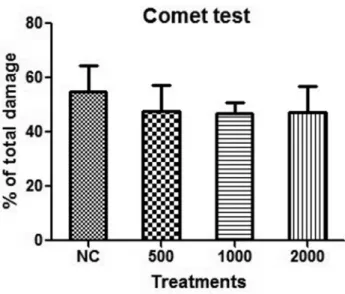
articulatus did not increase the frequency of DNA damage as measured by the comet assay and micronucleus test in treated mice, indicating a non-genotoxic and

guyanensis did not present genotoxic action for the index and frequency of damage evaluated by the comet assay, or for the formation of micronuclei on the bone marrow of male and

The probability of attending school four our group of interest in this region increased by 6.5 percentage points after the expansion of the Bolsa Família program in 2007 and

As doenças mais frequentes com localização na cabeça dos coelhos são a doença dentária adquirida, os abcessos dentários mandibulares ou maxilares, a otite interna e o empiema da

Este relatório relata as vivências experimentadas durante o estágio curricular, realizado na Farmácia S.Miguel, bem como todas as atividades/formações realizadas

Neste trabalho o objetivo central foi a ampliação e adequação do procedimento e programa computacional baseado no programa comercial MSC.PATRAN, para a geração automática de modelos

Ousasse apontar algumas hipóteses para a solução desse problema público a partir do exposto dos autores usados como base para fundamentação teórica, da análise dos dados

enfermagem estivesse mais próximo do viver cotidiano das pessoas, da sua cultura, do seu saber e das suas práticas de saúde, de forma mais humana e mais