ABSTRACT
Imaging plays a vital role in the evaluation of adrenal pathology. The most widely used modalities are computed tomography and magnetic resonance imaging. Alone or in conjunction with appropriate clinical and biochemical data, imaging can provide specific diagnoses that preclude the need for tissue sampling. This article reviews imaging fea-tures of normal and diseased adrenals, from both benign and malig-nant causes.(Arq Bras Endocrinol Metab 2004;48/5:592-611)
Keywords:Adrenal glands; Imaging; Computed tomography; Magnet-ic resonance imaging
RESUMO
Procedimentos de Imagem na Patologia Adrenal.
Procedimentos de imagem têm um papel vital na avaliação da patologia adrenal. As modalidades mais amplamente empregadas são a tomografia computadorizada e a imagem por ressonância magnética. Isoladas ou em combinação com dados clínicos e bio-químicos apropriados, a imagem pode prover diagnósticos específi-cos que dispensam a necessidade de amostras de tecido por biopsia. Este artigo revisa os achados de imagem da adrenal normal e patológica, incluindo tanto causas benignas como malignas. ( A r q Bras Endocrinol Metab 2004;48/5:592-611)
Descritores:Glândulas adrenais; Imagem; Tomografia computadoriza-da; Ressonância magnética
INTRODUCTION AND IMAGING TECHNIQUE
T
HE ADRENAL GLANDS CAN BE imaged through ultrasound, computed tomography or magnetic resonance imaging.Sonography (US) of the adrenal glands is not performed routinely, either because of the deep and often times inaccessible location of the gland or because of the lack of specific imaging patterns, leading to non-diagnostic studies. Adrenal pathology is therefore usually an incidental finding in an US study performed for other reasons, and patients are referred for further evaluation.
Computed Tomography (CT) is the primary diagnostic imaging method for evaluation of adrenal disorders. When optimal CT scan-ning technique is used, the normal or pathologic adrenal glands can be well visualized in virtually 100% of patients (1). Large and small mass-es and hyperplasia can be readily detected when prmass-esent. With modern CT scanners, imaging of the adrenals can be achieved even in the most emaciated patients, and in patients who are unable to suspend respira-t i o n .
Suzan M. Goldman
Rafael Darahem Coelho
Edison de O. Freire Filho
Nitamar Abdala
Denis Szejnfeld
Juliano Faria
Paola L.P. Judice
Viviane Vieira Francisco
Philip J. Kenney
Jacob Szejnfeld
Genitourinary Imaging Service, Department of Diagnostic Imaging, Universidade Federal de São Paulo, São Paulo, SP, Brasil; and Department of Radiology, University of Alabama (PJK), Birmingham, Alabama, USA
ent techniques will be discussed later. For most patients, the following technique for a helical CT study is appropriate: 2.5 – 3mm slice thickness, with 1.5 – 3mm reconstruction interval through the adrenal region, following oral contrast, performed before and after intravenous injection 100 – 150ml low osmolar iodinated contrast medium. Imaging delays after con-trast injection are routine “portal venous phase” (60 – 90 seconds) and “delayed-phase” (15 minutes), when appropriate. With a 4 multidetector-row CT scanner, using HS mode (pitch 1.5 or “6”) with 1.25mm detectors, 2.5mm slice thickness with table speed 7.5mm/rotation (1.5 pitch x 1.25mm detector x 4 row). Alternatively, in HQ mode (pitch 0.75 or “3”), 2.5mm detectors, slice thickness of 2.5mm with table speed 7.5mm/rotation (0.75 pitch x 2.5mm detector x 4 row). Reformatted images (in sagital or coronal plane) can sometimes be useful in determining whether a mass arises from the adrenal, liver or kidney. Great strides have been made in magnetic reso-nance imaging (MRI) of the abdomen; and with cur-rent techniques, the adrenals also can be delineated in nearly all patients with MRI (2-4). Patients who are claustrophobic, are unstable or erratic breathers, exceed the size limits, or have pacemakers cannot be evaluated with MRI. State-of-the-art MRI scanners
lipid (such as in/out-phase and fat suppression) are most useful in differentiating between subacute hem-orrhage and fat-containing adrenal masses, both of which can appear as high signal lesions on T1-weight-ed spin echo images.
An MRI evaluation of the adrenals usually con-sists of: T1 gradient refocused echo (GRE) weighted sequences in and out of phase in the axial and coronal planes, 3 to 5mm slice thickness; a T2-weighted TSE sequence with fat saturation in the axial plane, 3 to 5mm slice thickness; and a T2-weighted HASTE sequence in the coronal plane, slice thickness of 5mm. The use of intravenous contrast agents should be lim-ited to those cases where there rests a doubt, being, in those cases, used to differentiate solid lesions from hemorrhages, and in cases of pheochromocytoma. The use of such contrast media is not yet established in the diagnosis of adenomas.
Despite the many improvements in MRI that allow more rapid data acquisition, an MRI examina-tion of the adrenals is still a relatively lengthy proce-dure compared to CT. Therefore, it is most useful as an alternative to CT in patients who cannot tolerate intravenous iodinated contrast, in cases of suspected pheochromocytoma and to confirm a diagnosis of hemorrhage.
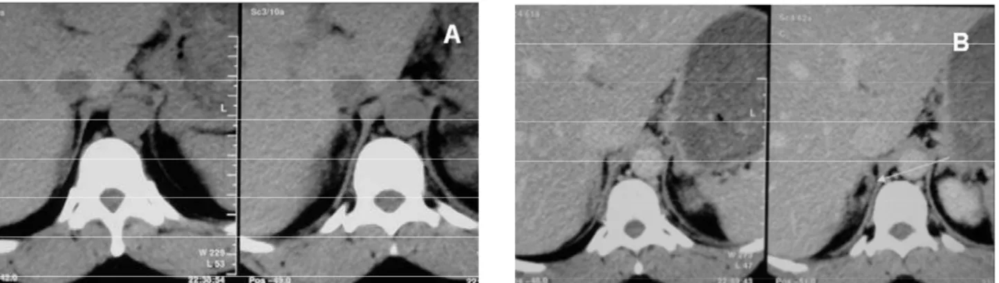
Figure 1.Use of thinner collimation.A:CT with 7mm contiguous sections demonstrates an equivocal right adrenal nodule.
NORMAL ANATOMY
The adrenal glands are paired retroperitoneal organs that lie in a suprarenal location and are enclosed with-in the perwith-inephric fascia. The right adrenal is usually seen directly superior to the upper pole of the right kidney with its most caudal portion just anterior to the upper pole. The right adrenal is directly posterior to the inferior vena cava and insinuates between the right lobe of the liver and the right crus of the diaphragm. The left adrenal may be seen at the same level as the right, but it often is slightly more caudal. It is antero-medial to the upper pole of the left kidney and is usu-ally seen on CT in the same slice as the left kidney. The left adrenal lies lateral to the aorta and the left diaphragmatic crus, and superior to the left renal vein. A portion of the pancreas or the splenic vessels may be immediately anterior to the left adrenal. When per-forming a dedicated (thin section) CT of the adrenals, it is important to obtain a sufficient number of images, because the adrenals each extend about 2 to 4cm in craniocaudal direction, and because masses may pro-trude superiorly or inferiorly.
The adrenal glands have medial and lateral limbs extending posteriorly from central ridges. Inverted V, Y, L, and other configurations may be seen. On precontrast CT, the adrenals have a soft-tis-sue density similar to that of the liver. If very early post-contrast scans are obtained, there is considerable enhancement, which fades quickly to moderate enhancement – slightly less than that of liver. The adrenal cortex and medulla cannot be reliably distin-guished by either CT or MRI. On T1-weighted MRI images, the adrenals have intermediate signal intensity, similar to that of the liver, somewhat greater than the diaphragmatic crus but much less than the surround-ing fat (5). On standard T2-weighted images, the adrenals are hypointense to fat and isointense to liver, but they are hyperintense to the crus. There is less dif-ference between adrenal and fat signal intensities on T2-weighted images, and significant chemical shift artifact may obscure details of the normal adrenals. On fat-suppressed T2-weighted images, however, the nor-mal adrenals appear somewhat brighter than liver, and are much brighter than the suppressed fat. Thus, nor-mal adrenals and snor-mall masses are best seen on T1-weighted or fat-suppressed T2-T1-weighted images.
There is considerable variability in the lengths of the limbs. The surface of the normal adrenal glands should be quite smooth, without protruding nodules, and the limbs should have uniform thickness. Although no strict measurements have been
standard-ized, any area thicker than 10mm is abnormal. A use-ful rough estimate of normal size is that the thickness of the gland’s limbs should not exceed the thickness of the ipsilateral diaphragmatic crus, at the same level. It must be recognized that in the face of stress (as may be seen in severely ill patients), the adrenals may become enlarged in response to physiologically high circulating adrenocorticotropic hormone (ACTH) levels.
PSEUDOTUMORS
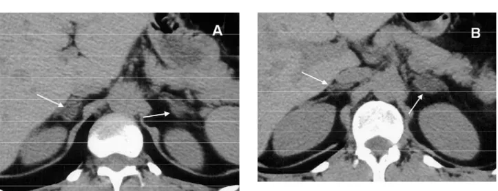
A variety of normal structures may simulate an adren-al mass if meticulous technique is not used. The rou-tine use of oral contrast and rapid scanning after bolus intravenous contrast will usually allow these pseudotu-mors to be distinguished from true adrenal masses. Pseudotumors are less common on the right, because there are fewer adjacent organs. Rarely, the duode-num, stomach or colon may produce a mass-like appearance (figure 2a). One should be careful when interpreting images of previously splenectomized patients, some of which develop splenosis and the small splenic tissue nodules may mimic tumors when located near to the adrenal space (figure 2b). Other potential mimickers of adrenal masses on the left are the pancreatic tail, tortuous vessels and small bowel.
PATHOLOGY
are isodense with adrenal tissue, they are detected as focal bulges on the otherwise smooth adrenal surface. Focal enlargement is a more important finding than any measurement. Inability to visualize a normal adre-nal is suggestive of adreadre-nal origin, but extra-adreadre-nal malignant masses can engulf or invade the adrenal. Large hepatic, renal, or retroperitoneal tumors may simulate an adrenal mass. In these cases, demonstration of a normal adrenal gland essentially excludes the pos-sibility that the mass is adrenal in origin. Conversely, the presence of adrenal hyperfunction confirms an adrenal origin. The pattern of displacement of organs likewise may be helpful in determining the center of origin, because most tumors grow centrifugally. Right adrenal masses typically displace the kidney inferiorly, and the inferior vena cava anteriorly, whereas hepatic masses rarely displace the vena cava anteriorly.
Adrenal hyperplasia may result in diffuse thick-ening of the adrenal glands, but retention of the nor-mal shape (figure 3). This enlargement is usually smooth, but there may be nodularity. However, bilat-eral enlargement, whether smooth or nodular, is indicative of hyperplasia. A significant number of patients with clinical and biochemical evidence of hyperplasia will have normal adrenals with CT. In part, this is because there may be significant hyperfunction of the adrenal cortex before enough thickening has occurred to be recognizable. Thus, a normal appear-ance of the adrenals does not exclude hyperplasia. Adrenal hyperplasia can be diagnosed when there are bilateral nodules or when there is nodularity associat-ed with bilateral adrenal thickening. With primary functioning adrenal tumors, the ipsilateral remaining adrenal tissue and contralateral adrenal gland should
be normal or atrophic. However, it is not always pos-sible to distinguish an adenoma from a solitary hyper-plastic nodule by CT.
On MRI, hyperplastic adrenals can be seen to be enlarged, but the signal intensity of the tissue is similar to that of normal adrenal gland. On occasion, the adrenals are found to be enlarged in patients with no evidence of adrenal dysfunction. These findings are not clinically significant. A number of conditions have been associated with such nonspecific hyperplasia, including acromegaly (100%), hyperthyroidism (40%), aortic aneurysm and dissection (55%), hypertension with arteriosclerosis (16%), diabetes mellitus (3%), and a variety of malignancies (6,7). In some cases, this adrenal enlargement is a response to physiologic stress. On imaging, the appearance is indistinguishable from hyperplasia producing adrenal hyperfunction.
CUSHING’S SYNDROME
Cushing’s syndrome results from excess production of cortisol by the adrenal cortex. In about 85% of cases, this is a result of excess ACTH production (an ACTH-dependent disorder) from a pituitary adenoma or hyperplasia (80%), or from an ectopic source (8). About 15% of cases of Cushing’s syndrome are ACTH independent, usually due to a cortical adenoma or adrenal carcinoma, rarely secondary to primary nodu-lar hyperplasia (8,9). Adrenal CT is performed in patients with Cushing’s syndrome to distinguish ACTH-dependent (hyperplasia) from ACTH-inde-pendent (focal mass) disorders, and to determine the location of the focal mass in the latter. In patients with
an adenoma or a carcinoma, the remaining ipsilateral and the contralateral adrenals are normal or atrophic. Surgical removal of an adenoma (or of a resectable car-cinoma) is curative. Biochemical testing can be extremely useful.
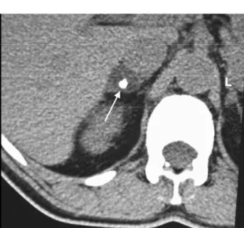
Computed tomography is virtually 100% accu-rate for detection of adrenal adenomas resulting in hypercortisolism. These adenomas are almost always over 2cm at the time of presentation, are usually in the range 2 to 5cm (10), and are readily seen on CT, espe-cially because these patients have abundant retroperi-toneal fat. The masses are smooth, round or oval, and relatively homogeneous, with little enhancement after intravenous contrast and rapid washout at 15 minutes, in excess of 60% from baseline attenuation (11). Most often they have a soft-tissue-attenuation value, but they may have near water attenuation because of rela-tively high lipid content. Calcification can be found in adenomas, although it is rare (12) (figure 4).
The contralateral adrenal is commonly visibly thinner than normal, indicating atrophy from ACTH suppression (figure 5). With these findings, and in the proper clinical setting, no further investigation is need-ed. It should be noted that no CT characteristic has been reported that distinguishes whether an adenoma produces excess cortisol or aldosterone, or whether it is not hyperfunctioning.
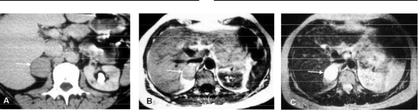
Magnetic resonance imaging is equally accurate in patients with cortisol producing adenomas, because of their size (9). Typically, the signal intensity of the ade-nomas is similar to that of liver on T1-weighted images, and similar or only slightly greater on T2-weighted images (3) (figure 6). The presence of intracellular lipid can be documented by opposed-phase imaging (17). Although adrenal adenomas and cysts may have similar
attenuation values on CT, they are readily distinguished on MRI, because cysts are very hyperintense on T2-weighted images and do not enhance.
A normal CT or MRI appearance does not exclude hyperplasia. In fact, in the absence of a focal mass, a patient with biochemical evidence of hypercor-tisolism and normal adrenals can confidently be given the diagnosis of hyperplasia as long as exogenous steroid use is excluded. Most often, both adrenals become smoothly thickened due to excess ACTH. The thickening may become massive, especially with ectopic ACTH production (10).
Adrenal nodules ranging from 6mm to 7cm may be seen in 12% to 15% of patients with ACTH-dependent Cushing’s syndrome (10). Although usual-ly bilateral, such nodules may be unilateral. Careful examination will usually show some evidence of bila-teral enlargement. Primary pigmented nodular adrenocortical disease is a rare disorder that causes Cushing’s syndrome. It tends to present in younger patients than the other types of Cushing’s syndrome. Elevated cortisol is found, with very low ACTH levels. On CT, multiple bilateral nodules up to 3cm are typi-cal. Unlike macronodular hyperplasia due to excess ACTH, the cortex between the nodules is atrophic. On MRI, the nodules are relatively low signal on both T1- and T2-weighted images.
PRIMARY ALDOSTERONISM
Primary aldosteronism (Conn’s syndrome) results from excess adrenal production of the mineralocorti-coid aldosterone. It is characterized by reduced plasma renin levels, hypokalemia, and hypertension, with as
many as 95% of cases resulting from an autonomous cortical adenoma (aldosteronoma). Most of the remaining cases result from primary idiopathic bilater-al hyperplasia. A few rare cases have been reported as a result of bilateral adenomas, unilateral hyperplasia, and adrenal carcinoma. Correct diagnosis is important, because surgical removal of an aldosteronoma is cura-tive, but partial and even bilateral total adrenalectomy commonly fails to cure hypertension in patients with hyperplasia. Adrenal CT should be performed first to evaluate a patient with biochemical evidence of prima-ry hyperaldosteronism. Certain biochemical features are strongly indicative of an aldosteronoma, and CT should be done to locate the tumor before surgical
removal. If the imaging study is inconclusive, adrenal venous sampling should be done and all diagnostic studies correlated prior to surgery.
On CT, aldosteronomas appear as round or oval lesions, similar to other adenomas, but typical-ly smaller than cortisol-producing adenomas. They are rarely larger than 3cm, with a median size of 16 to 17mm. Because of relatively high lipid content, they often (50%) have an attenuation value similar to that of water (-10 to +10). This propensity for low attenuation should be recognized, so that a low-attenuation mass discovered in a patient with documented hyperaldosteronism is not misdiag-nosed as a cyst.
Figure 4. Calcification on a right adrenal adenoma (arrow).
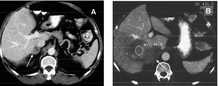
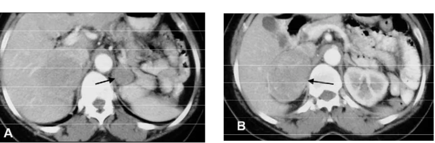
Figure 5.Cushing’s syndrome due to a cortical adenoma. The left adrenal (arrowhead) is atrophic in this patient with hypercortisolism. There is a 3cm right adrenal mass (arrow).
Because aldosteronomas are typically small, they present a greater challenge than cortisol-pro-ducing adenomas. Early publications reported a sensitivity of 70% with several false negative CTs because of inability to detect small aldosteronomas. With better quality, thin-section CT, a sensitivity of 80% to 90% has been achieved. With the 5mm-section technique, false negatives are uncommon (12% to 14%). Primary adrenal hyperplasia causing aldosteronism may be micronodular or macron-odular. The adrenals may appear normal or diffuse-ly thickened on CT. One or more discrete nodules ranging from 7 to 16mm may be seen. Diagnostic errors may occur because of a unilateral nodule that simulates an adenoma on CT. Conversely, tiny bilateral nodules that are present in 25% of patients with aldosteronomas may result in an erroneous diagnosis of hyperplasia. An accuracy of 80% for CT showing lack of lateralization (either both glands are normal, both are enlarged, or there are bilateral nodules) has been reported. Because of these potential difficulties, adrenal venous sam-pling still can be useful in certain patients. Although aldosteronomas can be shown with MRI, there is no advantage of MRI over CT. Because many aldosteronomas are small, the lesser spatial resolution of MRI is a theoretical disadvantage. Aldosteronomas are not distinguishable from other adenomas by any MRI feature. In fact, as with CT, no MRI feature has been described that indicates whether an adenoma is hyperfunctioning or non-h y p e r f u n c t i o n i n g .
ADRENAL CARCINOMA
Adrenal carcinoma is a highly malignant neoplasm that arises in the adrenal cortex. It is rare, with an incidence estimated at two cases per million people. It can occur at any age, with a median age of about 40 years. About 50% of adrenal carcinomas will produce an endocrine disorder. Cushing’s syndrome is commonest, seen in about 50% of adrenal carcinoma patients, and it accounts for 65% of the functional disorders. It may be seen alone or in combination with virilization. Viril-ization alone, feminViril-ization, and aldosteronism may be seen, in order of decreasing frequency.
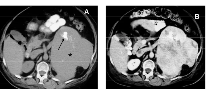
Adrenal carcinomas are often very large when first detected. This is especially true of nonfunctioning tumors, which remain clinically silent until advanced stages, they may be discovered because of flank pain, fatigue, palpable mass, or evidence of metastases. Even functioning tumors are usually large at presentation, which may be a result of relatively inefficient hormone production. Average size at presentation is 12cm (range, 3 to 30cm). Nowadays, however, with widespread use of imaging, some of these neoplasms are discovered inci-dentally and are smaller than previously reported. Ade-nomas are typically small, round or ovoid, homoge-neous, with less than 10 Hounsfield units attenuation on precontrast CT and signal loss on opposed phase images on MRI and show very slow growth. Adrenal carcinoma typically shows rapid growth (figure 7). As a result, if there is concern, a single 6-month follow-up CT can readily distinguish these entities; lack of growth effec-tively excludes adrenal carcinoma.
The histology of adrenocortical carcinoma is variable. It can be difficult to distinguish a well-differ-entiated carcinoma from an adenoma, even with a resected specimen. Needle biopsy may be nondiagnos-tic. Correlation of histology with radiological features, and sometimes with biologic behavior, is needed for diagnosis. The overall prognosis is very poor, with 5-year survival of 20% to 25%. However, prognosis is bet-ter (42% to 57%) for localized (stage I) adrenal carcino-mas if complete surgical resection can be accomplished. CT readily detects adrenal carcinomas. Most often they are seen as large, irregularly shaped, hetero-geneous masses in the adrenal region. Bilateral disease is seen in less than 10% of cases. Necrosis is common. Calcification is found in about 40% (12) (figure 8). Heterogeneous enhancement after intravenous con-trast is typical, with strong peripheral and little central enhancement. The tumors may be poorly marginated, or they may show local invasion. Invasion of the infe-rior vena cava, liver metastases, and retroperitoneal lymphadenopathy may be seen. In general, these fea-tures allow differentiation from adenoma or hyperpla-sia. However, an incidentally found carcinoma may be more difficult to discriminate from an adenoma on CT, as both may be less than 5cm, well circumscribed, and homogeneous.
On MRI, adrenal carcinomas are easily seen as large heterogeneous masses in the adrenal bed, with areas isointense or hypointense to liver on T1-weight-ed images, and isointense or hyperintense to fat on T2-weighted images (2) (figure 9). Areas of hemor-rhage may result in variable signal intensity depending on the age of the hemorrhage. With multiplanar
capa-bility and the high tissue contrast of T2-weighted images, MRI can be useful to define the adrenal origin and the extent of disease (figure 9). After injection of gadolinium, bright heterogeneous enhancement is seen (18). The high sensitivity of MRI for venous involvement and liver disease make it helpful for stag-ing; venous extension is a poor prognostic sign, and if metastases are present surgery is not indicated.
PHEOCHROMOCYTOMA
A pheochromocytoma is a neoplasm of the adrenal medulla that contains chromaffin cells and causes excess catecholamine production. When such a tumor arises outside the adrenal, it is labeled a paragan-glioma. Most are benign, although about 10% are malignant. Sporadic cases are usually unilateral, affect-ing the right adrenal slightly more frequently; about 5% are bilateral (figure 10). Most patients are hyper-tensive. However, pheochromocytoma is found in less than 1% of the hypertensive population, and in 0.3% of autopsies. There is an increased likelihood of pheo-chromocytoma in patients with neurofibromatosis, von Hippel-Lindau disease, and multiple endocrine neoplasia (MEN) syndromes (50% in MEN 2 and 90% in MEN 2b). In such syndromes and in children, mul-tiple or bilateral cases are more likely. In MEN 2b, bilateral tumors are so common that bilateral adrena-lectomy is recommended, because lesions may recur after unilateral surgery.
In most cases, diagnosis of pheochromocytoma can be established with biochemical testing. However,
biochemical tests are expensive, time consuming and fraught with difficulty, because such factors as episodic catecholamine production, concurrent medication, stress, inadequate urine collection for 24-hour samples, and other factors can contribute to both false positive and false negative results. Detection and localization are important because surgical resection is curative, and because there is no effective medical therapy.
Although 90% of pheochromocytomas arise in the adrenal, up to 10% are extra-adrenal (figure 11), with many such lesions (7%) in the infrarenal portion of the retroperitoneum, arising in the organ of
Zuck-erkandl (figure 11). Paragangliomas can be single or multiple, and they may have greater malignant poten-tial. Paragangliomas also can be found in the neck, the mediastinum, and the wall of the urinary bladder.
Pheochromocytomas are usually over 3cm at presentation and should be invariably identified by CT. When small, the tumors are round and have homogeneous soft-tissue attenuation values (figure 12). Because pheochromocytomas are hypervascular neoplasms, they have a propensity to undergo hemor-rhagic necrosis even when benign, accounting for the central low attenuation seen in large neoplasms.
tral necrosis may be so extensive as to simulate a cyst. Calcification is uncommon; when present, it may have an eggshell pattern. After intravenous administration of iodinated contrast medium, pheochromocytomas exhibit heterogeneous enhancement, a pattern indis-tinguishable from a malignant adrenal neoplasm. Cor-relation with biochemical function is required to establish the correct diagnosis.
Because pheochromocytomas are large, they can be detected even with unenhanced CT. Some cern has been raised about the use of intravenous con-trast in patients with pheochromocytoma. Plasma cat-echolamine levels can be raised by intravenous injec-tion of iodinated contrast medium, but symptomatic blood pressure elevations do not usually result. Only if a patient has had hypertensive episodes and no ade-quate pharmacologic adrenergic blockade is it neces-sary to avoid contrast. Contrast is especially useful for detection of extra-adrenal lesions. Although paragan-gliomas can usually be identified on CT (figure 11),
they have a nonspecific appearance. The CT features of malignant paragangliomas in particular overlap with those of other retroperitoneal malignancies. Radionu-clide metaiodobenzylguanidine (MIBG) scintigraphy can be useful to document whether a retroperitoneal mass is in fact a paraganglioma.
Pheochromocytomas have a rather characteris-tic appearance on MRI (2,3). They are readily detect-ed (with a sensitivity of 100% in one report), because they are several centimeters in diameter. When small, they are usually homogeneous and isointense to mus-cle, hypointense to liver on T1-weighted images, and have a distinctive marked signal hyperintensity relative to fat on T2-weighted images (3) (figures 12 and 13). As they grow and develop central necrosis, there may be central areas that are hyperintense on both T1- and T2-weighted images.
Persistent enhancement after intravenous gadolinium is typical (18). Because no lipid is found in pheochromocytoma, there is no decrease in signal on opposed phase images. Paragangliomas have similar distinctive imaging characteristics; as a result, MRI is superior to CT for diagnosis of paragangliomas, and nearly as sensitive as MIBG. Because most such tumors lie in the adrenal or retroperitoneum, coronal MRI can quickly and effectively show the area of abnormality.
Biopsy of a mass suspected to be a pheochro-mocytoma is not recommended, especially if adequate hypertensive control has not been achieved, as several episodes of severe hemorrhage and even death have resulted following percutaneous biopsy. MIBG has both high sensitivity and high specificity, and it can detect a paraganglioma in any part of the body. How-ever, it is an expensive test that requires up to 72 hours to complete and is not widely available. Furthermore, it does not provide sufficient anatomic detail for surgi-cal planning. It is most useful in evaluating patients
Figure 10.Multiple pheochromocytomas in MEN2b.AandB:CT shows bilateral heterogeneous adrenal tumors (arrows).
with a strong clinical suspicion and in whom CT or MRI is normal or equivocal, or for follow-up of malig-nant lesions.
The follow up for these patients should be very judicious and information on clinical presentations and surgical data should be included and taken into account during analysis of exam results, be they from a CT or from an MR. The presence of hypertension in this group of patients is not always indicative of persis-tence or recurrence of the disease. Other causes of hypertension should be evaluated, such as renal changes due to chronic disease.
NONHYPERFUNCTIONING NEOPLASMS
Nonhyperfunctioning adrenal neoplasms are clini-cally silent until they become very large, although they may present with pain if they bleed. Current-ly, most such masses are found incidentally on studies performed for other reasons. About 30% of
all adrenal masses are incidentally detected by CT. An adrenal mass is seen in about 4% of all abdomi-nal CT scans, with one third being serendipitous findings; the remainder are either metastases in patients with known malignancies, or they are functioning lesions. Most incidental adrenal masses are benign and of no clinical significance, especial-ly in patients with no known malignancy. In two large series, only 6.7% and 9% of serendipitous adrenal masses were subsequently proven malig-nant. Although historically size has been consi-dered an important factor, larger tumors having a greater likelihood of malignancy, size is an imper-fect criterion. Although malignant neoplasms were all larger than 6.5cm in one study, and most benign masses are less than 5cm, there is conside-rable overlap. The majority of incidental masses greater than 5cm are still benign in patients with no history of malignancy, and lesions as small as 1cm may be metastases (13). Thus, imaging crite-ria are required for differentiation.
Figure 12.Pheochromocytoma in a man with episodic hypertensive crises.A:Homogeneous right adrenal mass on CT image (arrow).B:On T1-weighted image (366/26), the mass (arrow) has signal intensity similar to liver.C:On T2-weighted image (2500/80), the mass (arrow) is homogeneous and more intense than fat.
except that contralateral atrophy is not present. They are smooth, round or oval, with a well-defined margin. CT densitometry can be used to accurately dif-ferentiate benign from malignant adrenal masses. Lee et al. reported the use of nonenhanced CT attenua-tion values for the characterizaattenua-tion of adrenal masses where most adenomas had attenuation values lower than those of malignant masses (15). Korobkin et al. and Boland et al., confirmed these findings. Boland pooled the data from 10 articles and showed that a sensitivity of 71% and a specificity of 98% result from choosing a threshold value of 10HU for the diagno-sis of adrenal adenoma. Ninety-eight percent of homogeneous adrenal masses with a nonenhanced CT attenuation value of 10HU or less will be benign (most will be adenomas), whereas 29% of adenomas will have an attenuation value of more than 10HU and will be indistinguishable from most nonadeno-mas, including metastases.
Chemical shift MR imaging can be used to dif-ferentiate adrenal nodules. A relative loss in signal intensity in an adrenal mass, when opposed-phase
relative change in signal intensity on opposed-phase chemical shift MR images.
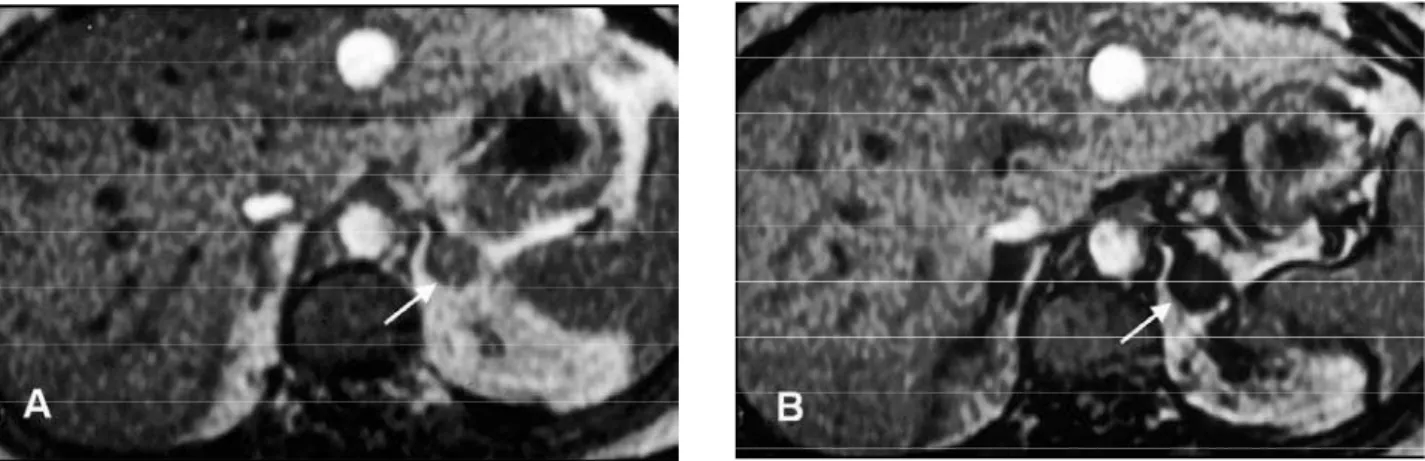
A substantial minority of adrenal adenomas is lipid-poor and cannot be characterized by means of their nonenhanced CT attenuation. Korobkin et al. established washout curve values for adenomas. The adenomas, notwithstanding the presence and quanti-ties of lipids in their composition, have the property of presenting rapid loss of enhancement and attenuation values at 15 minutes, which can be used to differenti-ate them from other masses (figure 15).
The established formula for this calculation includes the density of the lesion during the precon-trast, portal (60sec) and 15-minute delay phases. The exam should be performed with thin collimation and the attenuation measurements must have the region-of-interest (ROI) spanning 1/2 to 2/3 the area of the lesion, always in the same position. Calcified and necrotic nodules are not suitable for this calculation. Washout values of more than 60% are indicative of adenomas, whereas those less than 60% indicate inde-terminate masses (figure 15).
The percentage enhancement washout is calcu-lated as follows:
% enhancement washout= (E-D/E-U) x 100, where:
E= Enhancement: attenuation at portal phase D= delayed enhanced attenuation at 15min U= attenuation at pre contrast
In summary, the evaluation of a known adrenal mass starts by using nonenhanced CT. If the attenua-tion of the mass is 10HU or less the diagnosis is, most of the times, a lipid-rich adrenal adenoma and a small fraction of these will be cysts. In such a case, there is no need for further evaluation. If the attenuation is more than 10HU, the mass is considered to be inde-terminate and an enhanced and 15-minute-delayed enhanced CT scan should be done. If the enhance-ment washout is more than 60%, the most likely diag-nosis is of a lipid-poor adenoma. Again, there is no need for further evaluation. If the enhancement washout is less than 60%, the mass is considered inde-terminate. Percutaneous adrenal biopsy is recom-mended if the patient has a primary neoplasm with no other evidence of metastases. In a patient without can-cer, surgery is recommended if the mass measures more than 4 – 5cm. Follow-up CT, or adrenal scintig-raphy with the use of radioiodinated norcholesterol (NP-59) can also be done in this group of patients.
METASTATIC DISEASE
Metastases to the adrenals are common from a variety of primary malignancies, including thyroid, renal, gas-tric, colon, pancreatic, and esophageal carcinomas, and melanoma. Lung and breast cancer, however, are the most common sources, with adrenal metastases found on CT in approximately 19% of lung cancer patients. Because adrenal metastases are so common in lung
cancer and the adrenals may be the only site of metas-tasis, they should be included in the CT examination of all patients presenting with lung cancer. Even in patients with lung cancer, about one third of adrenal masses are benign. Thus, imaging features must be used to help make the correct diagnosis. If the imaging findings are equivocal, a percutaneous CT-guided biopsy should be performed to establish a histologic diagnosis. A baseline CT at the time of presentation of patients with lung cancer is a useful aid in follow-up. Detection of a new small adrenal mass on follow-up is clear evidence of metastasis if the baseline showed nor-mal adrenals.
Adrenal metastases can vary considerably on CT. Size can range from less than a centimeter to large masses; the size, however, overlaps with that of ade-nomas (13) (figure 16). Adrenal metastases may be unilateral or bilateral. When small (< 5cm), they com-monly are fairly well circumscribed, round or oval, and of soft-tissue density. They may have smooth or irreg-ular, lobulated contours. They may show local inva-sion, a sign of malignancy. Calcification is rare (12), and they may bleed (14).
Small adrenal metastases are solid tumors and thus usually have homogeneous soft-tissue-attenua-tion values, similar to or higher than that of muscle on noncontrast scans. Even if there is central necrosis, however, the density is not lower than that of water, because malignant tumors do not produce lipid (15).
Following intravenous administration of iodi-nated contrast material, there may be homogeneous enhancement, but commonly enhancement is hetero-geneous, especially with larger tumors. A thick, nodu-lar enhancing rim also may be seen (16).
Adrenal metastases can vary in size and appear-ance on MRI. On T1-weighted images, metastases usually have signal intensity similar to or lower than that of normal liver tissue, not distinctly different from
that of adenomas. They may be heterogeneous (2,3). On T2-weighted images, they are often heteroge-neous, and they are usually hyperintense compared to normal liver, often similar to or of higher intensity than fat, unlike the typical adenoma (2,3). Numerous calculations based on signal intensity ratios, or calcu-lated T2 values, have been investigated, but none have been found to be reliable in practice for distinguishing metastases from adenomas (2,5). Because metastases do not produce lipid, there is no decrease in signal on opposed phase images (17). This has been shown to be a more consistent finding than T2 values. After intravenous administration of gadolinium compounds, metastases exhibit exuberant and heterogeneous enhancement that persists for several minutes, an enhancement pattern quite different from that of ade-nomas (18). MRI is readily able to demonstrate or exclude local invasion because of the great contrast between neoplastic and normal tissue, especially on fat-suppressed T2-weighted images.
Computed tomography is the most cost-effec-tive method for screening and following patients with malignancies. In most cases, adrenal metastases can be diagnosed or excluded by a well-performed CT. MRI can be valuable in cases in which CT could not be per-formed. Percutaneous biopsy under CT guidance can be very effective, with accuracy and negative predictive value of over 90% (19). However, complications can occur, and biopsy is not needed if the imaging findings are diagnostic. Follow-up by CT can be diagnostic, because adenomas are very slow growing and will not change in size over a period of a few months, while metastases will show growth.
Neoplastic patients frequently show a diffuse enlargement of the adrenal glands but with no masses or changes in contours. Biochemical studies carried out with this group of patients have demonstrated a number of changes in the gland function, which are shown to be consistent with hyperplasia. A definite association between malignant neoplasms and adrenal gland hyperplasia is observed; there is a higher preva-lence of adrenal gland hyperplasia in tumors patients when compared to the general population (20).
ADRENAL LYMPHOMA
Lymphoma occasionally involves the adrenal glands, with diffuse non-Hodgkin’s being the commonest type. This may be found at presentation or at follow-up, with adrenal lymphoma reported in 1% to 4% of patients being followed up for lymphoma. Adrenal involvement is most commonly seen in conjunction with an extra-adrenal disease site. Primary adrenal lym-phoma is rare and is believed to arise from hematopoi-etic cells in the adrenal (21).
Lymphomatous adrenal masses are bilateral in one third of cases; when bilateral, the patient may develop Addison’s disease.
On CT, adrenal lymphomas usually are seen as large soft-tissue masses replacing the adrenal. Mild to moderate enhancement is seen after intravenous administration of iodinated contrast (figure 17). The lesions may be homogeneous, but they are often het-erogeneous with low attenuation areas even before therapy (21). Sometimes the growth pattern can
gest lymphoma, as it is more likely to infiltrate or insinuate around the upper pole of the kidney than displace it, as would be typical of carcinoma. There may be hemorrhage, and calcification can be found, especially after chemotherapy (21). On MRI, adrenal lymphomas are indistinguishable from other malig-nancies. They are usually heterogeneous, with low sig-nal on T1-weighted images (less intense than normal liver, but more intense than muscle) and more intense than fat on T2-weighted images (2,22).
MYELOLIPOMA
Myelolipoma is an uncommon, benign, nonfunction-ing neoplasm of the adrenal, found in less than 1% of autopsies. It is composed of variable amounts of fat and hematopoietic tissue, including myeloid and ery-throid cells and megakaryocytes. It affects men and women equally. Although this is a nonfunctioning tumor, in 10% of cases it is associated with endocrine disorders, including Cushing’s syndrome, congenital adrenal hyperplasia, and Conn’s syndrome. Most myelolipomas (80%) are asymptomatic and of no clin-ical significance. Some (10%) become large and cause vague symptoms or pain. Large myelolipomas may hemorrhage, which can be the cause of pain. Size ranges from 1 to 15cm, with a mean of about 4cm.
On CT, most myelolipomas are well-circum-scribed masses, sometimes with a discrete thin appar-ent capsule (figure 18). Occasionally, the mass may appear to extend into the retroperitoneum. Nearly all contain some definite fat density (< -20HU).
Howe-ver, the amount of fat is widely variable, ranging from nearly all fat, to more than half fat (50%), to only a few tiny foci of fat in a soft-tissue mass (10%) (23). Occa-sionally, the mass has an attenuation value between that of fat and water because the fat and myeloid ele-ments are diffusely mixed. Calcification is seen in 30%, often punctate. With hemorrhage, high-density areas can be seen. Bilateral myelolipomas occur in about 10% (23).
The presence of fat in an adrenal mass also can be recognized on MRI, because fat is typically bright on both T1- and T2-weighted images (figure 18). Decrease in signal with fat suppression or phase can-cellation is confirmatory (17) (figure 18). However, if the mass is nearly all mature fat, there will not be loss of signal with opposed phase images, because the loss of signal occurs only with phase cancellation in areas with an admixture of fat and water protons (24). Overall, myelolipomas are often heterogeneous because the nonfatty areas will have signal intensity similar to that of hematopoietic bone marrow. The lesions enhance brightly after intravenous administra-tion of gadolinium.
The presence of fat in an adrenal mass is the key to the diagnosis of myelolipoma, because virtually no other adrenal lesion contains fat. Teratoma and liposarcoma of the adrenals are extraordinarily rare. An angiomyolipoma of the upper pole of a kidney may be mistaken to be an adrenal myelolipoma. However, this is of no clinical significance, because both these lesions are benign. If necessary, diagnosis can be confirmed by percutaneous needle biopsy. If the biopsy reveals bone marrow elements and the mass contains fat, the
nosis is assured, because extramedullary hematopoiesis does not contain fat. The presence of megakaryocytes also is an important diagnostic histologic feature. A definite diagnosis is important because surgical resec-tion is not indicated unless there has been significant hemorrhage. In nearly all cases, a diagnosis of adrenal myelolipoma can be made confidently based on CT or MRI findings alone.
ADRENAL CYSTS
Adrenal cysts are rare, found in only 1 of 1,400 autop-sies. They are nonfunctional and usually found inci-dentally. The commonest (45%) type is endothelial cyst. These are predominantly lymphangiomatous cysts, typically small and asymptomatic. Thirty-nine percent are pseudocysts, which lack an endothelial lin-ing and are most often a sequelae of remote adrenal
hemorrhage. These are the type most often detected by CT. These can be quite large and may produce symptoms because of their size. The remaining cystic lesions of the adrenals are parasitic cysts (7%), caused by Echinococcus, and true epithelial cysts (9%).
On CT, adrenal cysts are large masses (5 to 20cm) that are well circumscribed and round. They are suprarenal in location, but it may be difficult on CT to recognize that they arise in the adrenal rather than the kidney or liver. Sonography or MRI may bet-ter show their true origin. They usually are of near-water attenuation, but they can have higher or mixed attenuation values resulting from old hemorrhage. The wall may be thick and can show contrast enhance-ment. Calcification is common (75%) (12), being usu-ally curvilinear in shape and often limited to the infe-rior aspect of the wall. (12). Although MRI may show typical cystic features, homogeneous low intensity on T1-weighted images and extreme hyperintensity on
T2-weighted images, signal intensity can vary depend-ing on the age of hemorrhage. There is no central enhancement on either CT or MRI. Adrenal cysts are of no clinical significance, and surgical removal is unnecessary if the diagnosis can be established by imaging. Concern about malignancy is reasonable only in complicated cysts, which may have high attenuation values, a thick enhancing wall, and septations. In such cases, percutaneous needle aspiration may be helpful for both diagnosis and treatment.
INFLAMMATORY DISEASE
Inflammatory processes in the adrenals are uncom-mon. Adrenal abscesses occur rarely; they sometimes represent adrenal hematomas that became infected. More often, adrenal inflammation is due to chronic granulomatous disease, with tuberculosis (TB) and histoplasmosis most common, although North Ame-rican blastomycosis has been reported to involve the adrenals. Paracoccidioidomycosis, or South American blastomycosis, is the most common systemic mycosis in South America, with Brazil responsible for about 70% of total world cases. Its etiologic agent is Para-coccidioides brasiliensis. This disease chiefly involves the respiratory tract and its presentation can vary from acute and self-limited to a progressive pulmonary dis-ease or extra pulmonary dissemination. Diagnosis is made by lesion biopsy and fungal culture, serological tests and chest radiographs. The organs that constitute the reticuloendothelial system, including the adrenals, are the most affected ones.
Granulomatous disease of the adrenals can cause a variety of appearances depending on the dis-ease stage. In most cases, there is bilateral enlargement of the adrenals (25). Bilateral adrenal enlargement in a
patient with a reactive tuberculin skin test, or with chest radiographic changes of TB, paracoccidioidomy-cosis or histoplasmosis, should suggest the diagnosis, even if pulmonary cultures are nondiagnostic.
With active adrenal TB or paracoccidioidomyco-sis, both glands are usually enlarged to some degree for a disease course of less than 3 months (acute stage) (fi-gure 19). The masses have frequently heterogeneous enhancement and low-attenuation central areas repre-senting caseous necrosis (26). Enlarged glands and inward atrophic areas are found in the subacute stage of the disease (disease course of 6 to 24 months) (27).
Calcification is present in nearly half of cases of adrenal TB or paracoccidioidomycosis (35,161). Atro-phy is seen at the chronic stage, when the disease exceeds 24 months. In some cases, at a late disease stage, dense calcification with no soft-tissue mass may be seen (28). Active adrenal histoplasmosis most often presents with mild to marked symmetrical enlarge-ment of both adrenals, which retain the normal shape (30). There is low attenuation in the center with high-er phigh-eriphhigh-eral density, because of caseous necrosis (30). Calcification is not usually seen in the acute phase, but it may be seen with healing.
With TB, paracoccidioidomycosis and histo-plasmosis, there may be associated lymphadenopathy. All may cause adrenal insufficiency. The diagnosis usu-ally is suggested because of the CT appearance of the glands and the clinical presentation, especially when there is adrenal insufficiency. Percutaneous biopsy can be done to confirm the diagnosis (28).
The MRI signal intensity of inflammatory adrenal masses is nonspecific. It has signal intensity similar to spleen on T1-weighted images, and similar to or higher than fat on T2-weighted images. Enhancement patterns have not been described. Calci-fication is difficult to recognize.
Figure 19.Adrenal Paracoccidioidomycosis. A:Acute stage – Bilateral adrenal enlargement with areas of necrosis (arrows).
Adrenal hemorrhage in the adult may be seen in the setting of severe illness, such as sepsis, including but not limited to meningococcemia, burns, hypotension, and other life threatening illnesses. In these circum-stances, it is likely that the stress-related hyperplasia makes the adrenal prone to spontaneous rupture. About one third of cases of adrenal hemorrhage are associated with anticoagulant therapy. Commonly, the bleeding occurs in the first 3 weeks of anticoagulation. On CT, adrenal hemorrhage results in unilateral or bilateral adrenal masses that are usually ovoid, about 3cm in diameter, and of about muscle density or high-er with poor enhancement, if any, afthigh-er intravenous administration of iodinated contrast. With larger hem-orrhages, the masses may be heterogeneous and may have ill-defined margins. The size and attenuation value decrease over time if followed with CT. Calcification may develop in several weeks to months. If the hemor-rhage is bilateral, adrenal insufficiency may occur.
The adrenal glands are well protected in the retroperitoneum. Posttraumatic adrenal hemorrhage is
retroperitoneal bleeding (14). Because these patients usually have advanced disease, neoplasm elsewhere is also usually evident.
On MRI, adrenal hemorrhage will produce a mass that can have varying signal intensities dependent on the age of the blood. They usually have a pattern distinct from that of an adenoma or malignancy. A subacute hematoma usually has high signal intensity on both T1- and T2-weighted images (figure 20). In some cases of subacute hematoma, on T1-weighted images the center may be low intensity because of the presence of intracellular deoxyhemoglobin, and the periphery may have high signal because of free methe-moglobin; because both substances are high signal on T2, the mass is all high signal on T2. In chronic adren-al hematoma, the center consists of methemoglobin and thus this is of high signal on T1- and T2-weight-ed images, whereas the periphery contains hemo-siderin, which is low signal on both T1- and T2-weighted images. This ring pattern is quite distinctive and suggestive of the diagnosis (33).
ADDISON’S DISEASE
Adrenal insufficiency may result from a variety of caus-es including bilateral hemorrhage, inflammatory dis-ease, and idiopathic autoimmune primary Addison’s disease. Although metastases to the adrenal are com-mon, they rarely lead to Addison’s disease. When it occurs, the disease is either in advanced stages or is associated with spontaneous hemorrhage. Another rare cause is hemochromatosis, which can be recog-nized on CT, as the adrenals are normal or small in size but have increased attenuation values (28).
CT is indicated in evaluation of patients with Addison’s disease. In one study, all cases of idiopathic Addison’s disease could be distinguished from other etiologies (29).
CT can also determine the stage of the dis-ease: acute, subacute and chronic, as discussed pre-viously (27).
Adrenal atrophy that results from autoimmune disease will result in small glands without calcification (28,29). This must be distinguished from adrenal atro-phy due to exogenous steroids, which has a similar appearance, by careful history taking. Conversely, either adrenal hemorrhage, neoplasms, or inflammato-ry disease will show either adrenal masses or calcifica-tion, as discussed previously. Small adrenals that are partly or completely calcified suggest old granuloma-tous disease (particularly TB and Pb), whereas very dense calcifications with no soft tissue component sug-gest remote adrenal hemorrhage.
CT AND OTHER IMAGING METHODS
Except in a pediatric population, ultrasound is not used as a primary imaging method for adrenal disease, because it has both lower sensitivity and specificity than CT or MRI. Adrenal angiography and venogra-phy have been replaced by CT and MRI. Adrenal venous sampling remains useful in certain cases.
Adrenal scintigraphy is useful in patients sus-pected of having primary extra-adrenal or malignant metastatic pheochromocytomas.
MRI is extremely useful in selected cases, par-ticularly in the evaluation of pheochromocytomas and paragangliomas. MRI is equivalent to CT in evaluation of Cushing’s syndrome. However, CT is preferred for evaluation of hyperaldosteronism or adrenal insuffi-ciency. MRI can be very useful to further characterize lesions detected on CT. It can demonstrate more clearly the extent of a mass, its exact organ of origin,
and vascular compromise. MRI can be useful in evalu-ating an adrenalectomy bed for possible recurrence of a malignancy, because spin echo images show less degradation by clip artifact than CT. Last, pregnant patients suspected of having adrenal disease can be more safely studied with MRI than with CT.
REFERENCES
1. Abrams HI, Siegelman SS, Adams DF. Computed tomog-raphy versus ultrasound of the adrenal gland: a prospective study.Radiology 1982;143:121-8.
2. Chang A, Glazer HS, Lee JKT, et al. Adrenal gland: MR imaging.Radiology 1987;163:123-8.
3. Falke TH, te-Strake L, Shaff MI, et al. MR imaging of the adrenals: correlation with computed tomography.J Comput Assist Tomogr 1986;10:242-53.
4. Schultz CL, Haaga JR, Fletcher BD, et al. Magnetic reso-nance imaging of the adrenal glands: a comparison with computed tomography.AJR 1984;143:1235-40. 5. Chezmar JL, Robbins SM, Nelson RC, et al. Adrenal
masses: characterization with T1-weighted MR imaging.
Radiology 1988;166:357-9.
6. Vincent JM, Morison ID, Armstrong P, et al. Computer tomography of diffuse, non-metastatic enlargement of the adrenal glands in patients with malignant disease.
Clin Radiol 1994;49:456-60.
7. Goldman SM, Palacio G, Borri ML, et al.Prevalence of adrenal gland enlargement in patients with aortic aneurysm or aortic dissection. Presented at SUR Annu-al Meeting,2000.
8. Kamilaris TC, Chrousos GP. Adrenal diseases. In: Moore WT, Eastman RC, eds. Diagnostic endocrinology. Philadelphia: BC Becker,1990. p. 79-109.
9. Perry RR, Nieman LK, Cutler GB, et al. Primary adrenal causes of Cushing’s syndrome: diagnosis and surgical management.Ann Surg 1989;210:59-68.
10. Doppman JL, Miller DL, Dwyer AJ, et al. Macronodular adrenal hyperplasia in Cushing’s disease.Radiology 1988;166:347-52.
11. Korobkin M. Combined unenhanced and delayed enhanced CT for characterization of adrenal masses.
Radiology 2002;222:629-33.
12. Kenney PJ, Stanley RJ. Calcified adrenal masses. Uro-logic Radiol 1987;9:9-15.
1 3 . Kobayshi S, Seki T, Nonomura K, et al. Clinical experience of incidentally discovered adrenal tumor with particular reference to cortical function.J Urol 1993; 1 5 0 : 8 - 1 2 . 14. Shah HR, Love L, Williamson MR, et al. Hemorrhagic
adrenal metastases: CT findings. J Comput Assist Tomogr 1989;13:77-81.
20. Goldman SM, Borri M L, Abbehusen C, Faiçal S, Szenjn-feld J, Ajzen S.Prevalence of non-metastatic adrenal glands’ enlargement in patients with malignant neo-plasms evaluated by Computed Tomography (CT). Presented at SUR/SGR meeting2000-Hawaii).
21. Falchook FS, Allard JC. Case report. CT of primary adrenal lymphoma. J Comput Assist Tomogr 1991;15:1048-50.
22. Lee FT, Thornbury JR, Grist TM, et al. MR imaging of adrenal lymphoma.Abdom Imaging 1993;18:95-6. 23. Rao P, Kenney PJ, Wagner BJ, et al. Imaging and
patho-logic features of myelolipoma. R a d i o G r a p h i c s 1997;17:1373-85.
24. Tsushima Y, Ishizaka H, Matsumoto M. Adrenal masses: differentiation with chemical shift, fast low-angle shot MR imaging.Radiology 1993;186:705-9.
25. Hauser H, Gurret JP. Miliary tuberculosis associated with adrenal enlargement: CT appearance. J Comput Assist Tomogr 1986;10:254-6.
30. Levine E. CT evaluation of active adrenal histoplasmo-sis.Urol Radiol 1991;13:103-6.
31. Burks DW, Mirvis SE, Shanmuganathan K. Acute adrenal injury after blunt abdominal trauma: CT findings.AJR 1992;158:503-7.
3 2 . Bowen A, Keslar PJ, Newman B, et al. Adrenal hemor-rhage after liver transplantation.Radiology 1990; 1 7 6 : 8 5 -8 .
33. Itoh K, Yamashita K, Satoh Y, et al. Case report. MR imaging of bilateral adrenal hemorrhage.J Comput Assist Tomogr 1988;12:1054-6.
Endereço para correspondência:
Suzan M. Goldman