Cop
yright
© ABE&M t
odos os dir
eit
os r
eser
vados
.
Giant adrenal myelolipoma
associated with 21-hydroxylase
deiciency: unusual association
mimicking an androgen-secreting
adrenocortical carcinoma
Mielolipoma adrenal gigante associado à deiciência da 21-hidroxilase: associação não usual simulando um carcinoma adrenocortical secretor de androgênios
Lívia Mara Mermejo1, Jorge Elias Junior2, Fabiano Pinto Saggioro3, Silvio Tucci Junior4, Margaret de Castro1, Ayrton Custódio Moreira1, Paula C. Lamparelli Elias1
summaRy
The objective of this study was to describe a case of giant myelolipoma associated with undiagnosed congenital adrenal hyperplasia (CAH) due to 21-hydroxylase (21OH) deiciency. Five seven year-old male patient referred with abdominal ultrasound revealing a left adrenal mass. Biochemical investigation revealed hyperandrogenism and imaging exams characterized a large heterogeneous left adrenal mass with interweaving free fat tissue, compatible with the diagnosis of myelolipoma, and a 1.5 cm nodule in the right adrenal gland. Biochemical correlation has brought concerns about differential diagnosis with adrenocortical carcinoma, and surgical excision of the left adrenal mass was indicated. Anatomopathologic indings revealed a myelolipoma and multinodular hyperplasic adrenocortex. Further investigation resulted in the diagnosis of CAH due to 21OH deiciency. Concluded that CAH has been shown to be associated with adrenocortical tumors. Although rare, myelolipoma associated with CAH should be included in the differential diagnosis of adrenal gland masses. Moreover, CAH should always be ruled out in incidentally detected adrenal masses to avoid unnecessary surgical procedures. Arq Bras Endocrinol Metab. 2010;54(4):419-24
sumáRio
O objetivo deste trabalho foi descrever um caso de mielolipoma gigante associado à hiperpla-sia adrenal congênita (HAC) por deiciência da 21-hidroxilase (21OH). Paciente do sexo mascu-lino, 57 anos de idade, encaminhado por achado ultrassonográico de massa adrenal esquerda. Investigação bioquímica revelou hiperandrogenismo e exames de imagem revelaram grande lesão sólida em adrenal esquerda de aspecto heterogêneo, entremeada de tecido gorduroso, compatível com diagnóstico de mielolipoma, e um nódulo de 1,5 cm na adrenal direita. Os achados bioquímicos sugeriam o diagnóstico de carcinoma adrenocortical, indicando cirurgia para retirada da massa adrenal esquerda. O anatomopatológico conirmou mielolipoma e hi-perplasia multinodular do córtex adrenal. A investigação subsequente diagnosticou HAC por deiciência da 21OH. Concluiu-se que a HAC tem sido descrita em associação com tumores adrenocorticais. Apesar de raro, o mielolipoma associado à HAC deve ser incluído nas possibi-lidades diagnósticas de massa adrenal. Adicionalmente, a HAC deve ser sempre afastada nos casos de massa adrenal de achado incidental, evitando cirurgias desnecessárias. Arq Bras Endocrinol Metab. 2010;54(4):419-24
1 Divisão de Endocrinologia,
Departamento de Clínica Médica, Faculdade de Medicina de Ribeirão Preto, Universidade de São Paulo (FMRP-USP), Ribeirão Preto, SP, Brazil
2 Divisão de Radiologia,
Departamento de Clínica Médica, FMRP-USP, Ribeirão Preto, SP, Brazil
3 Departamento de Patologia,
FMRP-USP, Ribeirão Preto, SP, Brazil
4 Departamento de Cirurgia
e Anatomia, FMRP-USP, Ribeirão Preto, SP, Brazil
Correspondence to:
Paula C. Lamparelli Elias Divisão de Endocrinologia, Departamento de Clínica Médica, FMRP-USP
Av. Bandeirantes, 3900 14048-900 − Ribeirão Preto, SP, Brazil
Cop
yright
© ABE&M t
odos os dir
eit
os r
eser
vados
.
inTRoDucTion
A
drenal myelolipomas are relatively uncommon be-nign tumors, usually small and unilateral. They are usually found incidentally and rarely present as giant my-elolipomas (1). They are composed of mature adipose cells and haematopoietic myeloid cells (2,3). Although usually asymptomatic, myelolipomas can cause pain upon the occurrence of hemorrhage, necrosis or compression. Occasionally, they can occur concomitantly with other lesions such as cortical adenoma (2-4), ganglioneuroma (5), carcinoma (6), pheochromocytoma (7), or CAH (8). Although characteristically non-functioning, there have been a few reports of myelolipomatous masses as-sociated with adrenocortical hypersecretion (8,9). On radiologic evaluation, they typically present a high fat content that gives them a pathognomonic appearance on CT and MRI images (10,11). On CT, the presence of low attenuation fat in the lesion, which has a density of -30 HU, is a speciic and diagnostic inding. On MRI, the fat components in the lesion demonstrate high sig-nal on T1- and T2-weighted images and lose sigsig-nal on T1-fat-saturated images resembling intra-abdominal fat (10,12). However, the presence of hemorrhage and necrosis features can emulate the radiological aspect of adrenocortical carcinoma (10).In this report, we describe an uncommon case of a 57-year-old man with a giant myelolipoma presented as an adrenal mass with a heterogeneous appearance on ra-diologic evaluation associated with hyperandrogenism resulting in preoperative diagnosis of adrenocortical carcinoma. The anatomopathologic indings revealed a myelolipoma, and further investigation resulted in the associated diagnosis of CAH due to 21OH deiciency.
case RePoRT
A 57-year-old Brazilian male patient was referred to the Endocrine Division in June 2008 with abdominal pain and an abdominal ultrasound revealing a left adrenal mass of 12 x 9 x 12 cm. He was father of two daughters aged 32 and 34 years. At admission, the patient wei-ghed 56.8 kg and was 160 cm tall, with a body mass index of 22 kg/m2. He had a blood pressure of 100/70
mmHg and a heart rate of 72 beats/min. Other phy-sical examination was unremarkable, except by slightly decreased and hard testis (3.5 x 2 cm bilaterally). There was no clinical evidence of Cushing’s syndrome, hype-raldosteronism or pheochromocytoma. A laboratory screening for adrenal incidentaloma demonstrated
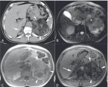
su-ppressed plasma cortisol levels after 1 mg overnight dexamethasone (33 nmol/l; normal < 50); normoka-lemia (4.3 mmol/L; normal range 3.5-5); normal uri-nary metanephrines (502 nmol/24h; normal range 31-1167) and urinary normetanephrines (1147 nmol/24h; normal range 240-2459), and plasma catecholamines (3.6 nmol/L; normal range 0.7-3.9); normal DHEAS (4 µmol/L; normal range 6.5-9.1); normal testostero-ne (13 nmol/L; normal 8.7-31.2 range), and elevated androstenedione (74 nmol/L; normal range 2.1-8.7). CT and MRI exams have characterized the lesion as a large heterogeneous left adrenal soft tissue mass with substantial amount of interweaving free fat tissue. The-re was also a 1.5 cm nodule with similar featuThe-res in the right adrenal gland (Figure 1).
Although the imaging features were typical for mye-lolipoma, the biochemical indings showing androstene-dione hypersecretion associated with a large adrenal mass led to a preoperative diagnosis hypothesis of androgen secreting adrenocortical carcinoma. Patient underwent open left adrenalectomy. The intraoperatory indings demonstrated a 15 x 10 cm left adrenal circumscribed mass which was completely excised, and a small pig-mented right adrenal mass of around 1.5 cm, that was not removed due to its benign appearance. Pathological indings revealed an enlarged left adrenal gland with a yellow and tan circumscribed mass of 15 cm in its lar-gest diameter that weighed 585 g. At the microscopy the tumor was characterized as a myelolipoma that rose on a multinodular hyperplasic adrenocortex (Figure 2).
The patient postoperative recovery was unremarka-ble, but the androgens remained elevated on the ifth day after surgery (Table 1). To rule out a potentially unsuccessful surgery and to diagnose a suspected CAH, measurement of basal and post ACTH1-24 stimulation test 17OHP was performed, followed by a 2 mg/day dexamethasone suppression test during 5 days. Basal 17OHP was 261 nmol/L, reaching 342 nmol/L 60 minutes after ACTH1-24 (Table 1). All androgens sho-wed a marked suppression after 5 days of the dexame-thasone suppression test, thus conirming the diagnosis of CAH (Table 1).
androste-Cop
yright
© ABE&M t
odos os dir
eit
os r
eser
vados
.
figure 1. Axial CT image (a), axial MR T2-weighted image (b), and in-phase (c) and out-of-phase (D) T1-weighted images show a large heterogeneous left adrenal soft tissue mass (arrowheads; a, b and c) with substantial amount of interweaving free fat tissue (*; b). The free fat tissue is seen as low CT density (a) and low MR intensity (b and c) areas within the lesion. There is also a 1.5 cm nodule with similar features in the right gland (white arrow; a and D).
figure 2.a. The gross pathology features of the left adrenal tumor show a yellowish to tan extradrenal mass that has grown beside the gland. b. The photomicrography shows the myelolipoma with mature adipose cells intermingled with the benign and cellular haematopoietic component (right side) that lies over a hyperplasic adrenal gland (left side). c. The photomicrography illustrates the micronodular pattern of the adrenocortical hyperplasia. D. A high-power field of the myelolipoma showing all haematopoietic lineages in maturation between the adipose cells. The arrow shows a normal megakaryocytic cell.
Discussion
The pathogenesis of adrenal myelolipomas remains unclear. A variety of mechanisms are proposed to un-derlie the etiology of adrenal myelolipomas, such as the presence of embryonic bone marrow in adrenal tissue (3). Some evidences indicate that ACTH may have a role in the development of these tumors, de-monstrated by an increase in the relative frequency of myelolipomas in patients with excessive ACTH secretion, such as in CAH (13,14), Nelson’s syn-drome (15), and Addison’s disease (3). Moreover, myeloid metaplasia in the adrenal cortex is observed in severely burned and cancer patients, two groups that are subject to long periods of intense stress (16). Indeed, myelolipomas have been associated with various forms of CAH like 21OH deiciency, 17-hydroxylase (17OH) deiciency and recently with 11β-hydroxylase deiciency (17). It is worth pointing out that most of these patients were either untreated or had stopped taking their medication for an extended period and were exposed for many years to chronically elevated ACTH and androgen levels which have been shown to induce transformation of adrenal cortical precursor cells into mature fat cells (17,18).
To date, approximately 25 cases of myelolipomas as-sociated with CAH have been reported. The mean age of these patients was 48 years (range 23-82 years). The characteristics of each patient and tumor are shown in table 2. The majority of them, 19 (76%), was associated with CAH due to 21OH deiciency, 4 (16%) with CAH due 17OH deiciency and 1 (4%) with CAH due to 11β-hydroxylase deiciency. Our patient presents CAH due to 21OH deiciency, with a genotype Q318X / Sp2. Considering size and localization, the mean tumor size was 12.4 cm (range 1-43 cm) and bilateral lesions were reported in ten cases (40%). Jaresch and cols. (37) previously described a positive correlation between the age of CAH patients and the age at onset of therapy, and adrenal size; with older patients and patients who were untreated for a long period presenting the most hyperplastic adrenal glands with no correlation between tumor size and serum 17OHP concentrations. In the-se cathe-ses, chronic ACTH excess could induce diffuthe-se or nodular adrenocortical hyperplasia which would later become autonomous because of oncogenic mutations in the tissue. Although under-diagnosis of CAH due to 21OH deiciency is more frequent in male patients, as observed in our case, there was no difference in gender prevalence of myelolipomas associated with CAH des-nedione level falling into normal range (Table 1). A low
Cop
yright
© ABE&M t
odos os dir
eit
os r
eser
vados
.
Table 1. Basal hormone concentrations before, after surgery and under treatment with prednisone (5 mg daily) associated with fludrocortisone (0.1 mg daily). Dexamethasone suppression test and ACTH1-24 stimulation test performed after surgery
before
surgery surgeryafter beforedexa after dexa acTHbasal1-24 acTH60 min1-24 after treatmentunder
DHEA-S [6.5-9.1 µmol/L] 3.9 1.9 4.6 0.5 _ _ 0.2
Testosterone [8.7-31.2 nmol/L] 13.4 10.7 13.5 0.7 _ _ 3.2
Androstenedione [2.1-8.7 nmol/L] 74 25.5 24.6 0.5 _ _ 4.9
Cortisol [138-552 nmol/L] 154 _ _ _ 231 259.3 _
17OHP [0.9-6 nmol/L] 218 _ _ _ 261 342.5 _
17OHP: 17α-hydroxyprogesterone; dexa: dexamethasone; normal range in brackets. The dexamethasone test was performed with 2 mg/day dexamethasone during 5 days at 40 days post surgery. The ACTH1-24 testwas performed with 25 ug ACTH at 35 days post surgery.
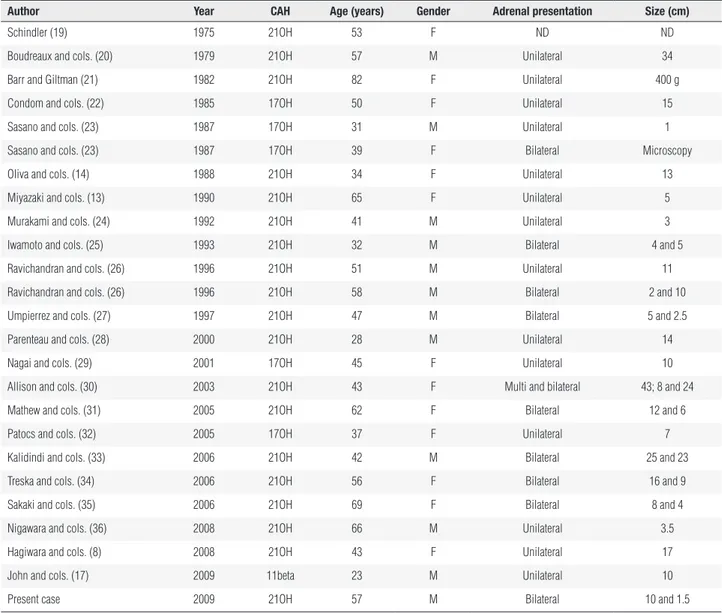
Table 2. Reported cases of adrenal myelolipoma associated with congenital adrenal hyperplasia (CAH)
author year caH age (years) Gender adrenal presentation size (cm)
Schindler (19) 1975 21OH 53 F ND ND
Boudreaux and cols. (20) 1979 21OH 57 M Unilateral 34
Barr and Giltman (21) 1982 21OH 82 F Unilateral 400 g
Condom and cols. (22) 1985 17OH 50 F Unilateral 15
Sasano and cols. (23) 1987 17OH 31 M Unilateral 1
Sasano and cols. (23) 1987 17OH 39 F Bilateral Microscopy
Oliva and cols. (14) 1988 21OH 34 F Unilateral 13
Miyazaki and cols. (13) 1990 21OH 65 F Unilateral 5
Murakami and cols. (24) 1992 21OH 41 M Unilateral 3
Iwamoto and cols. (25) 1993 21OH 32 M Bilateral 4 and 5
Ravichandran and cols. (26) 1996 21OH 51 M Unilateral 11
Ravichandran and cols. (26) 1996 21OH 58 M Bilateral 2 and 10
Umpierrez and cols. (27) 1997 21OH 47 M Bilateral 5 and 2.5
Parenteau and cols. (28) 2000 21OH 28 M Unilateral 14
Nagai and cols. (29) 2001 17OH 45 F Unilateral 10
Allison and cols. (30) 2003 21OH 43 F Multi and bilateral 43; 8 and 24
Mathew and cols. (31) 2005 21OH 62 F Bilateral 12 and 6
Patocs and cols. (32) 2005 17OH 37 F Unilateral 7
Kalidindi and cols. (33) 2006 21OH 42 M Bilateral 25 and 23
Treska and cols. (34) 2006 21OH 56 F Bilateral 16 and 9
Sakaki and cols. (35) 2006 21OH 69 F Bilateral 8 and 4
Nigawara and cols. (36) 2008 21OH 66 M Unilateral 3.5
Hagiwara and cols. (8) 2008 21OH 43 F Unilateral 17
John and cols. (17) 2009 11beta 23 M Unilateral 10
Present case 2009 21OH 57 M Bilateral 10 and 1.5
Type of CAH: 21-hydroxylase deficiency (21OH); 17-hydroxilase deficiency (17OH); ND: not described.
cribed thus far in the literature, with 13 women and 12 men accounting for the casuistics. The great majority of myelolipomas associated with CAH are localized in the adrenal gland, however there are descriptions of myelolipomas in other locations, such as the testis (38).
There was also an increase in reports of myelolipomas in more recent years (12 reports in the last decade ver-sus 13 from 1975 to 1997) explained by the widespread
Cop
yright
© ABE&M t
odos os dir
eit
os r
eser
vados
.
In conclusion, we describe a rare case of giant mye-lolipoma associated to CAH due to 21OH deiciency. The excessive ACTH and/or androgen secretion over a long period of time could have had a stimulatory role in the development of the adrenal myelolipoma in the present patient. However, the mechanism underlying the reduced 21OH reserve among incidentaloma pa-tients and the increased occurrence of adrenal tumors in simple virilizing or late-onset CAH forms remains a matter of speculation. Additionally, although surgery was mandatory in the present case due to tumor symp-toms and size, CAH should always be ruled out in in-cidentally detected adrenal masses to avoid unnecessary surgical procedures.
Acknowledgements: This work was supported by Fundação de Amparo à Pesquisa do Estado de São Paulo (Fapesp) (grant num-bers 08/09276-0 and 07/58365-3).
Disclosure: no potential conlict of interest relevant to this article was reported.
RefeRences
1. Kelekis NL, Alexopoulou E, Brountzos EN, Ladis V, Boussiotou A,
Kelekis DA. Giant adrenal myelolipoma with minimal fat content in a patient with homozygous beta-thalassemia: appearance on MRI. J Magn Reson Imaging. 2003;18(5):608-11.
2. Olsson CA, Krane RJ, Klugo RC, Selikowitz SM. Adrenal myeloli-poma. Surgery. 1973;73(5):665-70.
3. Plaut A. Myelolipoma in the adrenal cortex; myeloadipose struc-tures. Am J Pathol. 1958;34(3):487-515.
4. Manassero F, Pomara G, Rappa F, Cuttano MG, Crisci A, Selli C. Adrenal myelolipoma associated with adenoma. Int J Urol. 2004;11(5):326-8.
5. Merchant SH, Herman CM, Amin MB, Ro JY, Troncoso P. Myeloli-poma associated with adrenal ganglioneuroma. Arch Pathol Lab Med. 2002;126(6):736-7.
6. Sun X, Ayala A, Castro CY. Adrenocortical carcinoma with conco-mitant myelolipoma in a patient with hyperaldosteronism. Arch Pathol Lab Med. 2005;129(6):e144-7.
7. Ukimura O, Inui E, Ochiai A, Kojima M, Watanabe H.
Combi-ned adrenal myelolipoma and pheochromocytoma. J Urol. 1995;154(4):1470.
8. Hagiwara H, Usui T, Kimura T, Tagami T, Naruse M, Minamiguchi S, et al. Lack of ACTH and androgen receptor expression in a giant adrenal myelolipoma associated with 21-hydroxylase deiciency. Endocr Pathol. 2008;19(2):122-7.
9. Lamas C, Lopez LM, Lozano E, Atienzar M, Ruiz-Mondejar R, Alfa-ro JJ, et al. Myelolipomatous adrenal masses causing Cushing’s syndrome. Exp Clin Endocrinol Diabetes. 2009;117(8):440-5. 10. Lockhart ME, Smith JK, Kenney PJ. Imaging of adrenal masses.
Eur J Radiol. 2002;41(2):95-112.
11. McLoughlin RF, Bilbey JH. Tumors of the adrenal gland: indings on CT and MR imaging. AJR Am J Roentgenol. 1994;163(6):1413-8. 12. Ilias I, Sahdev A, Reznek RH, Grossman AB, Pacak K. The optimal
imaging of adrenal tumours: a comparison of different methods. Endocr Relat Cancer. 2007;14(3):587-99.
13. Miyazaki Y, Yoshida M, Doi J. [A case of adrenal myelolipo-ma associated with adrenogenital syndrome]. Hinyokika Kiyo. 1990;36(1):35-9.
14. Oliva A, Duarte B, Hammadeh R, Ghosh L, Baker RJ. Myelolipo-ma and endocrine dysfunction. Surgery. 1988;103(6):711-5. 15. Maschler I, Rosenmann E, Ehrenfeld EN. Ectopic functioning
adrenocortico-myelolipoma in longstanding Nelson’s syndrome. Clin Endocrinol (Oxf). 1979;10(5):493-7.
16. Delarue J, Monsaingeon A. [Myeloid metaplasia in the adrenal cortex of burned subjects.]. C R Seances Soc Biol Fil. 1950;144(11-12):777-8.
17. John M, Menon SK, Shah NS, Menon PS. Congenital adrenal hyperplasia 11beta-hydroxylase deiciency: two cases managed with bilateral adrenalectomy. Singapore Med J. 2009;50(2):e68-e70. 18. Selye H, Stone H. Hormonally induced transformation of adrenal
into myeloid tissue. Am J Pathol. 1950;26(2):211-33.
19. Schindler H. [Myelolipoma of the adrenal gland in adrenogenital syndrome]. Wien Med Wochenschr. 1975;725(48):695-7.
20. Boudreaux D, Waisman J, Skinner DG, Low R. Giant adre-nal myelolipoma and testicular interstitial cell tumor in a man with congenital 21-hydroxylase deiciency. Am J Surg Pathol. 1979;3(2):109-23.
21. Barr AB, Giltman LI. Congenital adrenal hyperplasia diagnosed in an 82-year-old: case report. Va Med. 1982;109(12):844-5.
22. Condom E, Villabona CM, Gomez JM, Carrera M. Adrenal mye-lolipoma in a woman with congenital 17-hydroxylase deiciency. Arch Pathol Lab Med. 1985;109(12):1116-7.
23. Sasano H, Masuda T, Ojima M, Fukuchi S, Sasano N. Congenital 17 alpha-hydroxylase deiciency: a clinicopathologic study. Hum Pathol. 1987;18(10):1002-7.
24. Murakami C, Ishibashi M, Kondo M, Ohshiro S, Fujita M, Sato S, et al. Adrenal myelolipoma associated with congenital adrenal 21-hydroxylase deiciency. Intern Med. 1992;31(6):803-6. 25. Iwamoto T, Yajima M, Tanaka H, Minagawa N, Osada T. [A case
re-port: reversible male infertility due to congenital adrenal hyper-plasia]. Nippon Hinyokika Gakkai Zasshi. 1993;84(11):2031-4. 26. Ravichandran R, Lafferty F, McGinniss MJ, Taylor HC.
Conge-nital adrenal hyperplasia presenting as massive adrenal inci-dentalomas in the sixth decade of life: report of two patients with 21-hydroxylase deiciency. J Clin Endocrinol Metab. 1996;81(5):1776-9.
27. Umpierrez MB, Fackler S, Umpierrez GE, Rubin J. Adrenal mye-lolipoma associated with endocrine dysfunction: review of the literature. Am J Med Sci. 1997;314(5):338-41.
28. Parenteau C, Mongeau CJ, Benard B, Maheux P. Pigmented adre-nal hyperplasia with myelolipomatous changes and bilateral testicular enlargement in an untreated man with 21-hydroxylase deiciency. Endocr Pract. 2000;6(3):260-3.
29. Nagai T, Imamura M, Honma M, Murakami M, Mori M. 17alpha-hydroxylase deiciency accompanied by adrenal myelolipoma. Intern Med. 2001;40(9):920-3.
30. Allison KH, Mann GN, Norwood TH, Rubin BP. An unusual case of multiple giant myelolipomas: clinical and pathogenetic implica-tions. Endocr Pathol. 2003;14(1):93-100.
31. Mathew J, Menon PS, Shah NS. An elderly lady in shock. J Post-grad Med. 2005;51(1):51-3.
appearan-Cop
yright
© ABE&M t
odos os dir
eit
os r
eser
vados
.
ces and change in the CT morphology following steroid treatment: a case report. Abdom Imaging. 2006; DOI: 10.1007/s00261-006-9096-x. 34. Treska V, Wirthova M, Hadravska S, Mukensnabl P, Kuntscher V, Kreu-zberg B, et al. [Giant bilateral adrenal myelolipoma associated with congenital adrenal hyperplasia]. Zentralbl Chir. 2006;131(1):80-3. 35. Sakaki M, Izaki H, Fukumori T, Taue R, Kishimoto T, Kanayama HO.
Bilateral adrenal myelolipoma associated with adrenogenital syndrome. Int J Urol. 2006;13(6):801-2.
36. Nigawara T, Kageyama K, Sakihara S, Takayasu S, Kawahara M, Imai A, et al. A male case of nonclassical 21-hydroxylase dei-ciency irst manifested in his sixties with adrenocortical inciden-taloma. Endocr J. 2008;55(2):291-7.
37. Jaresch S, Kornely E, Kley HK, Schlaghecke R. Adrenal incidenta-loma and patients with homozygous or heterozygous congenital adrenal hyperplasia. J Clin Endocrinol Metab. 1992;74(3):685-9. 38. Adesokan A, Adegboyega PA, Cowan DF, Kocurek J, Neal DE Jr.
Testicular “tumor” of the adrenogenital syndrome: a case report of an unusual association with myelolipoma and seminoma in cryptorchidism. Cancer. 1997;80(11):2120-7.