Case Report
136
Turk J Endocrinol Metab 2016;20:136-139
Address for Correspondence: Banu Aktaş Yılmaz MD, Gazi University Faculty of Medicine, Department of Endocrinology and Metabolism, Ankara, Turkey Phone: +90 312 202 58 29 E-mail: [email protected] Received: 12.05.2015 Accepted: 03/01/2016
Banu Aktaş Yılmaz, Müjde Aktürk, Erdal Kan, Aylar Poyraz*, Nil Tokgöz**, Nuri Çakır, Metin Arslan
Gazi University Faculty of Medicine, Department of Endocrinology and Metabolism, Ankara, Turkey *Gazi University Faculty of Medicine, Department of Pathology, Ankara, Turkey **Gazi University Faculty of Medicine, Department of Radiology, Ankara, Turkey
Abs tract
Öz
Adrenal incidentalomas are clinical dilemmas for the clinicians. The work up, to differentiate between malignant and benign lesions, and hyperfunctioning and nonfunctioning lesions is mandatory before the consideration of surgical resection. Ectopic thyroid tissue located in the adrenal gland (ETTAG) is a very rare condition. We report a case of ETTAG presenting with adrenal incidentaloma. A 57-year-old woman was admitted with incidental right adrenal mass. Hormone evaluation showed no hormonal activity. Magnetic resonance imaging revealed a 20x17 mm lobulated solid mass, which contained millimetric hypointense nodular areas consistent with calcifications. Loss of signal intensity on out-of-phase could not be evaluated because of the calcifications. Right adrenalectomy was performed to establish the histopathological diagnosis and to rule out malignancy. Histopathological diagnosis revealed ETTAG. Her medical history was positive for multinodular goiter and bilateral subtotal thyroidectomy 32 years ago. Thyroid ultrasonography showed residual thyroid tissue in both the right and left lobes, and colloid thyroid nodules. Fine needle aspiration biopsy from the nodules revealed benign nodules. The patient has been followed up for six years, and no change in thyroid nodule sizes and no evidence of metastatic foci have been detected. ETTAG would be considered in the differential diagnosis of adrenal mass showing no hormonal activity, especially when magnetic resonance images are not consistent with adrenal adenoma. Long follow-up duration of this case suggests that it was a benign condition.
Keywords: Ectopic thyroid, adrenal gland, adrenal incidentaloma, adrenal calcification
Adrenal insidentalomalar klinisyenlerin bulmacaları gibidir. Adrenal kitleler için cerrahi rezeksiyon kararı alınmadan evvel; kitlelerin benign-malign ayrımı, hormonal olarak fonksiyon gösterip göstermedikleri ayrıntılı olarak değerlendirilmelidir. Adrenal yerleşim gösteren ektopik tiroid dokusu (AYETD) oldukça nadir karşılaşılan durumdur. Bu yazıda adrenal insidentaloma olarak tespit edilen AYETD olgusu sunulmaktadır. Elli yedi yaşındaki kadın hasta sağda tespit edilen adrenal insidentaloma nedeni ile değerlendirildiğinde kitlenin hormonal olarak fonksiyon göstermediği; manyetik rezonans görüntülemede kalsifikasyonlarla uyumlu milimetrik hipointens alanları olan 20x17 mm boyutlarında lobule solid kitle özelliklerine sahip olduğu görüldü. manyetik rezonans görüntülemede kalsifikasyonlar nedeni ile kitlenin sinyal kayıp özellikleri net olarak değerlendirilemedi. Malignensiyi dışlamak ve histopatoljik tanıya ulaşmak amacı ile hastaya sağ adrenalektomi uygulandı. Histopatoljik tanı AYETD olarak raporlandı. Hastanın öyküsünden 32 yıl evvel multinodüler guatr nedeni ile bilateral subtotal tiroidektomi operasyonu geçirdiği öğrenildi. Tiroid ultrasonogrofisinde bilateral rezidu tiroid dokusunun ve kolloid nodüllerinin olduğu görüldü. Nodüllerden yapılan tiroid ince iğne aspirasyon biyopsilerinde benign özellikler raporlandı. Hastamız altı yıldır izlenmektedir. İzlem süresinde tiroid nodüllerinin boyut ve özelliklerinde fark olmadığı görüldü ve herhangi bir metastatik alan tespit edilmedi. AYETD tanısı, hormonal olarak fonksiyon göstermeyen ve MRG özellikleri tipik adrenal adenom ile uyumlu olmayan adrenal insidental kitlelerin ayırıcı tanısında akla gelmelidir. Hastamızın uzun izlem süresi ve tecrübemiz AYETD’sinin benign olay olduğunu düşündürmektedir.
Anahtar kelimeler: Ektopik tiroid, adrenal gland, adrenal insidentaloma, adrenal kalsifikasyon
Adrenal İnsidentaloma Olarak Tespit Edilen Adrenal Yerleşim Gösteren
Ektopik Tiroid Dokusu
Ectopic Thyroid in the Adrenal Presenting as an Adrenal
Incidentaloma
DOI: 10.4274/tjem.3153
137
Turk J Endocrinol Metab2016;20:136-139
Introduction
The prevalence of adrenal incidentaloma on abdominal computed tomography (CT) scans has been reported to be 0.4-4.4% (1,2). Benign adrenal masses contribute to approximately 90% of incidentalomas including nonfunctioning adenoma, functioning adenoma [subclinical Cushing’s syndrome (SCS), aldosteronoma and pheochromocytoma], ganglioneuromas, myelolipomas and benign cysts, whereas malignant lesions including adrenocortical carcinomas and metastatic lesions contribute to a small percentage of the incidentalomas. Patients with adrenal incidentalomas should be evaluated clinically, biochemically and radiographically to differentiate between malignant and benign lesions, and hyperfunctioning and nonfunctioning lesions. The goal of the imaging studies is differential diagnosis of adrenal adenoma, adrenal carcinoma, pheochromocytoma, and metastatic lesions. The term of ectopic thyroid refers to the presence of thyroid tissue apart from the pretracheal location, which is the final location of embryological migration route of the thyroid gland. The true incidence of ectopic thyroid tissue is not known because of the undiagnosed asymptomatic cases. Most of the ectopic thyroid cases are located in sublingual position along the route of thyroglossal duct that can be explained by the premature arrest of its migratory process. Though hard to explain how it develops, cases of ectopic thyroid tissue located in the thoracic region, ovaries, gallbladder, pancreas, duodenum and adrenals have been reported in the literature (1). Here, we report a case of ectopic thyroid tissue in the adrenal gland (ETTAG) presented as an adrenal incidentaloma.
Case Report
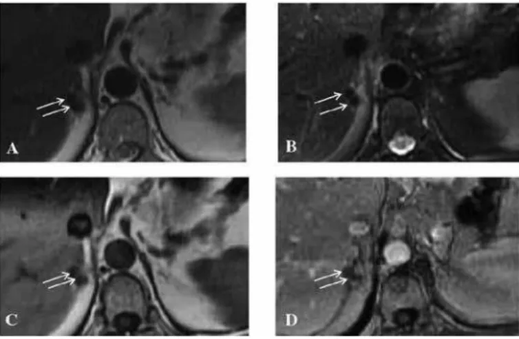
A 57-year-old woman was admitted to our clinic for right adrenal mass which was detected on thorax CT while evaluating her persistent cough. CT of the adrenal gland showed a right adrenal mass with a high attenuation value on early contrast (68 HU) and late contrast images (93 HU). Magnetic resonance imaging (MRI) revealed that a well-circumscribed 20x17 mm lobulated solid mass containing millimetric hypointense nodular areas consistent with calcifications. Loss of signal on out-of-phase could not be evaluated due to calcifications (Figure 1A, B, C, D). No lesion was identified in the left adrenal gland. She had a history of bilateral subtotal thyroidectomy 32 years ago due to multinodular goiter. The patient had hypertension for twelve years. She was on losartan 300 mg, thiazide 12.5 mg and amlodipine 5 mg therapies. The blood pressure was 130-70 mmHg and body mass index was 29.5 kg/m2. According to the laboratory analysis, cortisol was
suppressed with low dose (1 mg) overnight dexamethasone suppression test. Serum potassium levels were within the normal ranges despite thiazide diuretic use. Plasma renin activity was not suppressed and urinary catecholamine levels were slightly elevated. Radiological findings of the adrenal mass were not concordant with adenoma; and differential diagnosis of adrenal adenoma, adrenal carcinoma, metastatic lesion and pheochromocytoma could not be made. Right adrenalectomy was performed to achieve the histopathological diagnosis.
The size of the surgical specimen was 5x4x2 cm on gross examination. A 0.6x0.4x0.6 cm off-white solid lesion with partly cystic areas was identified on cut section. Histological sections of the lesion showed differentiated thyroid tissue composed of enlarged thyroid follicles filled with colloid, some in between adrenal cortical cells (Figure 2A, B). Immunohistochemically, epithelial cells of the follicles were positive for thyroid transcription factor-1 and thyroglobulin (Figure 2C, D). Although there was not any nuclear features suggestive of thyroid carcinoma, thyroid ultrasonography and fine needle aspiration (FNAB) of the thyroid nodules were performed to rule out thyroid malignancies. Thyroid ultrasonography revealed residual thyroid tissue in both right and left lobes; 45x19x20 mm and 27x21x25 mm, respectively. There were two colloid thyroid nodules measuring 28x17x17 mm and 18x23x17 mm in the right residue thyroid lobe. FNAB from both nodules and left lobe parenchyma revealed non-neoplastic
Aktaş Yılmaz et al. Ectopic Thyroid in the Adrenal
Figure 1. T2-weighted (A), fat-supressed T2-weighted (B), pğrecontrast (C) and postcontrast (D) T1-weighted magnetic resonance images in axial plane show a well-circumscribed lobulated solid mass (20 mm x 17 mm). The mass contains millimetric hypointense nodular areas consistent with calciications (arrows). The lesion demonstrates contrast enhancement on postcontrast image (D)
138
Aktaş Yılmaz et al.Ectopic Thyroid in the Adrenal
Turk J Endocrinol Metab 2016;20:136-139
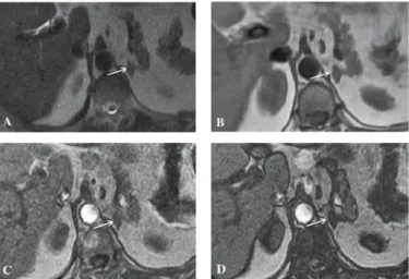
nodules. The thyroid scintigraphy showed diffuse activity in the right lobe and focal activity in the left lobe; and no activity in other parts of the body. The patient has been followed up for six years, and no change in thyroid nodule sizes and no evidence of metastatic foci have been detected. After six years of the first evaluation, repeated FNAB of the nodules were consistent with benign nodules. MRI of the adrenal glands which was performed after six years revealed a left adrenal mass measuring 20x10 mm, consistent with adrenal adenoma (Figure 3A, B, C, D). When the preoperative adrenal images were reevaluated, thickening in the medial part of the adrenal gland with 9 mm adenoma was considered. Hormone evaluation showed no hormonal activity suggesting SCS, hyperaldosteronism and pheochromocytoma. MRI revealed a decrease in signal intensity on out-of-phase images, which is consistent with intracytoplasmic fat content; and no calcification was identified. The patient is still being followed up for the nonfunctional adrenal adenoma located in the left adrenal gland.
Discussion
We report a case of ETTAG in the right adrenal gland which was discovered incidentally. Adrenal incidentalomas are the clinical dilemmas that we encounter more frequently as a result of the widespread use of radiological imaging methods. The clinician should perform a biochemical and radiological work up to identify adrenal masses which require surgical management and which may be followed up without surgery. Adrenalectomy is suggested for the lesions, which are malignant, larger than 4 cm in diameter, have hormonal activity and have indeterminate radiological characteristics (2).
Radiological work up of the adrenal mass in our patient revealed conflicting results since CT scans of the adrenal mass showed high attenuation values and intracellular lipid content could not be evaluated properly on MRI scans due to calcification of the mass. Variety of adrenal lesions, such as adrenal hemorrhage,
adrenocortical carcinoma, pheochromocytoma, mature adrenal teratoma, adrenal cysts, hemangioma, and metastatic disease may show calcification (3). Biochemical evaluation showed no hormonal activity. The patient had no history that could explain the reason of the adrenal calcification such as malignancy or anticoagulation medication. Because of the indeterminate radiological characteristics of the adrenal lesion, adrenalectomy was performed to get the proper histopathological diagnosis. Histopathological diagnosis of the adrenal mass revealed ectopic thyroid tissue unexpectedly. Considering the thyroid gland passage from the floor of the primitive foregut to pretracheal position during its embryogenesis, it is hard to explain how the thyroid tissue can be located in the adrenal gland. However, there is a limited number of case reports about ETTAG and in other organs located at the subdiaphragmatic area such as gallbladder, ovary, vagina, porta hepatis, perisplenic area (1) and adrenal gland (4,5,6,7,8,9). In the literature, most of the patients with ETTAG were female aged 38-67 years. The cases were identified during evaluation of hypertension, metastatic work-up for nonthyroidal malignancies, and incidentally for other unrelated situations such as hypochondriac pain or acute abdomen (4,5,6,7,8,9,10). Most of the lesions had the cystic appearance on CT scans, calcification was reported in some of the cases (9,10). Histopathological examination showed similar benign properties in all patients including our patient. To the best of our knowledge, thyroidectomy was performed in only one patient and papillary microcarcinoma 5 mm in diameter was identified. Neither the histopathological examination nor the immunophenotype features of both papillary microcarcinoma and thyroid tissue in the adrenal gland suggested metastasis in that case (5). Thyroidectomy was not performed in our patient since thyroid ultrasonography and FNAB of the thyroid nodules showed benign features. Besides, during more than six years of follow-up, no change in thyroid nodule size and appearance was detected, and the second FNAB that was performed six years after the adrenalectomy showed benign thyroid nodule. Metastasis of well differentiated thyroid cancer, teratoma, metaplasia, and ectopic thyroid tissue may be the possible explanations for ETTAG. Histopathological diagnosis in our patient was not consistent with teratoma; the lesion did not contain other tissue components typical of teratoma, such as skin appendages, cartilage and bone. Metaplasia does not seem as an appropriate explanation for ETTAG since the adrenal cortex originates from mesodermal, the adrenal medulla originates from ectodermal, and the thyroid glands form from the endodermal germ layers. Romero-Rojas et al. (6) showed that the thyroid-restricted transcription factors thyroid transcription factor-1, PAX8, and FDEX1, essential for regulating proliferation, survival, and/or migration of the medial thyroid primordium, were also expressed in the ETTAG in two patients. Specific mutation for thyroid migration has not been determined in human yet (11). It is still hard to explain the mechanism of aberrant migration of the thyroid tissue to the adrenal glands. Some authors suggested that congenital defects in diaphragm leading to diaphragmatic hernias would be the possible explanation of the aberrant location of thyroid tissue in the adrenal gland (4,6).
139
Aktaş Yılmaz et al.Ectopic Thyroid in the Adrenal Turk J Endocrinol Metab
2016;20:136-139
Our patient is still being followed up for the contralateral adrenal mass with no hormonal activity and consistent with adrenal adenoma. Appearance of another adrenal mass in the contralateral adrenal gland and/or mass enlargement have also been reported in patients with adrenal adenoma (12). Having the benign radiological properties corresponding to adrenal adenoma and no hormonal activity, we continue following up the patient.
As a conclusion, ectopic thyroid in the adrenal gland would be considered in the differential diagnosis of adrenal incidentalomas, especially in patients with indeterminate radiological findings. We do not have sufficient explanations for the aberrant migration of thyroid tissue. The experience about ETTAG is limited with few case reports. Adrenal masses with radiological images not consistent with adrenal adenoma and without hormonal activity would suggest ectopic thyroid tissue located in the adrenal gland. The long follow-up duration in our patient suggests that it is a benign condition.
Ethics
Informed Consent: Consent form was filled out by the patient. Peer-review: Externally and Internally peer-reviewed.
Authorship Contributions
Surgical and Medical Practices: Banu Aktaş Yılmaz, Erdal Kan, Müjde Aktürk, Concept: Banu Aktaş Yılmaz, Müjde Aktürk, Design: Banu Aktaş Yılmaz, Müjde Aktürk, Data Collection or Processing: Banu Aktaş Yılmaz, Müjde Aktürk, Erdal Kan, Analysis or Interpretation: Nuri Çakır, Metin Arslan, Aylar Poyraz (pathological examination), Nil Tokgöz (radiological examination), Literature Search: Banu Aktaş Yılmaz, Müjde Aktürk, Writing: Banu Aktaş Yılmaz, Müjde Aktürk.
Conflict of Interest: No conflict of interest was declared by the authors.
Financial Disclosure: The authors declared that this study received no financial support.
References
1. Noussios G, Anagnostis P, Goulis DG, Lappas D, Natsis K. Ectopic thyroid tissue: anatomical, clinical, and surgical implications of a rare entity. Eur J Endocrinol. 2011;165:375-382.
2. Zeiger MA, Thompson GB, Duh QY, Hamrahian AH, Angelos P, Elaraj D, Fishman E, Kharlip J, American Association of Clinical E, American Association of Endocrine S. The American Association of Clinical Endocrinologists and American Association of Endocrine Surgeons medical guidelines for the management of adrenal incidentalomas. Endocr Pract. 2009;15(Suppl 1):1-20.
3. Hindman N, Israel GM. Adrenal gland and adrenal mass calcification. Eur Radiol. 2005;15:1163-1167.
4. Shuno Y, Kobayashi T, Morita K, Shimizu S, Nishio Y, Ito A, Kobayashi K, Kawahara M, Teruya M. Ectopic thyroid in the adrenal gland presenting as cystic lesion. Surgery. 2006;139:580-582.
5. Bohinc BN, Parker JC, Hope WW, Kotwall C, Turner J, Cheng W, Lloyd RV. Micropapillary thyroid carcinoma and concomitant ectopic thyroid tissue in the adrenal gland: metastasis or metaplasia? Thyroid. 2011;21:1033-1038. 6. Romero-Rojas A, Bella-Cueto MR, Meza-Cabrera IA, Cabezuelo-Hernandez
A, Garcia-Rojo D, Vargas-Uricoechea H, Cameselle-Teijeiro J. Ectopic thyroid tissue in the adrenal gland: a report of two cases with pathogenetic implications. Thyroid. 2013;23:1644-1650.
7. Shiraishi T, Imai H, Fukutome K, Watanabe M, Yatani R. Ectopic thyroid in the adrenal gland. Hum Pathol. 1999;30:105-108.
8. Takao H, Doi I, Watanabe T. Ectopic thyroid in the adrenal gland: Computed tomography findings. J Comput Assist Tomogr. 2006;30:221-222. 9. Hagiuda J, Kuroda I, Tsukamoto T, Ueno M, Yokota C, Hirose T, Deguchi N.
Ectopic thyroid in an adrenal mass: a case report. BMC Urol. 2006;6:18. 10. Gourmaud J, Bongiovanni M, Triponez F, Pusztaszeri M. Ectopic thyroid
tissue in the adrenal gland. Endocr Pathol. 2014;25:353-355.
11. Szinnai G. Genetics of normal and abnormal thyroid development in humans. Best Pract Res Clin Endocrinol Metab. 2014;28:133-150.