Surgical management of adrenal cysts: a single-institution
experience
_______________________________________________
Xiao Lyu, Liangren Liu, Lu Yang, Liang Gao, Qiang Wei
Department of Urology, West China Hospital, Sichuan University, Chengdu, Sichuan, P.R. China
ABSTRACT
ARTICLE
INFO
______________________________________________________________ ______________________
Objective: To analyze surgical methods and evaluate treatment efficacy and safety for managing adrenal cystic lesions.
Materials and methods: All patients presenting with adrenal lesions of the West China Hospital were reviewed retrospectively from January 2003 to April 2013 and 47 were diagnosed as adrenal cysts. Basic information, clinical history, physical examination, laboratory investigations, abdominal ultrasound and enhanced computed tomography were detailed noted. Cysts with different surgical management were analyzed and surgery option operative time, postoperative complications and after-surgery hospital stay were all noted. The final diagnosis was judged by histopathology. Patients were followed from 3 month to 10 years.
Results: All the 47 patients with a mean age of 43.8 years were managed by surgical intervention. Compared laparoscopic technology with open technology, the laparosco-pic has the advantage of a shorter operation time, shorter hospital stay after surgery and enhanced cosmesis. The histopathologic result was: 23 (50%) were endothelial cysts and 16 (35%) were pseudocysts. One patient had evidence to recurrence at the followed-up stage.
Conclusion: Adrenal cysts are rare and with the development of imaging techniques many of these are diagnosed incidentally. CT has advantages in detecting the cysts with haemorrhage, intracystic debris, calcification and mixed adrenal mass. Minimally invasive surgery offers equivalent efficacy to traditional open procedures, while provi-ding a shorter operation time, shorter convalescence and improved cosmesis. Patients after surgical resection should be followed up closely especially if functional cysts and histopathology of cystic tumor are present.
Key words:
Laparoscopy; Cysts; Endothelium; Adrenal Rest Tumor
Int Braz J Urol. 2014; 40: 656-65
_____________________
Submitted for publication: December 19, 2013
_____________________
Accepted after revision: March 19, 2014
INTRODUCTION
Adrenal cystic lesions (ACL) are rare. Sin-ce the first report of Greiselius in 1670, over 600 adrenal cysts have been reported in literatures (1,2). The occurrence of ACL by autopsy is about 0.064~0.18% of general people (3).The incidence rate increases with the development of diagnostic
7% (2, 4-7). The pathogenesis is still unclear and several hypotheses have been proposed (3,7). The-re is controversy concerning the The-recommendation of adrenal cyst management because of low in-cidence, difficult pre-surgery pathologic diagno-sis and various surgical intervention ways (6-8). The previous questions made us to review patients with adrenal cysts with different clinical characte-ristics who were managed in different ways.
MATERIALS AND METHODS
We retrospectively reviewed the patients with adrenal lesions from the Urology Depart-ment, West China Hospital from January 2003 to April 2013. 47 patients were diagnosed with adrenal cysts whose basic information such as age, gender and location of cystic lesion were recorded. Each patient had a brief clinical his-tory and physical examination. Abdominal US was used as first choice to screen the cysts. Then abdominal enhanced CT scans were provided for all patients. MRI was used for two patients only. The diameter of cysts was measured by CT scans.
Serum cortisol at 8 am, 4 pm and 24 pm points, 24-hour urinary catecholamine levels and serum catecholamine, aldosterone in both lying position and standing position, serum metane-phrine and potassium were analyzed to evaluate the hormonally active adrenal cysts. 46 patients chose surgical removal and one underwent per-cutaneous FNA (perper-cutaneous fine-needle aspi-ration) mediated by CT. Surgical resection were taken in our series in patients with the following characteristics: hormonally active adrenal cysts, cystic lesion greater than 4cm, smaller cysts en-larged at least by 1 cm per year, cysts with image changing suggesting haemorrhage and calcifica-tion. Patients out of these standards were treated with watchful waiting with abdominal enhanced CT scans provided at the urological outpatient service every 6 months. Surgery option, opera-tive time, postoperaopera-tive complications and after--surgery hospital stay were all detailed noted. In all open cases the procedure was performed using waist incision methods and all laparoscopy cases were submitted to retroperitoneal laparoscopy. The final diagnosis was obtained according to
the histopathologic result of specimen obtained from surgical resection of surgical patients.
All the patients after surgery were follo-wed from 3 months to 10 years as urological outpatients every 3 months in the first year. If patients showed no evidence of recurrence, we changed it to every 6 months in the second year. After that, if there was no positive found, we changed it to every one year in the next years.
SPSS19.0 (IBM, Chicago, USA) was used for statistical analysis. Independent samples t--test was used to compare the laparoscopy sur-gery with open sursur-gery.
RESULTS
Among 922 patients operated on for adre-nal lesions in the ten years period in the Urology Department, West China Hospital, cystic lesions were found in 47 patients, accounting for 5.1% of adrenal lesions. The age ranged from 18 to 77 with a mean age of 43.8. 29 (61.7%) of them were female and the rest 18 (38.3%) were male. All cysts were unilateral with 26 (55.3%) at left side and 21 (44.7%) at right side (Table-1). 12 of them presented with lower back pain and 2 complained of abdominal pain with abdominal symptoms. 3 masses could be obviously palpa-ted. 6 cysts were discovered incidentally by non--specific symptoms like headache, palpitations, fever, hypertension and ureteral calculi. 27 of them were asymptomatic and detected by con-ventional medical examination. US detected 38 adrenal cysts, 3 adrenal masses, 2 kidney cysts and 1 hepatic cyst with 3 patients not reported. CT detected 42 adrenal cysts, 4 adrenal masses and 1 hepatic cyst. MRI detected 2 adrenal cysts. The mean diameter of the cysts measured by CT was 5.0 cm, ranging from 1.4 cm to 20 cm. Among 4 patients with functional tumors, 2 had hypertension with elevated level of 24-hour uri-nary catecholamine and 2 had hypertension and hyperaldosteronism.
or decortication, 21 had laparoscopic cysts ex-cisions and 4 had laparoscopic adrenalectomies. One patient proposed to receive laparoscopic cyst excision was immediately replaced by open cyst excision, as there were difficulties in building re-troperitoneal laparoscopic passageway. Another patient who had a laparoscopic cyst excision was found the cyst reappeared (about 2 cm) in the next 3 months. Thus an open adrenalectomy was cho-sen, and during the operation the inferior vena cava was inadvertently injured and was
success-fully repaired. One patient who had adrenal cyst with ureteral calculi had laparoscopic adrenalec-tomy and holmium laser lithotripsy simultaneou-sly. Therefore, the 3 patients were excluded in the comparison of the open and laparoscopic tech-nique (Table-2). The mean cysts size, operative time and after-surgery hospital stay between the open and laparoscopic techniques were 6.55 and 4.66 cm (p=0.081), 95 and 62.44 min (p<0.001), and 6.09 and 4.47 days (p=0.006) respectively. There were no significant complications in the postoperative period in all 47 patients.
Histopathologic result of the 46 patients was as follow: 23 was endothelial cysts, 16 pseu-docysts, 2 cystic pheochromocytomas, 2 cystic adenomas, 1 epithelial cyst, 1 bronchogenic cyst and 1 pulmonary sequestration cyst (Table-1). Calcifications within the cysts were found in 7 patients. Only one patient had evidence of recur-rence at the followed-up stage. 10 patients pre-sented with lower back pain and 2 complained of abdominal pain and both disappeared after sur-gery. However, 2 patients complained their lower back pain still existed, which we suspected was due to lumbar muscle strain.
DISCUSSION
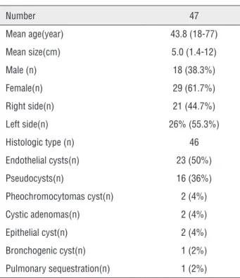
Cystic lesions in the adrenal gland are uncommon (a reported incidence at autopsy of 0.064% to 0.18%). Only few large series have been reported and our study is one of the largest studies with 47 cases (3, 6, 9, 10). Incidence of adrenal cysts in adrenal disease was 5.1%, accor-ding to various clinical series of 5.4% (11, 12).The average age of patients was 43.8 years, slightly Table 1 - Features of Patients with Adrenal cysts and
pathological examination result.
Number 47
Mean age(year) 43.8 (18-77)
Mean size(cm) 5.0 (1.4-12)
Male (n) 18 (38.3%)
Female(n) 29 (61.7%)
Right side(n) 21 (44.7%)
Left side(n) 26% (55.3%)
Histologic type (n) 46
Endothelial cysts(n) 23 (50%)
Pseudocysts(n) 16 (36%)
Pheochromocytomas cyst(n) 2 (4%)
Cystic adenomas(n) 2 (4%)
Epithelial cyst(n) 2 (4%)
Bronchogenic cyst(n) 1 (2%)
Pulmonary sequestration(n) 1 (2%)
Table 2 - The demographic and clinical data of surgical intervention patients.
Variable Open surgery Laparoscopy p 95% CI
n 11 32
age 44.64 44.84 0.967 [-9.72,10.14]
Cyst size,cm 6.55 4.66 0.081 [-4.02,0.25]
Operative time, min 95.00 62.44 <0.001 [-49.28,-15.84]
after-surgery hospital stay 6.09 4.47 0.006 [-2.74,-0.50]
higher than previously reported (6, 12) because our center deals only with adult patients. Adrenal cysts have been reported to be more common in women. In our study, the male to female ratio was 1:1.6 (1, 13). All cysts were unilateral and appeared almost equally in both sides although epidemiology indicated 8~15% are bilateral. 27 of the 47 cases (57%) were discovered asympto-matic by health examination. 6 cysts were disco-vered incidentally by non-specific symptoms like headache, palpitations, fever, hypertension and ureteral calculi. Abdominal or flank pain, gas-trointestinal symptoms and a palpable mass were the main clinical manifestations. Large adrenal cysts adjacent to abdominal structures are typical examples. 14 patients (30%) complained of abdo-minal pain or gastrointestinal symptoms, whe-rein 3 cases of adrenal cysts could be palpated obviously. The mean size of symptomatic cysts was larger than that of asymptomatic cysts (7.2 cm vs. 4.4 cm). Large cysts have a tendency to develop such complications as intracystic hemor-rhage and rupture that can manifest as a surgical emergency; however, no case was observed in our study (14-17). Functioning cysts of the adre-nal cortex and medulla have been reported (9). Two cystic pheochromocytomas and two cystic adenomas were found. Cystic pheochromocyto-mas presented hypertension, headache and pal-pitations. Laboratory test showed high values of 24-hour urinary catecholamine. Cystic adenomas presented hypertension and hyperaldosteronism.
US, CT and MRI are major modalities used in the evaluation of adrenal cysts. In the pre-sent series, the sensitivity of US for adrenal cyst was 89% and of CT was 98%. US can easily find the borders of the cysts and identify uncompli-cated cysts, however, it had limitation in distin-guishing the source of retroperitoneal cysts and adrenal mass accompanied with cysts (18). In our series the US mislead adrenal cysts as renal cysts or hepatic cysts. And it had low sensitivity for detection of small masses compared with CT and MRI. The advantage of CT was to detect the cysts with haemorrhage, intracystic debris and calcifi-cation (18). Meanwhile, CT was good for mixed adrenal mass (7). On CT, uncomplicated benign adrenal cysts are of water density (0-20 HU).
Higher attenuation values can be observed due to haemorrhage, intracystic debris and calcifica-tion (18). The CT misdiagnosed an adrenal cyst as a hepatic cyst due to its 20 cm diameter and unclear border with the liver. Some literatures found that CT may suggest malignancy through calcification, thickness and enhancement of the vesicle wall (Figure-1) (18, 19). It requires more samplings and thorough histopathologic exami-nation. We only had two cases diagnosed by MRI so it proved with low reference value.
Cysts of the adrenal gland have been clas-sified traditionally into four categories by origin: endothelial cyst, 45%; pseudocyst, 39%; epithe-lial cyst, 9%; parasitic cyst, 7% (Figure-2) (6, 17, 20). However, Neri and Nance found pseudocyst represented the most common subtype (2). Among the 46 cases, 23 (50%) were endothelial cysts, 16 (35%) were pseudocysts and 1 (2%) was an epi-thelial cyst. No parasitic cysts were identified. Endothelial cysts can also be subcategorized by histology into lymphangiomatous and angioma-tous cysts. They are distinguished from tumors by the absence of proliferating endothelium. The pathogenesis of adrenal pseudocyst is unclear. Most scholars think pseudocysts result from the organization of a previous traumatic hematoma or infectious process (7, 9, 21).The epithelial cyst is rare and only one example was found. Its pa-thogenesis is still unclear and can be subdivided into glandular or retention cysts, cystic adenomas and embryonal cysts (7, 22). In our series, a bron-chogenic cyst and a pulmonary sequestration cyst were found in the adrenal region. Bronchogenic cyst is a rare developmental abnormality of the primitive foregut that arises during early embryo-genesis from abnormal budding of the developing tracheobronchial tree (23). Pulmonary sequestra-tion cyst is extremely rare and is characterized by a mass of nonfunctional lung tissue without com-munication with the bronchopulmonary tree and fed from an aberrant systemic artery (24). In both bronchogenic cyst and pulmonary sequestration cyst, histology still remains the routine method of diagnosis and surgery remains the predominant therapeutic option (23, 24).
surgical techniques. Only one of the cases un-derwent FNA without sclerotherapy mediated by CT. Although FNA patients may avoid surgery when they present no clinical alteration in the punctured fl uid, some experts argued that FNA has limited effect on determining cyst histopa-thology and the puncture fl uid may reaccumula-te (2, 3). Without histopathology diagnosis, it is hard to judge the cystic lesions malignancy. Sur-gical resection enabled us to obtain histopatho-logic diagnosis. Surgical resection were taken in our series in patients with the following charac-teristics: hormonally active adrenal cysts, cystic lesion greater than 4 cm, smaller cysts enlarged
at least by 1cm per year, cysts with image chan-ges like haemorrhage and calcifi cation. Recently, multiple studies have described the minimally invasive approaches had shorter hospital stays, less blood loss, and enhanced cosmesis (6, 25-27). In the present series, most open excisions were managed in the early time and great majo-rity were submitted to laparoscopic excisions in recent years. Compared laparoscopic technology with open technology, the former has the ad-vantage of a shorter operation time (p<0.001), a shorter after-surgery hospital stay (p=0.006) and enhanced cosmesis. According to our experience, we preferred open surgery for those re-operation Figure 1 - A) Adrenal endothelial cyst. Contrast-enhanced computed tomography scan of abdomen showing a cystic lesion measuring 7.9x9.3 cm in size at the left adrenal area. B) Adrenal pseudocyst. Contrast-enhanced computed tomography scan of abdomen showing a cystic lesion surrounded by calcifi cation measuring 3.3x3.0 cm in size at the left adrenal area. C) Adrenal lymphangiomatous cyst. Contrast-enhanced computed tomography scan of abdomen showing a cystic lesion with several punctuate calcifi cation measuring 1.8x1.3 cm in size at the right adrenal area. D) Recurrence of adrenal cyst. Contrast-enhanced thinner computed tomography scan of abdomen showing a cystic lesion measuring 2x1.5 cm in size at the right adrenal area.
A
C
B
patients and cystic lesions with severe adhesion detected by imaging examination. We preferred retroperitoneal laparoscopy of the remaining patients with few complications being relative-ly safe. As operations performed from a single abdominal incision was popular, there have been no detailed randomized studies of ACL (4). Some authors chose laparoscopic adrenalectomy in all patients with adrenal cysts and other multiple centers prefer adrenal-sparing resections, even for functional cystic tumors (11). In our series, adrenal-sparing resections were managed in 37 (80%) patients and adrenalectomies in 9 (20%) patients. We preferred to preserve the adrenal gland except for the functional cysts or when the border between cysts and adrenal gland were unclear, accompanied with severe adhesion. One
patient underwent laparoscopic adrenal-sparing resection presented a 2 cm cyst 3 months later. An open adrenalectomy was then performed and with long term follow-up no recurrence was found. The pathology showed the presence of a cystic adenoma.
CONCLUSIONS
Adrenal cysts are rare. Abdominal or fl ank pain and gastrointestinal symptoms are the main clinical manifestations. However, with the development of imaging techniques, adrenal cysts are always diagnosed incidentally. CT has advantages in detecting the cysts with haemor-rhage, intracystic debris, calcifi cation and mixed adrenal mass. Minimally invasive surgery offers Figure 2 - A) The endothelial cyst was lined by a single layer of fl attened endothelial cells, x200. B) The pseudocyst presented the absence of a single layer of fl attened endothelial cells, x200. C) The adrenal lymphangiomatous cyst showed ectasia of lymphatic vessels in adrenal glands, x200. D) the pulmonary sequestration cyst was lined by benign pseudo-stratifi ed ciliated columnar epithelium, x200.
A
C
B
equivalent efficacy to traditional open procedu-res, while providing a shorter operation time, shorter convalescence and improved cosmesis. However, surgical intervention is not recommen-ded for asymptomatic patients with incidental cysts, for the fact that the pathological data of our series revealed no malignant cases. Patients after surgical resection should be followed up closely especially in functional cysts and when histopathology showed cystic tumor.
ACKNOWLEDGMENT
This research was funded by the National Natural
Science Foundation of China (Grant no. 30772171).
ABBREVIATIONS
ACL = Adrenal cystic lesions
US = ultrasound
CT = computed tomography
MRI = magnetic resonance imaging
FNA = percutaneous fine-needle aspiration
CI = confidence interval
REFERENCES
1. Tagge DU, Baron PL: Giant adrenal cyst: management and review of the literature. Am Surg. 1997; 63: 744-6.
2. Neri LM, Nance FC: Management of adrenal cysts. Am Surg. 1999; 65: 151-63.
3. Bellantone R, Ferrante A, Raffaelli M, Boscherini M, Lombardi CP, Crucitti F: Adrenal cystic lesions: report of 12 surgically treated cases and review of the literature. J Endocrinol Invest. 1998; 21: 109-14.
4. Major P, Pedziwiatr M, Matłok M, Ostachowski M, Winiarski M, Rembiasz K, et al.: Cystic adrenal lesions - analysis of indications and results of treatment. Pol Przegl Chir. 2012; 84: 184-9.
5. Kuruba R, Gallagher SF: Current management of adrenal tumors. Curr Opin Oncol. 2008; 20: 34-46.
6. El-Hefnawy AS, El Garba M, Osman Y, Eraky I, El Mekresh M, Ibrahim el-H: Surgical management of adrenal cysts: single-institution experience. BJU Int. 2009; 104: 847-50.
7. Wedmid A, Palese M: Diagnosis and treatment of the adrenal cyst. Curr Urol Rep. 2010; 11: 44-50.
8. Erickson LA, Lloyd RV, Hartman R, Thompson G: Cystic adrenal neoplasms. Cancer. 2004; 101: 1537-44.
9. Chien HP, Chang YS, Hsu PS, Lin JD, Wu YC, Chang HL, et al.: Adrenal cystic lesions: a clinicopathological analysis of 25 cases with proposed histogenesis and review of the literature. Endocr Pathol. 2008; 19: 274-81.
10. Erbil Y, Salmaslioglu A, Barbaros U, Bozbora A, Mete O, Aral F, et al.: Clinical and radiological features of adrenal cysts. Urol Int. 2008; 80: 31-6.
11. Castillo OA, Litvak JP, Kerkebe M, Urena RD: Laparoscopic management of symptomatic and large adrenal cysts. J Urol. 2005; 173: 915-7.
12. Pradeep PV, Mishra AK, Aggarwal V, Bhargav PR, Gupta SK, Agarwal A: Adrenal cysts: an institutional experience. World J Surg. 2006; 30: 1817-20.
13. Lal TG, Kaulback KR, Bombonati A, Palazzo JP, Jeffrey RB, Weigel RJ: Surgical management of adrenal cysts. Am Surg. 2003; 69: 812-4.
14. Stimac G, Katusic J, Sucic M, Ledinsky M, Kruslin B, Trnski D: A giant hemorrhagic adrenal pseudocyst: case report. Med Princ Pract. 2008; 17: 419-21.
15. da Silva EC, Viamontez F, Silva VS, Andrade A, Júlio Neto G, Gomes Cde P, et al.: Hemorrhagic adrenal cyst. Einstein (Sao Paulo). 2012; 10: 96-9.
16. Suh J, Heimann A, Cohen H: True adrenal mesothelial cyst in a patient with flank pain and hematuria: a case report. Endocr Pathol. 2008; 19: 203-5.
17. Favorito LA, Lott FM, Cavalcante AG: Traumatic rupture of adrenal pseudocyst leading to massive hemorrhage in retroperitoneum. Int Braz J Urol. 2004; 30: 35-6.
18. Sanal HT, Kocaoglu M, Yildirim D, Bulakbasi N, Guvenc I, Tayfun C, et al.: Imaging features of benign adrenal cysts. Eur J Radiol. 2006; 60: 465-9.
19. Lockhart ME, Smith JK, Kenney PJ: Imaging of adrenal masses. Eur J Radiol. 2002; 41: 95-112.
20. Lunca S, Romedea NS, Roata C, Bouras G: Cystic lymphangioma of the adrenal gland. Chirurgia (Bucur). 2004; 99: 255-8. 21. Takeuchi Y, Ozaki Y, Suyama T, Sawada Y, Kuroda K,
Nakajima K, et al.: A case of giant hemorrhagic adrenal pseudocystectomy with a flank incision. Hinyokika Kiyo. 2011; 57: 315-8.
22. Limaiem F, Korbi S, Jedidi S, Aloui S, Hassan F, Arfa N, et al.: Adrenal epithelial cyst: a case report. Pathologica. 2012; 104: 82-4.
23. Powell G, Burrows C, Houghton A, Otter M: An incidental peri-adrenal cystic lesion. BMJ Case Rep. 2012; 2012. 24. Liu L, Han P, Zhu Y, Gong J, Xu Y, Wei X, et al.:
Intra-abdominal pulmonary sequestration: a case report and literature review. Urol Int. 2012; 88: 121-4.
26. Disick GI, Munver R: Adrenal-preserving minimally invasive surgery: update on the current status of laparoscopic partial adrenalectomy. Curr Urol Rep. 2008; 9: 67-72.
27. Lee J, El-Tamer M, Schifftner T, Turrentine FE, Henderson WG, Khuri S, Hanks JB, et al.: Open and laparoscopic adrenalectomy: analysis of the National Surgical Quality Improvement Program. J Am Coll Surg. 2008; 206: 953-9; discussion 959-61.
_______________________ Correspondence address:
Qiang Wei, MD Department of Urology,
EDITORIAL COMMENT
Adrenal cysts are uncommon lesions. Most of them are unilateral and can shown a re-markable variation in size. The reported inciden-ce in post-mortem and clinical series has been 0.06–0.18% and up to 5.4%, respectively (1, 2). We congratulate the authors for reporting in this manuscript one of the world’s largest experiences on the treatment of adrenal cysts.
However, we would like to comment on the critical points of greatest controversy con-cerning adrenal cysts evaluation: 1) The preope-rative differentiation between benign and malign lesions 2) The precise criteria for surgical indi-cation 3) Is adrenal-sparing surgery feasible and appropriate? 4) The modern developments on mi-nimally invasive techniques for adrenal surgery.
Primary adrenal gland cysts have tradi-tionally been classified as pseudocysts, endothe-lial cysts (lymphangiomatous/angiomatous), and epithelial cysts and parasitic cyst. Although most of these lesions present a benign character, a wide histologic spectrum has been described (3). In this regard, adrenal cystic lesions have also been found harboring benign or malign adre-nal cortical-medullar neoplasms foci on the cyst wall. Histopathological association with foci of adrenal cortical adenoma, cortical carcinoma or pheochromocytoma has already been previous-ly reported. Parallel, adrenal solid neoplasms are also able to course presenting foci of cystic de-generative changes inside mimicking a primary adrenal cystic lesion. In addition, functionali-ty can also be associated demanding endocrine investigation whereas metastatic tumors may eventually present as adrenal cysts complicating even more the clinical evaluation scenario (4). Therefore, attention must be given when treating these adrenal benign-appearing cysts, differen-tiating them from neoplastic or functional cysts. In the modern era, adrenal cysts have been identified more incidentally and have sho-wn a dosho-wnward trend in diagnosis at sympto-matic stages, especially due to an increased frequency of imaging studies performed and to developments achieved on imaging technology. In this context, CT scan is the preferred primary
modality for imaging evaluation of the adrenal glands playing an important role in malignancy risk evaluation for adrenal solid or cystic lesions. Adrenal CT low risk features for solid lesions are described as less than 10HU on unenhanced CT, less than 30HU on enhanced CT, 10-minute de-layed CT washout greater than 50%, whereas wa-ter density up to 20HU, thin smooth walls and lack of enhancement are characteristics attribu-ted to benign cystic lesions. Additionally, cysts wall thickness greater than 5mm, wall enhance-ment, thick rim or stippled central calcification may further suggests malignancy (5). Although cysts size is not considered criteria for malig-nancy, large cysts are related to an increased risk of massive hemorrhage, infection and mass effect symptoms. Cysts presenting hemorrhage or in-fection can lead to attenuation misinterpretation on CT scan what turns harder to rule out malig-nancy and therefore MRI scan can be helpful on differential diagnosis at this setting (6).
In this context, it is clear that adrenal cysts represent a heterogeneous group of patho-logical entities and that the precise preoperative diagnosis is a tough work. Consequently, adre-nal cyst best management and treatment is often controversy, turning the management decision whereas surgical or conservative a paradigm. In general, surgical approach is recommended for all symptomatic or functional cysts, cysts greater than 5cm due to risk of bleeding and for cysts with hemorrhagic or heterogeneous nature sug-gesting malignancy. On the other hand, although asymptomatic low suspicious small cysts can be followed conservatively, no surveillance protocol has been clearly defined (7). Management con-troversy relies on borderline lesions in which due to invasiveness and morbidity of open surgical adrenalectomy many cysts used to be treated conservatively (4). However, current develop-ments on surgical field especially regarding to the minimally invasive techniques have changed this scenario, providing a remarkable reduction on surgical morbidity (8).
adre-nal-sparing resection is feasible allowing gland preservation when treating benign lesions (10). Advancements on laparoscopic surgical accesses have also been proposed for adrenalectomy. The laparoendoscopic single site (LESS) posterior re-troperitoneoscopic access has risen as an emer-ging technique providing similar results to con-ventional laparoscopy with better aesthetics (11). Percutaneous fine needle aspiration and cyst decortication were reported as treatment op-tions, although no studies have investigated the potential risks of malignant spread due to cys-tic fluid leakage. Other important aspect is that cyst aspiration presents increased rates of recur-rence and that complete surgical excision is the only option that allows cyst extensive sampling through histological examination (12).
In summary, minimally invasive techni-ques have determined a trend shift toward earlier surgical treatment of borderline cysts due to re-duced surgical related morbidity.
REFERENCES
1. Castillo OA, Litvak JP, Kerkebe M, Urena RD: Laparoscopic management of symptomatic and large adrenal cysts. J Urol. 2005; 173: 915-7.
2. Bellantone R, Ferrante A, Raffaelli M, Boscherini M, Lombardi CP, Crucitti F: Adrenal cystic lesions: report of 12 surgically treated cases and review of the literature. J Endocrinol Invest. 1998; 21: 109-14.
3. Pradeep PV, Mishra AK, Aggarwal V, Bhargav PR, Gupta SK, Agarwal A. Adrenal cysts: an institutional experience. World J Surg. 2006; 30: 1817-20.
4. Wedmid A, Palese M. Diagnosis and treatment of the adrenal cyst. Curr Urol Rep. 2010; 11: 44-50.
5. Sanal HT, Kocaoglu M, Yildirim D, Bulakbasi N, Guvenc I, Tayfun C, Ucoz T: Imaging features of benign adrenal cysts. Eur J Radiol. 2006; 60: 465-9.
6. Guo YK, Yang ZG, Li Y, Deng YP, Ma ES, Min PQ, et al.: Uncommon adrenal masses: CT and MRI features with histopathologic correlation. Eur J Radiol. 2007; 62: 359-70. 7. Lal TG, Kaulback KR, Bombonati A, Palazzo JP, Jeffrey RB,
Weigel RJ: Surgical management of adrenal cysts. Am Surg. 2003; 69: 812-4.
8. Tobias-Machado M, Lasmar MT, Zambon JP, Tristão R, Forseto PH Jr, Juliano RV, et al.: Laparoscopic adrenalectomy: a prospective study comparing transperitoneal and retroperitoneal approaches. Rev Assoc Med Bras. 2006; 52: 208-13.
9. Tobias-Machado M, Rincón Ríos F, Tulio Lasmar M, Tristão R, Hermínio Forseto P Jr, Vaz Juliano R, et al.: Laparoscopic retroperitoneal adrenalectomy as
10. a minimally invasive option for the treatment of adrenal tumors. Arch Esp Urol. 2006; 59: 49-54.
11. Walz MK, Peitgen K, Diesing D, Petersenn S, Janssen OE, Philipp T, et al.: Partial versus total adrenalectomy by the posterior retroperitoneoscopic approach: early and long-term results of 325 consecutive procedures in primary adrenal neoplasias. World J Surg. 2004; 28: 1323-9. 12. Tobias-Machado M, Matos P, Juliano C, Junior R, Carlos
A, Pompeo A. Adrenalectomia retroperitoneal posterior single port em paciente com feocromocitoma. Congresso Brasileiro de Urologia - 2013, Resumos Pubicados em Anais de Eventos 2013.
13. Neri LM, Nance FC. Management of adrenal cysts. Am Surg. 1999; 65: 151-63.
Igor Nunes-Silva, MD & Marcos Tobias-Machado, MD