*Corresponding author.
E-mail: [email protected] (B.G.Marinho) A R T I C L E I N F O
Article history: Received 18 June 2013 Accepted 30 October 2013
Keywords Malvaceae Nociception
Sidastrum micranthum Inflammation
A B S T R A C T
Sidastrum micranthum (A. St.-Hil.) Fryxell, Malvaceae, grows in the northeastern region of Brazil, where the leaves of this species are traditionally used to treat coughs, bronchitis or asthma. Male Swiss mice (20-22 g) were tested in models of acute pain (acetic acid-induced abdominal writhing, tail flick and formalin test), oedema assessment test (paw oedema test) and model for evaluation of spontaneous motor performance (open field test). The hydroethanolic extract of S. micranthum was administered orally at doses of 50-500 mg/kg. In addition were administered water, vehicle, morphine 5.01 mg/kg (evaluation of pain and motor performance) and dexamethasone 2.25 mg/kg (evaluation of oedema formation). The extract showed a significant effect at all doses in the acetic acid-induced abdominal writhing test and at the second phase of the formalin test, while in the first phase of this test and in the paw oedema test only at the highest dose (500 mg/kg). In the formalin and paw oedema tests, the extract had a potentiation of the anti-nociceptive and anti-inflammatory effects by pretreatment with L-NAME and reduction of the effect by pretreatment with L-arginine. The extract was not toxic after oral administration (LD50 > 2000 mg/kg).
© 2013 Elsevier Editora Ltda. All rights reserved.
Original Article
Anti-nociceptive and anti-oedematogenic properties of the
hydroethanolic extract of
Sidastrum micranthum
leaves in
mice
Gabriela Mastrangelo Gonçalves
a, Diogo Guimarães Marinho
b, Carlos Cesar Jorden Almança
c,
Bruno Guimarães Marinho
a,*aLaboratório de Farmacologia, Departamento de Ciências Fisiológicas, Universidade Federal Rural do Rio de Janeiro, Seropédica, RJ, Brazil bInstituto de Ciências Biomédicas, Universidade Federal do Rio de Janeiro, Rio de Janeiro, RJ, Brazil
cDepartamento de Medicina Veterinária, Universidade Federal do Espirito Santo, Alegre, ES, Brazil
Introduction
Non-steroidal anti-inflammatory drugs (NSAID) are used worldwide for the treatment of inflammation, pain and fever. However, the side effects of currently available anti-inflammatory drugs include gastric ulcer, renal damage, bronchospasm and cardiac abnormalities, which have limited their use (Burke et al., 2006). Opioids are one of the most powerful drugs for the
anti-inflammatory, analgesic and antipyretic activities with lesser side effects (Shukla et al., 2010).
Traditional medicine is used throughout the world and has rapidly gained growing economic importance, where modern health services are limited, particularly in developing countries. In these countries traditional medicine represents the only accessible treatment (Agra et al., 2007).
According to estimates by the World Health Organization (WHO, 1999), in many developed countries, a high proportion of the population uses traditional health practices, especially medicinal plants. Although access to modern medicine is available in these countries, the use of medicinal herbs has retained popularity for historical and cultural reasons.
The Malvaceae family consists of 243 genera and 4225 species distributed around the world, predominantly in South America (Gomes et al., 2011b). Sidastrum micranthum is an erect shrub, 2-3 m high with a stem up to 4 cm in diameter at base (Shimpale et al., 2009). In Brazil, Sidastrum micranthum (A. St.-Hil.) Fryxell, Malvaceae, appears mostly in the northeast region, where the leaves of this species are traditionally used to treat coughs, bronchitis, asthma and rheumatism, among other conditions (Fryxell, 1997). Rural communities in this area do not have access to immediate medical treatment and for this reason the hydroethanolic extract obtained from the leaves of S. micranthum is extensively used. In Brazil, S. micranthum
is commonly known by several names: malva-preta, relógio, vassoura-de-relógio, vassourinha, malva, vassoura-do-campo, guanxuma and mata-pasto (Wagner et al., 1999).
Gomes et al. (2011a) reported the isolation of the triterpene isoarborinol β-sitosterol and the glycosylated steroid 3-O-β
-D-glucopyranoside, from S. micranthum leaves. β-sitosterol and
stigmasterol mixture, isolated from S. paniculatum, showed a significant anti-inflammatory activity (Cavalcante et al., 2010). Triterpenes and steroids may be responsible for anti-inflammatory and anti-nociceptive effects, properties extensively documented in the literature (Diaz et al., 2000; Gaertner et al., 1999; Geetha and Varalakshmi, 2001; Santos et al., 1995).
The species Sidastrum micranthum, despite its popular use, has not been presented with scientific evidence of its effects, especially those related to the control of pain and inflammation. Based on the results of an ethnobotanical survey conducted among traditional medicine practitioners, the aim of this study is to evaluate the anti-nociceptive and anti-oedematogenic activity of the hydroethanolic extract of
Sidastrum micranthum leaves in mice.
Materials and methods
Plant material
The leaves of Sidastrum micranthum (A. St.-Hil.) Fryxell, Malvaceae, were collected in the city of Muniz Freire, ES, Brazil (20º 26’ 19’’ S e 41º 23’ 44’’ W) in March 2009. The plant was authenticated by Érika Von Sohsten de Souza Medeiros (Departamento de Botânica Sistemática, Jardim Botânico do Rio de Janeiro, Brazil) and a voucher specimen was deposited in the Herbarium of the Botanical Garden under the number RB492872.
Preparation of the plant extract
The leaves (200 g) were grounded and extracted with 70% hydroethanolic solution for 72 h, filtered and subjected for further extraction for the same amount of time. The filtrates were concentrated in a rotary vacuum evaporator, to obtain the crude hydroethanolic extract. The yield of the extract was about 23% (w/w). The material was stored at -20°C until use.
Phytochemical analysis
The material was partitioned with n-hexane, chloroform, ethyl acetate and n-buthanol. The n-hexane phase was subjected to column chromatography with 7734 silica gel 60, and eluted with n-hexane, ethyl acetate and then methanol, gradually increasing the polarity. The identification of the chemical constituents was performed through spectra analysis by infrared and nuclear magnetic resonance 1H and 13C spectroscopy using uni and bi-dimensional techniques, and compared results with the literature.
Chemicals
The following substances were used: acetic acid (Vetec, Rio de Janeiro, Brazil), formalin (Merck, Darmstadt, Germany), dexamethasone, L-NAME, L-arginine, mecamylamine, atropine,
λ-carrageenan, 70% ethanol and dimethyl sulfoxide
(Sigma-Aldrich, St. Louis, MO, USA), morphine and naloxone (Cristália, São Paulo, Brazil).
Animals
Male Swiss mice (20-22 g) were obtained from the housing facilities of the Department of Physiological Sciences of the Federal Rural University of Rio de Janeiro. The animals were maintained in a room with controlled temperature (22 ± 2°C) and a 12 h light/dark cycle with free access to food and water. Twelve hours before each experiment, the animals received only water, to avoid possible interference of food with the absorption of the drug. The experimental protocol was approved by the Ethics Committee for Animal Research of the Federal Rural University of Rio de Janeiro (COMEP-UFRRJ) (23083.004724/2011-16).
Treatments
Distilled water mixed with dimethyl sulfoxide (solubilizing agent) was used as a vehicle (5%), for the preparation of different doses of the extract. The control group consisted of mice that received only distilled water.
To evaluate the participation of particular systems (opioid, muscarinic, nicotinic and nitrergic) on the effect shown by
Sidastrum micranthum extract, naloxone (5 mg/kg), atropine (10 mg/kg), mecamylamine (10 mg/kg), L-NAME (20 mg/kg) and L-arginine (40 mg/kg) were administered intraperitoneally (i.p.) 15 min before the oral administration of the extract. Antagonists’ doses were chosen by comparison of increasing doses of the antagonist against a single Sidastrum micranthum
extract concentration. Antagonists were administered before standard reference drugs to verify the specificity and selectivity of these substances to the test and doses used. The doses were chosen for the highest ability to elicit the anti-nociceptive effects produced by extract without producing activity.
Acetic acid-induced abdominal writhing test
In order to evaluate the anti-nociceptive effect of the plant extract, different groups were treated orally with vehicle, water, morphine or extract (at increasing doses) 60 min before the i.p.
acetic acid injection. The behavioural protocol was performed as previously described by Koster et al. (1959). In brief, the total number of writhes after intraperitoneal (i.p.) administration of 1.2% (v/v) acetic acid (0.01 ml/g) was recorded over a period of 30 minutes, beginning immediately after injection.
The formalin test
In order to discriminate between inflammatory and non-inflammatory activities of the plant extract, different groups were treated orally with vehicle, water, morphine or extract (at increasing doses) 60 min before the subplantar injection of formalin. Formalin-induced behaviour was assessed as previously described by Hunskaar et al. (1986). Mice received a subplantar injection of 0.02 ml of formalin (2.5% v/v) at the dorsal surface of the left hind paw. The mice were immediately placed into an individual observation chamber, and the time (s) that the animal spent licking the injected paw was recorded. The nociceptive response includes two phases. The first phase is the neurogenic pain response, and it was recorded during the first 5 min after formalin injection. The second phase is the inflammatory response, and it was recorded 15-30 min after formalin injection. In order to evaluate the contribution of the different endogenous systems over the anti-nociceptive activity of the extract, experimental groups that received specific antagonists prior to administration of the extract were used.
The tail flick test
In order to evaluate the central anti-nociceptive effect of the plant extract, different groups were treated orally with vehicle, water, morphine or extract (at increasing doses). The test was performed as described by D’amour & Smith (1941). The animals were immobilized on the tail flick apparatus. A heat beam was focused at about 2 cm from the tip of the tail, and the latency before the tail flick, the reaction time (RT),
was recorded automatically. The intensity of the heat source was adjusted to achieve baseline RT values after 3-5 s and was maintained for the remainder of the experiment. Animals presenting baseline values outside these limits were excluded from the experiment. Two measurements were recorded before substances (water, morphine, vehicle and extract) were administered, and six measurements were recorded at 20 min intervals after the application of substances. In order to evaluate the contribution of different endogenous systems on the anti-nociceptive activity of the extract experimental groups that received specific antagonists prior to administration of the extract were used. The mean RT obtained before administration of substances was considered the baseline (BL). Anti-nociception was quantified as the percentage increase over the baseline shown in each measurement time, calculated using the following formula:
Increase in baseline (%) = RT x 100 -100 BL
The paw oedema test
In order to evaluate the anti-oedematogenic effect of the plant extract, different groups were treated orally with vehicle, water, dexamethasone or extract (at increasing doses). Mouse paw oedema was induced by subplantar injection of 0.02 ml of carrageenan (2%) onto one of the hind paws. As a control, 0.02 ml of distilled water was injected into the contralateral paw. Oedema was measured plethysmographically (Ferreira, 1979). Vehicle, water or extract were administered (p.o.) 60 min before the injection and dexamethasone was administered (s.c.) 20 min before the injection. Oedema paw was measured at 1, 2, 3 and 4 h after the injection of carrageenan, and the results were expressed as the increase in carrageenan-injected paw volume (µl) minus the volume of the water-injected paw. In order to evaluate the contribution of different endogenous systems on the anti-oedematogenic activity of the extract experimental groups that received specific antagonists prior to administration of the extract were used.
The open-field test
In order to evaluate the motor impairment induced by plant extract, different groups were treated orally with vehicle, water, morphine or extract (at increasing doses). Five days before behavioural testing, each animal was handled daily for a few minutes. The procedure followed was similar to the method described by Barros et al. (1991). The mice received vehicle, water, morphine or extract orally and were placed individually in an observation chamber (60 min after oral administration) the floor of which was divided into 50 squares (5 × 5 cm). The total number of squares crossed by the animals in a 5 min interval was counted.
In vivo toxicological evaluation
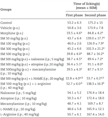
Groups
Time of licking(s) (mean ± SEM)
First phase Second phase
Control 53.2 ± 6.3 175.2 ± 15
Vehicle (p.o.) 50.8 ± 3.6 173.9 ± 7.8
Morphine (p.o.) 33.5 ± 4.6a 84.8 ± 4.2a
SM 50 mg/kg (p.o.) 43.7 ± 6.4 139.0 ± 17.7a SM 100 mg/kg (p.o.) 46.0 ± 2.6 126.9 ± 7.9a SM 300 mg/kg (p.o.) 45.2 ± 6.6 102.3 ± 21.2a SM 500 mg/kg (p.o.) 39.8 ± 4.0a 88.7 ± 16.2a SM 500 mg/kg (p.o.) + naloxone (i.p.; 5 mg/kg) 38.7 ± 4.5a 89.4 ± 7.2a SM 500 mg/kg (p.o.) + atropine (i.p.;10 mg/kg) 36.4 ± 5.1a 91.1 ± 8.0a SM 500mg/kg (p.o.) + mecamylamine
(i.p.; 10 mg/kg)
39.5 ± 4.3a 87.7 ± 9.1a
SM 500 mg/kg (p.o.) + L-NAME (i.p.; 20 mg/kg) 22.8 ± 4.0a,b 53.7 ± 6.2a,b SM 500 mg/kg (p.o.) + L-arginine
(i.p.; 40 mg/kg) 52.7 ± 6.0
,b 138.3 ± 16.3b
Naloxone (i.p.; 5 mg/kg) 54.1 ± 5.2 176.4 ± 18.4 Atropine (i.p.; 10 mg/kg) 50.3 ± 4.7 172.4 ± 18.0 Mecamylamine (i.p.; 10 mg/kg) 48.7 ± 4.1 169.7 ± 8.7 L-NAME (i.p.; 20 mg/kg) 48.6 ± 5.8 165.9 ± 12.1 L-Arginine (i.p.; 40 mg/kg) 50.7 ± 6.1 167.4 ± 14.0
ap < 0.05, when compared with the control group.
bp < 0.05, when compared with SM-treated group (500 mg/kg), calculated by One-way ANOVA followed by Bonferroni’s test. Table 1
Effect of hydroethanolic extract of Sidastrum micranthum
(SM) by individual oral administration or in combination with antagonists, over formalin-induced licking.
Development (OECD) guidelines for chemical testing (2001). The hydroethanolic extract of Sidastrum micranthum was administered orally at increasing doses up to 2000 mg/kg. Animal behaviour was observed starting 5 h after a single administration of the compound and monitored daily until the 14th day. Acute toxicity was expressed by the required dose in g/kg body weight to cause death in 50% of the animals tested (LD50).
Statistical analysis
All experimental groups consisted of 7-10 animals. The results are presented as the mean ± SEM. Statistical significance between groups was determined by a one-way analysis of variance (ANOVA) followed by Bonferroni’s test for acetic acid-induced abdominal writhing, formalin and open field tests; and statistical significance between groups was determined by a two-way analysis of variance (ANOVA) followed by Bonferroni’s test for tail-flick and paw oedema tests. A p value of less than 0.05 was considered to be statistically significant.
Results
Phytochemical analysis
The results of the chemical tests performed in the preliminary phytochemical screening revealed the presence of flavonoids, triterpenes, steroids and phenolic acids in the hydroethanolic leaf extract of Sidastrum micranthum.
Effect of Sidastrum micranthum extract on the acetic acid-induced writhing test
Intraperitoneal injection of acetic acid (1.2%) induced an average of 54.2 ± 6.1 writhes in a period of 30 min. The extract induced a dose-dependent anti-nociceptive effect on the writhing results. Doses of 50, 100, 300 and 500 mg/kg inhibited writhing by 49.8, 65.9, 71.9 and 83.4%, respectively. Morphine (5.01 mg/kg) inhibited the number of writhes by approximately 50% in relation to control group (Fig. 1).
Effect of Sidastrum micranthum extract on the formalin-induced licking test
Pre-treatment with S. micranthum extract significantly reduced the time subjects spent licking the injected paw, in both the first and the second phase after formalin injection. In the first phase, the inhibitory effect was observed only with the highest dose (500 mg/kg), whereas the second phase was inhibited at all doses (Table 1).
In the first phase, the extract showed 25.24% inhibition at dose of 500 mg/kg. In the second phase, the percentage of inhibition at the doses of 50, 100, 300, 500 mg/kg was 20.66, 27.57, 41.61, 49.38%, respectively (Table 1). Morphine (5.01 mg/kg) inhibited the number of licks by approximately 50% in relation to control group in both the first and second phases.
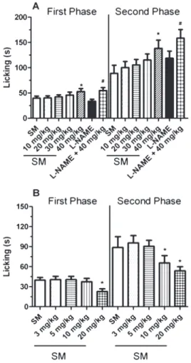
Evaluation of mechanism of action on the formalin-induced licking test
In Fig. 2, S. micranthum extract was administered in association with increasing doses of L-NAME and L-arginine to determine the lowest dose capable of producing a change in the anti-nociceptive activity of extract. The same procedure was adopted for naloxone, mecamylamine and atropine (data not shown).
In both phases (Table 1) the isolated use of the antagonists naloxone, atropine and mecamylamine produced similar
results to those obtained with the control and vehicle groups, whereas the concomitant use of these antagonists with S. micranthum extract produced results similar to those obtained with extract alone.
Table 1 shows that L-arginine and L-NAME, administered alone, did not produce significant effects relative to the control and vehicle groups. However, L-NAME administered with S. micranthum extract enhanced the anti-nociceptive effect of extract, and L-arginine reduced the anti-nociceptive effect of extract, in both phases.
Effect of Sidastrum micranthum extract on the tail-flick test
Fig. 3A shows the anti-nociceptive effect of the extract on the tail-flick test. An effect of the extract was observed only at higher doses (300 and 500 mg/kg), with a percentage of maximal effect of 30.26% at a dose of 300 mg/kg and 36.03% at 500 mg/kg.
In this model, the effects of L-arginine and L-NAME, used alone, were not significantly different from those of control and vehicle groups. The previous administration of L-arginine partially reduced the effect of S. micranthum extract, whereas the previous administration of L-NAME increased the anti-nociceptive effect of extract (Fig. 3B and 3C).
Effect of Sidastrum micranthum extract on the paw oedema test
In the paw oedema test induced by carrageenan, the effect was observed only with the highest dose (500 mg/kg) of S. micranthum extract and dexamethasone (2.25 mg/kg, s.c.). (Table 2)
The effects of L-arginine and L-NAME, used alone, were not significantly different from those of control and vehicle groups. The previous administration of L-arginine reduced the effect of S. micranthum extract on the first, second, and fourth hour of the model, whereas the previous administration of L-NAME increased the anti-nociceptive effect of extract in all times analysed (Table 2).
Effect of Sidastrum micranthum extract on the open-field test
The extract had no significant effect on locomotor activity, relative to the control and vehicle groups at dose of 500 mg/kg (Fig. 4) or with another dose tested (data not shown), whereas morphine significantly decreased locomotor activity (Fig. 4).
Toxicological evaluation of Sidastrum micranthum extract
The hydroethanolic extract of S. micranthum described in this paper was evaluated for acute toxicity in mice. No intoxication symptoms (disorientation, hyperactivity, piloerection or hyperventilation) were observed in the animals. The extract was not toxic after oral administration (LD50 > 2000 mg/kg).
Discussion
The present study aimed to evaluate the anti-nociceptive activity of the hydroethanolic extract of Sidastrum micranthum Fig. 2 - Evaluation of the antagonist dose used concurrently
with oral administration of Sidastrum micranthum extract in the formalin test. A. The mice were pretreated intraperitoneally with L-arginine (10, 20, 30 and 40 mg/kg) 15 min before the administration of extract or L-NAME. The dose of extract used was 500 mg/kg, dose of L-NAME was 40 mg/kg. B. The mice were pretreated intraperitoneal with L-NAME (3, 5, 10 and 20 mg/kg) 15 min before the administration of extract. The dose of extract used was 500 mg/kg. The results are presented as the mean ± SEM (n = 7-10). The statistical significance was calculated using One-way ANOVA, followed by Bonferroni’s test. *p < 0.05, when comparing the group administered S. micranthum
(A. St.-Hil.) Fryxell, Malvaceae, in models of acute pain and oedema formation in mice. These studies provide a foundation for the pharmacological study of this plant species in treating pain and inflammation.
The acetic acid-induced abdominal writhing test has been used as a screening tool for assessing analgesic or anti-inflammatory agents. Acetic acid induces an anti-inflammatory response in the abdominal cavity, with subsequent activation of nociceptors (Collier et al., 1968), and acetic acid- induced constriction is considered a non-selective anti-nociceptive model (Bighetti et al., 1999; Sanchez-Mateo et al., 2006). The extract reduced the number of writhes, implying that it had a significant anti-nociceptive effect. However, this test was unable to ascertain whether the antinociception was related to inflammatory or non-inflammatory effects.
The extract also produced anti-nociceptive effects in both phases of the formalin test in mice, but only with the highest dose (500 mg/kg) during the first phase. The advantage of the formalin model of nociception is that it can discriminate between inflammatory and non-inflammatory components of pain.
This first phase lasts for a few minutes and reflects the neurogenic component of nociception, being reduced mainly by opioid-like drugs. The inflammatory component of the nociceptive response (second phase) starts after a silent period of 10-15 min and is produced by the release of mediators, such as bradykinin, histamine, sympathomimetic amines, tumour necrosis factor- and interleukins (Hama and Menzaghi, 2001; Hong and Abbott, 1996; Jourdan et al., 1997; Parada et al., 2001). In addition, local levels of prostaglandins are responsible for nociception progress and are the target of most non-steroidal anti-inflammatory drugs (NSAID) (Ferreira, 2002).
The reduction of the time of licking in both phases of measurement indicates that the extract exerts anti-nociceptive effects on both inflammatory and non-inflammatory pain. This is consistent with the action of centrally acting analgesics while peripherally acting analgesics which act only during the second phase of the model (Le Bars et al., 2001).
Naloxone acts by competitively binding to opiate receptors. Naloxone shows the maximum affinity towards the µ receptor, but also shows antagonistic activity at the κ and δ receptors
(Handal et al., 1983). The previous administration of naloxone did not alter the effect produced by the extract, precluding the participation of opioid system.
Several lines of evidence show that the cholinergic system has broad therapeutic potential for the treatment of many clinically relevant pain states including inflammatory, neuropathic, visceral pain and pain because of arthritis (Jones and Dunlop, 2007). The previous administration of mecamylamine (a nonselective nicotinic acetylcholine receptor) and atropine (a nonselective muscarinic acetylcholine receptor) was used to evaluate the role of the nicotinic and muscarinic receptors, respectively, in the anti-nociceptive activity shown by S. micranthum extract. The results show that the muscarinic and nicotinic receptors are not involved in the effect of the extract, because pretreatment with atropine and mecamylamine did not alter the anti-nociceptive activity of the extract.
NO is an important mediator of nociception and it is unequivocally involved in central sensitization; it is also a signaling molecule that plays an important role in acute (Toriyabe et al., 2004) and chronic (Chen et al., 2010) pain states at both the central (Freire et al., 2009) and the peripheral (Omote et al., 2001) levels. However, experimental and clinical evidences have demonstrated that NO is also capable of inducing analgesia. Taken together, these data indicate that NO plays a complex and diverse role in the modulation of nociceptive processing.
Chen et al. (2010) recently demonstrated that pretreatment with L-NAME (non-selective NOS inhibitor), 7-nitroindazole (selective nNOS inhibitor), aminoguanidine hydrochloride (selective iNOS inhibitor), but not L-N(5)-(1-iminoethyl)-ornithine (L-NIO, selective eNOS inhibitor), significantly attenuated thermal hyperalgesia induced by subplantar injection of complete Freund’s adjuvant (CFA) in mice.
The subplantar (s.p.) injection of carrageenan and formalin causes the production and release of NO at the injured site (Omote et al., 2001). Previous administration of L-NAME (an NO synthase inhibitor) or L-arginine (a substrate of NO synthase) in the formalin test potentiated or inhibited the anti-nociceptive effect of S. micranthum extract, respectively. The use of an NO synthase inhibitor (L-NAME) enhances the effect of the compound by reducing the synthesis of NO, which acts as a retrograde messenger at the spinal level to increase the stimulatory neurotransmitters released (Oess et al., 2006). In contrast, L-arginine increases the concentration of NO at the spinal level and thus reduces the anti-nociceptive effect of the extract. These results show that the production and release of NO is important to the effect of the extract. On the basis of these results, we believe that S. micranthum extract can reduce the concentration of NO at the spinal level.
Tail flicking is predominantly a spinal reflex and is considered to be selectively sensitive to centrally acting analgesic compounds (Ramabadran et al., 1989). In the tail-flick test, the thermal stimulation activates peripheral nociceptors, leading to reflexive removal of the tail (Kuraishi et al., 1983). The anti-nociceptive effect of the extract was also observed in the tail-flick test with the highest doses (300 and 500 mg/kg) confirming its central activity.
This later peak effect of morphine can be explained by the irregular absorption of oral morphine (Tassinari et al., 1995). The profiles of the 300 and 500 mg/kg doses show two distinct phases of action, suggesting the possibility of existence of an active metabolite.
Although the oral morphine presents fourth to one-sixth of bioavailability obtained in parenteral administration (Tassinari et al., 1995), the oral route was chosen to be the same route of administration used to the hydroethanolic extract of S. micranhum and thereby establish a better comparison between the standard drug (morphine) and test substance (S. micranhum extract).
acid-induced abdominal writhing, formalin and tail-flick tests, where the stimulus is harmful.
To evaluate a possible anti-inflammatory action of the extract, paw oedema was induced by carrageenan. Three phases have been postulated for carrageenan-induced oedema: histamine and 5-hydroxytryptamine (5-HT) release in the early phase (first hour), kinin release in the second phase (second hour) and prostaglandin release in the third phase (third and fourth hours) (Olajide et al., 1999). In this test, the extract inhibited the induction of paw oedema in all phases, thus demonstrating its anti-oedematogenic activity.
The L-arginine-NO pathway has been proposed to play an important role in the carragenan-induced inflammatory response (Cuzzocrea et al., 1996), and the expression of the inducible isoform of NO synthase has been proposed as an important mediator of inflammation (Pan et al., 2009). The administration of L-NAME or L-arginine in the paw oedema test, potentiated or inhibited the carragenaan-mediated paw oedema formation from the administration of S. micranthum
extract, respectively.
We suggest that the anti-inflammatory mechanism of the extract may be through the L-arginine-NO pathway due to the influence of NO production on the anti-oedematogenic effect of the extract, as demonstrated by pretreatment with L-NAME and L-arginine.
The open field test was used to exclude the possibility that the anti-nociceptive action of extract could be related to nonspecific disturbances in the locomotor activity of the animals. We observed that with effective anti-nociceptive dose, the hydroethanolic extract of S. micranthum did not alter the motor performance of mice.
The anti-nociceptive and anti-oedematogenic action of the hydroethanolic extract of S. micranthum is reinforced by the results showing that acute administration of the extract inhibited carragenaan-mediated paw oedema formation and demonstrated activity in the tail flick test, formalin-induced paw licking test and acetic acid-induced abdominal writhing test. These data provide initial evidence that the traditional use of S. micranthum is effective at reducing pain and oedema formation.
It is important to state that there are no descriptions of adverse effects or intoxication in humans following the use of S. micranthum extract in folk medicine. This lack of adverse effects and intoxication is corresponded in our study by the lack of physiological complications in the animals that received the extract orally.
Authorship
CCJA collected plant samples for exsiccate preparation, performed phytochemical screenings and prepared hydroethanolic extract. GMG and DGM participated in the animal experiments, performed toxicity test and participated in statistical analysis and manuscript edition. BGM performed the statistical analysis and prepared the manuscript. All the authors have read the final manuscript and approved the submission.
Acknowledgments
CNPq by financial support.
R E F E R E N C E S
Agra, M.F., Freitas, P.F., Barbosa-Filho, J.M., 2007. Synopsis of the plants known as medicinal and poisonous in Northeast of Brazil. Rev. Bras. Farmacogn. 17, 114-140.
Barros, H.M.T., Tannhauser, M.A.L., Tannhauser, S.L.,
Tannhauser, M., 1991. Enhanced detection of hyperactivity after drug-withdrawal with a simple modification of the open-field apparatus. J. Pharmacol. Method. 26, 269-275. Bighetti, E.J.B., Hiruma-Lima, C.A., Gracioso, J.S., Souza Brito,
A.R.M., 1999. Anti-inflammatory and anti-nociceptive effects in rodents of the essential oil of Croton cajucara Benth. J. Pharmacol. Method. 51, 1447-1453.
Burke, A., E., S., Fitzgerald, G.A., 2006. Analgesic-antipyretic agents: pharmacotherapy of gout in: Brunton, L.L., Lazo, J.S., Parker, K.L. (Eds.), Goodman and Gilmans The Pharmacological Basis of Therapeutics. McGraw Hill, New York.
Butler, S.F., Budman, S.H., Fernandez, K., Jamison, R.N., 2004. Validation of a screener and opioid assessment measure for patients with chronic pain. Pain 112, 65-75.
Cavalcante, J.M.S., Nogueira, T.B.S.S., Tomaz, A.C.D., Silva, D.A.E., Agra, M.D., de Souza, M.D.V., Carvalho, P.R.C., Ramos, S.R., do Nascimento, S.C., Goncalves-Silva, T., 2010. Steroidal and phenolic compounds from Sidastrum paniculatum (L.) Fryxell and evaluation of cytotoxic and anti-inflammatory activities. Quim. Nova. 33, 846-849.
Chen, Y., Boettger, M.K., Reif, A., Schmitt, A., Uceyler, N., Sommer, C., 2010. Nitric oxide synthase modulates CFA-induced thermal hyperalgesia through cytokine regulation in mice. Mol. Pain. 6.
Collier, H.O., Dinneen, L.C., Johnson, C.A., Schneider, C., 1968. The abdominal constriction response and its suppression by analgesic drugs in the mouse. Br. J. Pharmacol. Chemother. 32, 295-310.
Cowan, D.T., Allan, L., Griffiths, P., 2002. A pilot study into the problematic use of opioid analgesics in chronic non-cancer pain patients. Int. J. Nurs. Stud. 39, 59-69.
Cuzzocrea, S., Zingarelli, B., Calapai, G., Nava, F., Caputi, A.P., 1996. Zymosan-activated plasma induces paw oedema by nitric oxide and prostaglandin production. Life Sci. 60, 215-220.
D’Amour, F.E., Smith, D.L., 1941. A method for determining loss of pain sensation. J. Pharmacol. Exp. Ther. 72, 74-79. Diaz, A.M., Abad, M.J., Fernandez, L., Recuero, C., Villaescusa, L.,
Silvan, A.M., Bermejo, P., 2000. In vitro anti-inflammatory activity of iridoids and triterpenoids compounds isolated from Phillyrea latifolia. Biol. Pharm. Bull. 23, 1307-1313. Ferreira, S.H., 1979. Oedema and increased vascular permeability,
in: Vane, J.R., Van Arman, C.G. (Eds.), Handbook of
experimental pharmacology. Springer-Verlag, New York, pp. 75-91.
Ferreira, S.H., 2002. Peripheral analgesic sites of action of anti-inflammatory drugs. Int. J. Clin. Pract. Supplement, 2-10. Freire, M.A., Guimaraes, J.S., Leal, W.G., Pereira, A., 2009. Pain
modulation by nitric oxide in the spinal cord. Frontiers in neuroscience 3, 175-181.
Gaertner, M., Muller, L., Roos, J.F., Cani, G., Santos, A.R., Niero, R., Calixto, J.B., ., Yunes, R.A., Delle Monache, F., Cechinel-Filho, V., 1999. Analgesic triterpenes from Sebastiana schottiana roots. Phytomedicine. 6, 41-44.
Geetha, T., Varalakshmi, P., 2001. Anti-inflammatory activity of lupeol and lupeol linoleate in rats. J. Ethnopharmacol. 76, 77-80.
Gilson, A.M., Ryan, K.M., Joranson, D.E., Dahl, J.L., 2004. A reassessment of trends in the medical use and abuse of opioid analgesics and implications for diversion control: 1997-2002. J. Pain Symptom Manag. 28, 176-188.
Gomes, R.A., Agra, M.F., Souza, M.F.V., 2011a. Constituintes químicos de Sidastrum micranthum (Malvaceae), 34ª Reunião Anual da Sociedade Brasileira de Química, Florianópolis, Brazil.
Gomes, R.A., Ramirez, R.R.A., Maciel, J.K.D., Agra, M.D., de Souza, M.D.V., Falcao-Silva, V.S., Siqueira, J.P., 2011b. Phenolic compounds from Sidastrum micranthum (A. St.-Hil.) Fryxell and evaluation of acacetin and
7,4’-di-O-methylisoscutellarein as motulator of bacterialdrug resistence. Quim. Nova. 34, 1385-U1279.
Hama, A., Menzaghi, F., 2001. Antagonist of nicotinic
acetylcholine receptors (nAChR) enhances formalin-induced nociception in rats: tonic role of nAChRs in the control of pain following injury. Brain Res. 888, 102-106.
Handal, K.A., Schauben, J.L., Salamone, F.R., 1983. Naloxone. Ann Emerg Med 12, 438-445.
Hong, Y., Abbott, F.V., 1996. Contribution of peripheral alpha 1A-adrenoceptors to pain induced by formalin or by alpha-methyl-5-hydroxytryptamine plus noradrenaline. Eur. J. Pharmacol. 301, 41-48.
Hunskaar, S., Berge, O.G., Hole, K., 1986. Dissociation between antinociceptive and antiinflammatory effects of acetylsalicylic acid and indomethacin in the formalin test. Pain. 25, 125-132.
Jones, P.G., Dunlop, J., 2007. Targeting the cholinergic system as a therapeutic strategy for the treatment of pain. Neuropharmacology. 53, 197-206.
Jourdan, D., Ardid, D., Bardin, L., Bardin, M., Neuzeret, D., Lanphouthacoul, L., Eschalier, A., 1997. A new automated method of pain scoring in the formalin test in rats. Pain. 71, 265-270.
Koster, R., Anderson, M., Debeer, E.J., 1959. Acetic acid for analgesic screening. Fed. Proc. 18, 412-412.
Kuraishi, Y., Harada, Y., Aratani, S., Satoh, M., Takagi, H., 1983. Separate involvement of the spinal noradrenergic and serotonergic systems in morphine analgesia - the differences in mechanical and thermal algesic tests. Brain Res. 273, 245-252.
Le Bars, D., Gozariu, M., Cadden, S.W., 2001. Animal models of nociception. Pharmacol. Rev. 53, 597-652.
OECD, 2001. Guideline for Testing of Chemical: 420 Acute oral toxicity, in: Development, T.O.o.E.C.-o.a. (Ed.). OECD, France.
Oess, S., Icking, A., Fulton, D., Govers, R., Muller-Esterl, W., 2006. Subcellular targeting and trafficking of nitric oxide synthases. Biochem. J. 396, 401-409.
Olajide, O.A., Makinde, J.M., Awe, S.O., 1999. Effects of the aqueous extract of Bridelia ferruginea stem bark on carrageenan-induced oedema and granuloma tissue formation in rats and mice. J. Ethnopharmacol. 66, 113-117. Omote, K., Hazama, K., Kawamata, T., Kawamata, M., Nakayaka,
Y., Toriyabe, M., Namiki, A., 2001. Peripheral nitric oxide in carrageenan-induced inflammation. Brain Res 912, 171-175. Pan, M.H., Lai, C.S., Dushenkov, S., Ho, C.T., 2009. Modulation
of Inflammatory Genes by Natural Dietary Bioactive Compounds. J. Agr. Food Chem. 57, 4467-4477.
Parada, C.A., Tambeli, C.H., Cunha, F.Q., Ferreira, S.H., 2001. The major role of peripheral release of histamine and 5-hydroxytryptamine in formalin-induced nociception. Neuroscience 102, 937-944.
Ramabadran, K., Bansinath, M., Turndorf, H., Puig, M.M., 1989. Tail immersion test for the evaluation of a nociceptive reaction in mice - methodological considerations. J. Pharmacol. Method. 21, 21-31.
Sanchez-Mateo, C.C., Bonkanka, C.X., Hernandez-Perez, M., Rabanal, R.M., 2006. Evaluation of the analgesic and topical anti-inflammatory effects of Hypericum reflexum L. fil. J. Ethnopharmacol. 107, 1-6.
Santos, A.R.S., Filho, V.C., Yunes, R.A., Calixto, J.B., 1995. Further-Studies on the antinociceptive action of the hydroalcoholic extracts from plants of the genus Phyllanthus. J. Pharm. Pharmacol. 47, 66-71.
Shimpale, V.B., Sutar, S.P., Yadav, S.R., 2009. Sidastrum
(Malvaceae): A new genus record for Asia. Rheedea. 19, 50-52. Shukla, S., Mehta, A., Mehta, P., Vyas, S.P., Shukla, S., Bajpai,
V.K., 2010. Studies on anti-inflammatory, antipyretic and analgesic properties of Caesalpinia bonducella F. seed oil in experimental animal models. Food Chem. Toxicol. 48, 61-64. Tassinari, D., Masi, A., Sartori, S., Nielsen, I., Ravaioli, A., 1995.
A typical absorption of morphine sulphate through oral mucosa: an usual case of acute opioid poisoning. J pain symptom mana 10.
Toriyabe, M., Omote, K., Kawamata, T., Namiki, A., 2004. Contribution of interaction between nitric oxide and cyclooxygenases to the production of prostaglandins in carrageenan-induced inflammation. Anesthesiology. 101, 983-990.
Wagner, W.L., Herbst, D.R., Sohmer, S.H., 1999. Manual of the flowering plants of Hawaii. Revised edition. University of Hawaii Press/Bishop Museum Press, Honolulu.
WHO, 1999. Monographs on selected medicinal plants. World Health Organization Switzerland.