Protein-Coupled Receptors on the Yeast Cell Surface
Jun Ishii1, Nobuo Yoshimoto2,3, Kenji Tatematsu2, Shun’ichi Kuroda2,3, Chiaki Ogino4, Hideki Fukuda4, Akihiko Kondo4*
1Organization of Advanced Science and Technology, Kobe University, 1-1 Rokkodai, Nada, Kobe, Japan,2Department of Structural Molecular Biology, Institute of Scientific and Industrial Research, Osaka University, 8-1 Mihogaoka, Ibaraki, Osaka, Japan,3Graduate School of Bioagricultural Sciences, Nagoya University, Furo, Chikusa, Nagoya, Japan,4Department of Chemical Science and Engineering, Graduate School of Engineering, Kobe University, 1-1 Rokkodai, Nada, Kobe, Japan
Abstract
G-protein-coupled receptors (GPCRs) regulate a wide variety of physiological processes and are important pharmaceutical targets for drug discovery. Here, we describe a unique concept based on yeast cell-surface display technology to selectively track eligible peptides with agonistic activity for human GPCRs (Cell Wall Trapping of Autocrine Peptides (CWTrAP) strategy). In our strategy, individual recombinant yeast cells are able to report autocrine-positive activity for human GPCRs by expressing a candidate peptide fused to an anchoring motif. Following expression and activation, yeast cells trap autocrine peptides onto their cell walls. Because captured peptides are incapable of diffusion, they have no impact on surrounding yeast cells that express the target human GPCR and non-signaling peptides. Therefore, individual yeast cells can assemble the autonomous signaling complex and allow single-cell screening of a yeast population. Our strategy may be applied to identify eligible peptides with agonistic activity for target human GPCRs.
Citation:Ishii J, Yoshimoto N, Tatematsu K, Kuroda S, Ogino C, et al. (2012) Cell Wall Trapping of Autocrine Peptides for Human G-Protein-Coupled Receptors on the Yeast Cell Surface. PLoS ONE 7(5): e37136. doi:10.1371/journal.pone.0037136
Editor:Debra Kendall, University of Connecticut, United States of America
ReceivedDecember 16, 2011;AcceptedApril 13, 2012;PublishedMay 18, 2012
Copyright:ß2012 Ishii et al. This is an open-access article distributed under the terms of the Creative Commons Attribution License, which permits unrestricted use, distribution, and reproduction in any medium, provided the original author and source are credited.
Funding:This work was supported in part by a Grant-in-Aid for Scientific Research on Priority Areas (Life surveyor) from the Ministry of Education, Culture, Sports, Science and Technology (MEXT), the Regional Innovative Consortium Project of the Ministry of Economy, Trade and Industry (METI) and a Special Coordination Funds for Promoting Science and Technology, Creation of Innovation Centers for Advanced Interdisciplinary Research Areas (Innovative Bioproduction Kobe; iBioK) from the MEXT of Japan, and funded in part by AS ONE Corporation and the Naito Foundation. The funders had no role in study design, data collection and analysis, decision to publish, or preparation of the manuscript.
Competing Interests:This work was supported in part by AS ONE Corporation. This does not alter the authors’ adherence to all the PLoS ONE policies on sharing data and materials.
* E-mail: [email protected]
Introduction
G-protein-coupled receptors (GPCRs) constitute a large super-family of cell surface receptors [1]. In humans, these 7-trans-membrane proteins respond to external stimuli to regulate various cellular processes including taste, smell, vision, heart rate, blood pressure, neurotransmission and cell growth [2]. All members of the guanine nucleotide binding protein family (G-proteins) share a common mechanism for signal transmission following GPCR-agonist binding [3]. This universal signaling mechanism has become a central tenet in G-protein research, and GPCRs have become major pharmaceutical targets for drug discovery [4].
The eukaryotic unicellular yeast, Saccharomyces cerevisiae, also shares the G-protein-mediated signal transmission mechanism with higher mammalian cells [3]. It is notable thatS. cerevisiaeoffers a crucial advantage to simplify the study of GPCR signaling because it expresses only one kind of G-protein, which thereby avoids potential problems such as signaling cross-talk in mamma-lian cells [5–8]. Therefore,S. cerevisiaeis a suitable host cell for the screening of functional residues in GPCRs [5,9,10].
Yeast cell-surface display technology is a powerful platform that enables proteins expressed in yeast to be tethered onto the cell surface [11–15]. This is accomplished by the use of ‘‘anchor’’ proteins that naturally localize on the cell surface in yeast cells. Typically, the gene encoding the target protein is fused to the
anchor protein together with a secretion signal sequence at the N-terminus to both enable secretion of the fusion protein and to tether it firmly to the cell surface. As typical anchor proteins, the C-terminal domains of truncateda-agglutinin (Sag1p; a manno-protein involved in sexual adhesion) and truncated Flo1p (a lectin-like cell-wall protein involved in flocculation) containing the glycosyl-phosphatidylinositol (GPI) anchor attachment signal sequence at the C-terminus are fused to the target protein at their N-termini [16,17]. Regarding other anchor proteins, the Flo1p flocculation functional domain without the GPI anchor attachment signal (FS anchor) permits the fusion of the target protein to both its N- and C-termini [18]. These anchor proteins are used to display the target proteins on the yeast cell wall. In contrast, periplasmic invertase (Suc2 anchor) can be fused to both the N- and C-termini of a target protein, enabling it to localize into the periplasmic space [19]. To date, yeast cell-surface display technology has been adopted for a broad range of applications including enzymatic catalysis, immune adsorption and protein engineering [16–18,20–23].
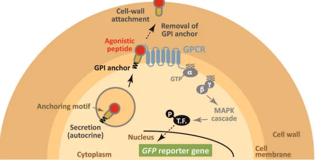
a secretion signal sequence and an anchoring motif. Agonistic peptides are capable of binding cell surface GPCRs that transduce the signal into the cell. Finally, the yeast traps the signaling peptide on its cell wall (Figure 1). Here, we use a yeast strain that is engineered to express a green fluorescent protein (GFP) reporter gene in response to GPCR activation. Therefore, stimulation by agonistic peptides can be recognized by the generation of a green fluorescence signal [3]. In principle, because signaling peptides are unable to diffuse to surrounding cells, our strategy has the potential to build autonomous signaling complexes on a cell-by-cell basis. Our peptide trapping method (cell-by-cell wall trapping of autocrine peptides (CWTrAP) system) will allow the identification of lead peptides from combinatorial peptide libraries as starting points for drug screening.
Results and Discussion
To corroborate the viability of cell-surface display technology to track agonistic activity for GPCRs (CWTrAP system), we useda -factor pheromone, a natural ligand for the endogenous yeast 7-transmembrane GPCR, Ste2, which is specifically expressed in the a-type-strain [24]. In nature,a-type yeast strains secretea-factor to induce mating signal transduction in the a-type strain by binding to the Ste2 receptor on its cell surface [25]. The ability of several types of protein motifs to anchor and transduce the autocrine a-factor were tested in the recombinant a-type yeast cells, which can express a GFP reporter gene in response to pheromone signaling (Figure 1). All constructs of fusion proteins that displayeda-factor peptides were designed to contain a Flag tag between thea-factor peptides and anchor proteins (Figure 2A and Table 1).
We used the IMG-4 yeast strain to displaya-factor pheromone on its cell surface because this strain can monitor signaling levels through its endogenous Ste2 receptor via a GFP reporter gene
(Table 1). To test our concept, we evaluated the C-terminal 320 aa of Sag1p (C-terminal half ofa-agglutinin; AG) [16] and various lengths of truncated Flo1p derivatives (C-terminal 42, 102, 146 and 318 aa of Flo1p; Flo42, Flo102, Flo146 and Flo318) [17] as anchor proteins with GPI anchoring motifs (Figure 2A and Table 1). A recombinant yeast strain, engineered to express thea -factor autocrine peptide with a secretion signal sequence but lacking an anchor motif, robustly generated a higher green fluorescence signal than a strain harboring a mock plasmid (Figure 3A, Mock and Sec). Immunofluorescence staining of Flag-taggeda-factor peptide revealed no fluorescence on the surface of engineered yeast cells (Figure 3B, Sec). These results suggest that secreteda-factor could bind the endogenous Ste2 receptor and transduce the signal inside the yeast cells.
Next, we tested the cell wall trapping (CWTrAP) strategy fora -factor peptide with GPI anchoring motifs. All engineered yeast strains expressinga-factor peptides fused to the N-termini of the anchor proteins (AG and Flo42–318) with an inserted Flag tag (Figure 2A) successfully generated a green fluorescence signal (Figure 3A), confirming that the fusion peptide is able to activate signal transduction in yeast. Using GFP fluorescence intensity as an indicator of signaling strength, shorter anchor peptides appeared more capable of activating the GPCR (Figure 3A). The a-factor peptide fused to Flag and Flo42 exhibited higher responsiveness compared toa-factor lacking the anchor protein. This interesting result may arise from the transient enrichment of the GPI-anchored peptide on the yeast cell membrane, although the GPI-anchored peptide should be cleaved from the plasma membrane by phosphatidylinositol-specific phospholipase C (PI-PLC) and tethered on the cell wall [11–13].
Although shorter peptides tend to make detection of the Flag tag more difficult, due to the report that shorter peptides can bury the tag within the cell wall [17], we were able to confirm an anchor-dependent association with the yeast cell wall by
immunofluores-Figure 1. Schematic illustration of our concept using yeast cell-surface display technology to selectively track eligible agonistic peptides for human GPCRs by assembling the autonomous signaling complex within individual cells (cell wall trapping of autocrine peptides (CWTrAP) strategy).The candidate autocrine peptides fused with the anchoring proteins are processed via secretion pathways in engineered yeast cells. Their agonistic activities for heterologously-expressed human GPCRs are discerned on yeast cell membranes. Only when the peptide possesses objective agonistic activity, membrane-peripheral G-proteins promote intracellular signaling and induce transcription of the GFP reporter gene. Because the autocrine peptides are automatically trapped onto individual yeast cell walls, the captured peptides are unable to diffuse toward surrounding yeast cells that express the target human GPCR and any other peptides. T.F. indicates transcription factor.
doi:10.1371/journal.pone.0037136.g001
cence staining (Figure 3B). Because peptides anchored to the cell wall are unable to diffuse to surrounding cells, this result emphasizes the viability of our concept for the assembly of the autonomous signaling complex within individual yeast cells. Additionally, we verified that a subset of Flo42 was highly glycosylated (Figure S1); however, the agonistic activity of thea -factor peptide was unlikely to be affected by the posttranslational glycosylation of the anchor protein.
Next, we tested additional motifs that permit peptides to be fused to both the N- and C-termini of the anchor proteins. We replaced the GPI anchor proteins with the FS anchor [18] and the Suc2 anchor [19] (Figure 2A, Table S2 and Document S1). Signal transduction was more efficient when using the FS anchor, compared to the Suc2 anchor (Figure S2). These results show that agonistic peptides can be fused to both the N- and C-termini of anchor proteins. Even though the FS anchor (1099 aa) served as an efficient motif for transducing a-factor peptide signaling, we used the Flo42 anchor motif, whose molecular mass is much lower (Figure 2A), in all following experiments in order to minimize the possibility of steric hindrance.
To further demonstrate the viability of our concept, the IMFD-70 yeast strain, which can monitor signaling levels from recombinantly expressed heterologous GPCRs by aGFPreporter
gene [5] (Table 1), was used to test if signal transmission from human GPCRs expressed on the yeast cell surface was possible. For these experiments, human somatostatin receptor subtype 5 (SSTR5), and the natural intramolecular-cross-linked cyclic peptide ligand, somatostatin 14 (S-14), were used [26,27].
To express the autocrine somatostatin and trap it on the yeast cell wall, we designed the S-14 peptide with an N-terminal secretion signal sequence and a C-terminal Flo42 anchor protein with a Flag tag (Figure 2B and Table 1). We constructed several negative controls by eliminating the S-14 peptide or by replacing it with agonistic peptides for other GPCRs (Figure 2B and Table 1). We expressed hemagglutinin (HA)-tagged human SSTR5 on the yeast cell surface using previously reported plasmids [5,6] (Table 1). We used these expression and mock plasmids to investigate the ability of the S-14–Flag–Flo42 autocrine peptide to activate GPCR signaling (Figure 4).
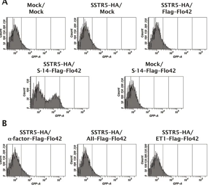
As shown in Figure 4A, the engineered yeast strain concom-itantly expressing SSTR5–HA and S-14–Flag–Flo42 successfully inducedGFPreporter gene expression, whereas the other control strains possessing either SSTR5–HA or S-14–Flag–Flo42 did not. Similarly, a control strain expressing SSTR5–HA and the autocrine Flag–Flo42 fusion protein lacking the S-14 peptide was unable to express a green fluorescence signal (Figure 4A).
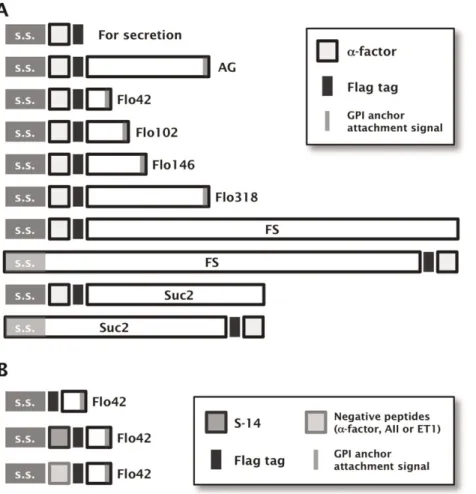
Figure 2. Schematic illustration of the fusion protein constructs used to display agonistic peptides on the yeast cell-surface.(A) Constructs for displayinga-factor peptides. AG: C-terminal half ofa-agglutinin anchor. s.s.: secretion signal sequence. The pre-pro-region derived froma-factor was used as s.s. For the fusion of FS and Suc2 anchors to thea-factor peptides at their C-termini, the original s.s. encoded in the N-termini of Flo1p or Suc2p were used, respectively. The uppermost construct for secretion ofa-factor peptide contains no anchoring motifs. All constructs contain the Flag tag. (B) Constructs for displaying S-14 by the Flo42 anchor. The upper construct displaying only Flag and Flo42 peptides was used as a negative control for the SSTR5 signaling assay. The middle and lower constructs displaying, respectively, eligible peptide (S-14) and negative control peptides (a-factor, AII and ET1) by Flag–Flo42 fusion proteins were also used for the SSTR5 signaling assay.
These results demonstrate that autocrine activation of recombi-nant SSTR5 by binding of the S-14 peptide fused to the Flo42 anchor mediates pheromone signaling via endogenous peripheral G-proteins in yeast [5]. Furthermore, we were able to confirm the specificity of the S-14 peptide because three control peptides in which the S-14 peptide was replaced with the yeast Ste2 receptor agonist, a-factor, the human angiotensin receptor agonist, angiotensin II (AII), or the human endothelin receptor agonist, endothelin-1 (ET1), did not generate a green fluorescence signal (Figure 4B).
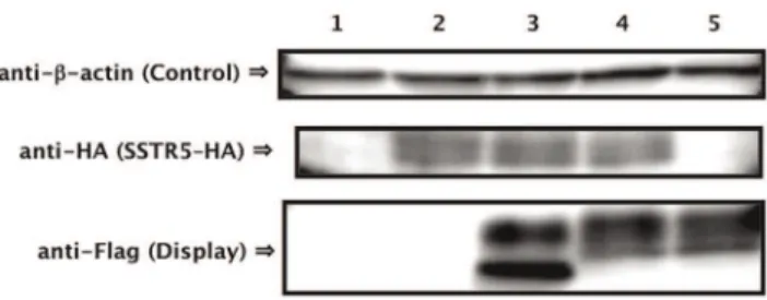
We confirmed the expression of SSTR5–HA receptor and S-14–Flag–Flo42 fusion protein by western blot analysis (Figure 5). Equal loading of the sodium lauryl sulfate (SDS)-extracted cell lysate fraction from each pellet was confirmed using anti-b-actin. SSTR5–HA receptor (anti-HA; lanes 2–4) and Flag–Flo42 anchor or S-14–Flag–Flo42 fusion proteins (anti-Flag; lanes 3–5) were successfully detected in the extracts of each appropriate transfor-mant. The two unequal bands detected by the anti-Flag antibody in the Flag–Flo42 and S-14–Flag–Flo42 transformants likely represent the signal-cleaved and -uncleaved proteins, because the pre-pro-region derived from a-factor was used as the secretion signal sequence. We therefore tested the ability of the other active somatostatin isoform S-28 [26] and other secretion signal sequences (pre-region of a-factor and signal sequences derived from S. cerevisiae Suc2p and Rhizopus oryzae glucoamylase) to mediate signal transduction in the IMG-50 yeast strain. This strain
has a slightly different genetic background to IMFD-70 (FAR1 -intact strain [28], the description of thefar1Dallele can be found in Materials and Methods; Table 1), but the expression profiles of the
GFPreporter genes remained essentially unchanged (Figure S3). Also, the insertion of GS linkers (GGGGS and GGGSGGGGS) between the S-14 peptide and Flag–Flo42 did not improveGFP
expression (Figure S4). Because GPCR signaling has been reported to decrease plasmid retention even in the far1D yeast strain [28], false-negative signals (non-signaling cell cluster; Figure 4A, SSTR5–HA/S-14–Flag–Flo42) may be caused by plasmid loss. Because other secretion signal sequences and the insertion of GS linkers had no effect on expression of theGFP
reporter gene, it is unlikely that a false-negative signal would be caused by steric hindrance of the S-14 peptide (Figure S3 and S4). Nevertheless, the presence of false-negative cells within an identical cell cluster implies that peptides captured on the cell wall have little influence on the surrounding cells (Figure 4, S3 and S4). Therefore, we demonstrated that peptides captured on the cell wall did not induce false-positive signals in surrounding non-target cells, even when two types of cells, one expressing the S-14–Flag– Flo42 (target cells) and the other expressing the Flag–Flo42 anchor lacking S-14 (non-target cells or surrounding cells), were mixed (Figure S5). Additionally, we successfully enhanced the weaker green fluorescence signal of the IMFD-70 strain expressing SSTR5–HA and S-14–Flag–Flo42 (Figure 4A) by concurrently introducing a multi-copy plasmid harboring theGFPreporter gene Table 1.Yeast strains and plasmids used in this study.
Strain or plasmid Relative feature Source
Yeast strain
BY4741 MATahis3D1 leu2D0 met15D0 ura3D0 [31]
IMG-4 BY4741fus1::FUS1-EGFP-TGAPDH-HIS3 bar1D::LEU2 far1D::kanMX4 This study
IMG-50 BY4741fus1::FUS1-EGFP-TGAPDH-HIS3 sst2D::AUR1-C ste2D::LEU2 [28]
IMFD-70 BY4741fig1D::EGFP his3D::PFIG1-EGFP far1Dsst2D::AUR1-C ste2D::LEU2 [5]
Plasmid
pESC-URAa Expression vector containing
GAL1-GAL10divergent promoter, 2morigin andURA3marker Agilent Technologies
pUESCasf pESC-URA,a-factor–Flag peptide expression (for secretion) This study
pUESCaf-AG pESC-URA,a-factor–Flag–AGbfusion protein expression (for display) This study
pUESCaf-FLO42 pESC-URA,a-factor–Flag–Flo42 fusion protein expression (for display) This study
pUESCaf-FLO102 pESC-URA,a-factor–Flag–Flo102 fusion protein expression (for display) This study
pUESCaf-FLO146 pESC-URA,a-factor–Flag–Flo146 fusion protein expression (for display) This study
pUESCaf-FLO318 pESC-URA,a-factor–Flag–Flo318 fusion protein expression (for display) This study
pGK421a Expression vector containingPGK1promoter, 2
morigin andMET15marker [5,6]
pGK-SSTR5-HA pGK421, SSTR5-HA human receptor expression [5,6]
pGK426a Expression vector containing
PGK1promoter, 2morigin andURA3marker [36]
pGK42 pGK426, Flag–Flo42 anchor protein expression (for display) This study
pGK-S1442 pGK426, S-14–Flag–Flo42cfusion protein expression (for display) This study
pGK-alpha42 pGK426,a-factor–Flag–Flo42 fusion protein expression (for display) This study
pGK-AII42 pGK426, AII–Flag–Flo42dfusion protein expression (for display) This study
pGK-ET142 pGK426, ET1–Flag–Flo42efusion protein expression (for display) This study
pMHG-FIG1 Multi-copy reporter plasmid containingFIG1promoter,GFPreporter gene, 2morigin andHIS3marker [6]
All transcription products for display or secretion contain the secretion signal sequence ofa-factor.
aThe indicated vectors were used as mock controls. bAG indicates C-terminal half ofa-agglutinin anchor protein. cS-14 encodes somatostatin 14 mature peptide.
dAII encodes angiotensin II mature peptide. eET1 encodes endothelin-1 mature peptide.
doi:10.1371/journal.pone.0037136.t001
cassette (pMHG-FIG1 [6]) (Figure 6). These results strongly support the feasibility of our conceptual CWTrAP system to identify eligible agonistic peptides for human GPCRs.
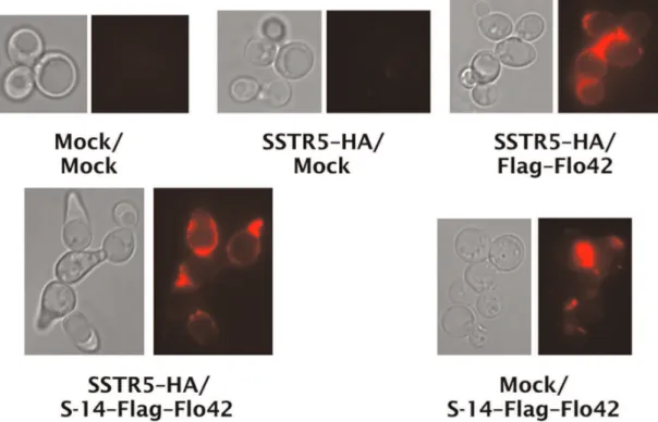
Finally, to examine whether the yeast cell wall did indeed trap the autocrine peptide fused to the Flo42 anchor, transformants were analyzed by immunofluorescence staining with anti-Flag primary antibody and Alexa Fluor 594 conjugated secondary antibody (Figure 7). We observed red fluorescence on the cell surfaces of appropriate transformants that expressed Flag–Flo42 anchor or S-14–Flag–Flo42 fusion proteins. In addition, we only
observed a morphology change [29] on cells expressing both SSTR5–HA and S-14–Flag–Flo42, supporting our hypothesis that the autocrine S-14 peptide specifically triggered signal trans-duction via the SSTR5 receptor in the recombinant yeast cells. Thus, we successfully verified that the S-14 autocrine peptide fused to the Flo42 anchor protein was trapped on the yeast cell wall.
In this study, we have demonstrated how a strategy for cell wall trapping of autocrine peptides (CWTrAP system) functions to discern agonistic activity for human GPCRs expressed in yeast
Figure 3. Evaluation of the CWTrAP system usinga-factor peptide for yeast endogenous Ste2 receptor.(A) Pheromone signaling assays ofa-factor-displaying yeast strains. Error bars represent the standard deviation of three independent experiments. (B) Immunofluorescence staining ofa-factor displaying yeast strains. Anti-Flag antibody and Alexa Fluor 546-conjugated secondary antibody were used for detection of secreteda -factor ora-factor-anchor fusion proteins. IMG-4 was used as the host strain. The transformants used in these experiments are listed in Table S3. Sec: free, secreted form ofa-factor. AG: C-terminal half ofa-agglutinin anchor.
cells, by using the intramolecular-cross-linked cyclic peptide S-14 and its specific receptor as our model. Our motivation was to selectively track eligible agonistic peptides for human GPCRs by assembling an autonomous signaling complex within individual cells. By combining cell-surface display technology and established yeast combinatorial genetic engineering technology with flow cytometric single-cell screening [30], we aim to identify eligible peptides from peptide libraries. Here, the feasibility of our concept is demonstrated by peptide capture, and subsequent signal transduction, by heterologously-expressed human GPCRs, which prevent the captured peptides from diffusing to surrounding yeast cells and eliciting a false-positive response. Therefore, the captured peptides are successfully presented by yeast cell-surface display technology.
Materials and Methods
Media
Synthetic raffinose (SR) media contained 6.7 g/l yeast nitrogen base without amino acids (YNB) (BD-Diagnostic Systems, Sparks, MD, USA) and 20 g/l raffinose. For SRGC media, 20 g/l galactose and 20 g/l casamino acids (BD-Diagnostic Systems) were added into SR media. Synthetic dextrose (SD) media contained 6.7 g/l YNB and 20 g/l glucose. For SDM71 media, SD media was adjusted to pH 7.1 with 200 mM MOPSO buffer (Nacalai Tesque, Kyoto, Japan). Amino acids and nucleotides (20 mg/l histidine, 60 mg/l leucine, 20 mg/l methionine or 20 mg/l uracil) were supplemented into each medium to provide the relevant auxotrophic components.
Figure 4. Evaluation of the CWTrAP system using somatostatin peptide for the human SSTR5 receptor.(A) SSTR5 signaling assays of the cyclic somatostatin peptide displaying yeast strain and control strains. (B) SSTR5 signaling assays of non-target peptide displaying yeast strains. IMFD-70 was used as the host strain. The transformants used in these experiments are listed in Table S3. S-14 indicates 14 aa of somatostatin cyclic peptide, a-factor indicates 13 aa of yeast pheromone peptide, AII indicates 8 aa of angiotensin II peptide, and ET1 indicates 21 aa of endothelin-1 peptide. doi:10.1371/journal.pone.0037136.g004
Yeast Strains
Yeast strains used for assays were generated from BY4741 [31] as a parental backbone strain and are listed in Table 1. The transformation procedure using linear DNA fragments followed the lithium acetate method [32]. All primers used for the strain constructions are listed in Table S1. Thebar1Dalleles that relieve the degradation of a-factor pheromone [33] were conferred to BY4741far1D (obtained from Saccharomyces Genome Deletion Project [34]) by homologous recombination with the amplified
LEU2 fragments, producing the IM-4 strain. The FUS1-GFP
reporter gene was integrated into theFUS1genomic loci of IM-4 with a fragment prepared by digestion of pUC119-FUS1-EGFP-HIS3 [28] with EcoRI and SphI, producing the IMG-4 strain. The PFUS1-FUS1-GFP or PFIG1-GFP reporter gene was used to monitor signal transduction promoted by stimulating GPCRs in yeast (IMG-4, IMG-50 or IMFD-70 [5]).far1Dalleles were used to avoid G1 arrest and promote cell-cycle progression during signal activation [5,28,35] (IMG-4 and IMFD-70). sst2D and
ste2D alleles were used to obtain hypersensitivity for ligand stimulation and to inhibit competitive expression of endogenous yeast GPCRs [5,28] (IMG-50 and IMFD-70).
Plasmids
All plasmids used for assays are listed in Table 1. All primers used for plasmid constructions are listed in Table S1. The amplified pre, pro (containing secretion signal sequence, s.s.) and first mature sequences ofa-factor peptide including a C-terminal Flag tag and stop codon were inserted into the pESC-URA yeast expression vector (Agilent Technologies, Santa Clara, CA, USA) at the BamHI and XhoI sites, creating pUESCasf. As the backbone for a-factor-displaying plasmids, pUESCaf and pUESCaf(AG) without stop codons were constructed in essen-tially the same manner. The amplified genes encoding Flo42, Flo102, Flo146 and Flo318 anchors were inserted into pUESCaf at the XhoI and NheI sites, resulting in pUESCafFLO42, -FLO102, -FLO146 and -FLO318, respectively. pUESCaf-AG was produced in a similar procedure by inserting the gene encoding the C-terminal 320 aa of Sag1p (C-terminal half ofa -agglutinin anchor, AG) into pUESCaf(AG) at the XhoI and NheI sites. As the backbone for somatostatin-displaying plasmids, we constructed pGK426-tgFLO42 by inserting the amplified
FLO42anchor gene withFLAGat the N-terminus into pGK426 at the SalI and BglII sites [36]. The DNA fragment containing s.s. of a-factor and S-14 mature peptide was amplified by overlapping PCR and inserted into pGK426-tgFLO42 at the NheI and SalI sites, producing S1442. We generated pGK-alpha42 as ana-factor peptide-displaying control plasmid, using essentially the same procedure. As other peptide-displaying control plasmids, the gene containing s.s. of a-factor and the mature peptide sequences of angiotensin II (AII) or endothelin-1 (ET1) was inserted into pGK426-tgFLO42 at the NheI and SalI sites, generating pGK-AII42 and pGK-ET142, respectively. As a peptide-non-displaying control plasmid, pGK42 was created in a similar procedure by using the DNA fragment containing s.s. of a-factor without the peptide sequence. pGK-SSTR5-HA [5,6] was used to express human SSTR5 receptor fused to a C-terminal HA tag. Transformation of plasmids was performed using the lithium acetate method. All transformants used for assays are listed in Table S3.
Pheromone Signaling Assay
To assay signal activation from the endogenous Ste2 pheromone receptor, the IMG-4 yeast strains harboring the
Figure 5. Confirmation of protein expression.Western blots of extracts from somatostatin displaying yeast strains. Lane 1: Mock/Mock, 2: SSTR5/Mock, 3: SSTR5/Flag–Flo42, 4: SSTR5/S-14–Flag–Flo42, 5: Mock/ S-14–Flag–Flo42. Anti-b-actin antibody was used as loading control. Anti-HA antibody was used for detection of SSTR5 receptor. Anti-Flag antibody was used for detection of Flag–Flo42 anchor or S-14–Flag– Flo42 fusion proteins. IMFD-70 was used as the host strain. The transformants used in these experiments are listed in Table S3. doi:10.1371/journal.pone.0037136.g005
Figure 6. Improved fluorescence signal in the CWTrAP system using somatostatin peptide for the human SSTR5 receptor.SSTR5 signaling assays of the cyclic somatostatin peptide displaying yeast strain and the non-displaying control strain, which contain the multi-copy plasmid harboring aGFPreporter gene cassette (pMHG-FIG1). IMFD-70 was used as the host strain. The transformants used in these experiments are listed in Table S3.
pESC-URA-based plasmids were grown in SR media at 30uC, and cells were then inoculated into 100 ml of SRGC media to give an initial optical density of 0.03 at 600 nm. Cultures were grown at 30uC with shaking at 150 opm for 72 h. The cells were collected and diluted into test tubes containing sheath solution and GFP fluorescence was measured using a BD FACSCalibur flow cytometer (BD Biosciences, San Jose, CA, USA). The green fluorescence signal from 10,000 cells was excited with an argon laser and collected through a 530/30 nm band-pass (FL1) filter. The data were analyzed using BD CELLQuest software (BD Biosciences). The ‘‘relative fluorescence unit’’ was defined using the FL1-H geometric mean of IMG-4 harboring mock plasmid (pESC-URA) as the benchmark.
SSTR5 Signaling Assay
To assay signal activation from human SSTR5 receptor, the IMFD-70 yeast strains harboring the pGK-SSTR5-HA and pGK426-based plasmids were grown in SD media at 30uC, and cells were then inoculated into 20 ml of SDM71 media to give an initial OD600 of 0.03. Cultures were grown at 30uC with
shaking at 150 opm for 15 h. The cells were collected and diluted into test tubes containing sheath solution and GFP fluorescence was measured using a BD FACSCanto II flow cytometer (BD Biosciences). The green fluorescence signal from 10,000 cells was excited with a blue laser and collected through a 530/30 nm band-pass (GFP) filter. The data were analyzed using BD FACSDiva software (BD Biosciences).
Western Blotting
Collected cells were suspended in 10 mM Tris-HCl (pH 7.8) containing 1 mM phenylmethylsulfonyl fluoride (PMSF) to give an OD600of 5, and 200ml of cell suspension was disrupted using
a Multi-beads shocker (Yasui Kikai, Osaka, Japan) with 0.5 mm glass beads. Cell lysates were centrifuged at 1,0006g for 5 min and the pellet was then washed three times with 10 mM Tris-HCl containing 1 mM PMSF. The pellet was resuspended in 200ml of SDS solubilization buffer (50 mM Tris-HCl [pH 7.8],
2% SDS [w/v], 100 mM ethylene diamine tetraacetic acid [EDTA], 40 mM 2-mercaptoethanol [2-ME]), and the suspen-sion was boiled at 95uC for 5 min and then centrifuged at 10,0006g for 5 min. The supernatant was collected and diluted with an equivalent volume of 26sample buffer (25 mM Tris-HCl [pH 6.8], 4% SDS [w/v], 20% glycerol [w/v], 10% 2-ME [v/v], 0.1 mg/ml bromophenol blue [BPB]). Twenty microliters of each sample was loaded onto a 12.5% SDS-polyacrylamide gel and proteins were separated by electrophoresis and then transferred to polyvinylidene fluoride (PVDF) membrane (Im-mobilon-FL; Millipore, Billerica, MA, USA) by electroblotting. Western blots were performed as follows: mouse anti-b-actin monoclonal antibody (Abcam, Cambridge, UK) as loading control, rabbit anti-HA antibody (Bethyl Laboratories, Mon-tgomery, TX, USA) for HA-tagged SSTR5 receptor, and mouse anti-Flag M2 monoclonal antibody (Sigma-Aldrich, St. Louis, MO, USA) for fusion proteins with S-14 peptide, Flag tag and Flo42 anchors were primarily used at dilutions of 1:5,000 in TBST (10 mM Tris-HCl [pH 8.0], 150 mM NaCl, 0.05% Tween-20 [v/v]). Anti-mouse or anti-rabbit secondary antibodies conjugated with alkaline phosphatase (Promega, Madison, WI, USA) were used at dilutions of 1:5,000 in TBST. Chemilumi-nescent visualization was performed with Amersham CDP-Star Detection Reagent (GE Healthcare, Buckinghamshire, UK) and the signal was detected using a lumino-image analyzer LAS-1000mini system (Fujifilm, Tokyo, Japan).
Figure 7. Confirmation of peptide trapping on yeast cell surfaces.Immunofluorescence staining of somatostatin displaying yeast strains. Anti-Flag antibody and Alexa Fluor 594-conjugating secondary antibody were used for detection of Flag–Flo42 anchor or S-14–Flag–Flo42 fusion proteins. Red fluorescence images are shown in false-color. IMFD-70 was used as the host strain. The transformants used in these experiments are listed in Table S3.
doi:10.1371/journal.pone.0037136.g007
Immunofluorescent Staining
For a-factor displaying yeasts (IMG-4), collected cells were diluted to give an OD600= 10 with distilled water and the cell
suspension was used for immunofluorescence staining by in-cubating with mouse anti-Flag M2 monoclonal antibody (Sigma-Aldrich) at a dilution of 1:500 for 1 h at room temperature. After washing in triplicate, anti-mouse secondary antibody conjugated with Alexa Fluor 546 (Invitrogen Life Technologies, Carlsbad, CA, USA) at a dilution of 1:500 was incubated with the cell suspensions for 1 h at room temperature. After washing in triplicate, cells were resuspended in distilled water and observed on a fluorescence microscope with a monochrome CCD camera. To obtain micrographs of better clarity, essentially the same procedure was used for somatostatin displaying yeasts (IMFD-70), but the density of the collected cells was adjusted to OD600= 5.
Antibodies were used at a dilution factor of 1:100. Anti-mouse IgG conjugated with Alexa Fluor 594 (Invitrogen Life Technologies) was used as the secondary antibody.
Supporting Information
Figure S1 Western blotting of SDS-extracted fractions from the IMG-4/pUESCaf-FLO42 yeast strain. EndoHf (Endoglycosidase H) was used to confirm glycosylation of the Flo42 anchor. Anti-Flag M2 monoclonal antibody and anti-mouse secondary antibody conjugated with alkaline phosphatase were used to detect thea-factor–Flag–Flo42 fusion protein. NBT (nitro blue tetrazolium) and BCIP (5-bromo-4-chloro-3-indolyl-phos-phate) were used for the colorimetric reaction.
(TIF)
Figure S2 Pheromone signaling assays of a -factor-displaying yeast strains with various anchor motifs (color histograms). Gray histograms show the data from control strains (mock). IMG-4 was used as the host strain. The transformants used in this experiment are listed in Table S3. (TIF)
Figure S3 SSTR5 signaling assays of somatostatin-displaying yeast strains with various secretion signal sequences (color histograms).The Flo42 anchor was used for somatostatin display. S-28 indicates the 28 aa active isoform of somatostatin peptide. Gray histograms show the data from control strains (mock). Cultures were grown in SDM71 media for 22 h. IMG-50 was used as the host strain. The transformants used in this experiment are listed in Table S3.
(TIF)
Figure S4 SSTR5 signaling assays of somatostatin-displaying yeast strains with different length GS linkers (color histograms).The S-14 peptide and Flo42 anchor were used for display. Gray histograms show the data from control strains (mock). Cultures were grown in SDM71 media for 12 h. IMG-50 was used as the host strain. The transformants used in this experiment are listed in Table S3.
(TIF)
Figure S5 SSTR5 signaling assays of somatostatin-displaying yeast strain (target cells) mixed with somato-statin-non-displaying strain (non-target cells).S-14–Flag– Flo42 and Flag–Flo42 fusion proteins were used as target and non-target cells, respectively. R1 regions in the dot plots show the gates for FACS sorting. The ratio of initial cell densities was adjusted to 10:1 (non-target cells : target cells), and the cultures were grown in SDM71 media. IMG-50 was used as the host strain. The transformants used in this experiment are listed in Table S3. (TIF)
Table S1 List of primers. (PDF)
Table S2 Plasmids used in Supplementary data. (PDF)
Table S3 List of strains and transformants used for assays.
(PDF)
Document S1 Supplementary Materials and Methods (Plasmid constructions for supporting information). (PDF)
Acknowledgments
We thank Dr. Ikuo Fujii, Dr. Takeshi Tsumuraya (Osaka Prefecture University), and Dr. Mitsuyoshi Ueda (Kyoto University) for helpful discussion.
Author Contributions
Conceived and designed the experiments: JI NY KT SK CO HF AK. Performed the experiments: JI. Analyzed the data: JI. Contributed reagents/materials/analysis tools: JI. Wrote the paper: JI AK.
References
1. Rasmussen SG, Choi HJ, Rosenbaum DM, Kobilka TS, Thian FS, et al. (2007) Crystal structure of the human b2 adrenergic G-protein-coupled receptor. Nature 450: 383–387.
2. Vo¨gler O, Barcelo´ JM, Ribas C, Escriba´ PV (2008) Membrane interactions of G proteins and other related proteins. Biochim Biophys Acta 1778: 1640–1652. 3. Ishii J, Fukuda N, Tanaka T, Ogino C, Kondo A (2010) Protein-protein
interactions and selection: yeast-based approaches that exploit guanine nucleotide-binding protein signaling. FEBS J 277: 1982–1995.
4. Heilker R, Wolff M, Tautermann CS, Bieler M (2009) G-protein-coupled receptor-focused drug discovery using a target class platform approach. Drug Discov Today 14: 231–240.
5. Togawa S, Ishii J, Ishikura A, Tanaka T, Ogino C, et al. (2010) Importance of asparagine residues at positions 13 and 26 on the amino-terminal domain of human somatostatin receptor subtype-5 in signalling. J Biochem 147: 867–873. 6. Iguchi Y, Ishii J, Nakayama H, Ishikura A, Izawa K, et al. (2010) Control of signalling properties of human somatostatin receptor subtype-5 by additional signal sequences on its amino-terminus in yeast. J Biochem 147: 875–884. 7. Fukuda N, Ishii J, Kaishima M, Kondo A (2011) Amplification of agonist
stimulation of human G-protein-coupled receptor signaling in yeast. Anal Biochem 417: 182–187.
8. Ryo S, Ishii J, Iguchi Y, Fukuda N, Kondo A (2012) Transplantation of the GAL regulon into G-protein signaling circuitry in yeast. Anal Biochem 424: 27–31.
9. Li B, Scarselli M, Knudsen CD, Kim SK, Jacobson KA, et al. (2007) Rapid identification of functionally critical amino acids in a G protein-coupled receptor. Nat Methods 4: 169–174.
10. Baranski TJ, Herzmark P, Lichtarge O, Gerber BO, Trueheart J, et al. (1999) C5a receptor activation. Genetic identification of critical residues in four transmembrane helices. J Biol Chem 274: 15757–15765.
11. Ueda M, Tanaka A (2000) Genetic immobilization of proteins on the yeast cell surface. Biotechnol Adv 18: 121–140.
12. Kondo A, Ueda M (2004) Yeast cell-surface display-applications of molecular display. Appl Microbiol Biotechnol 64: 28–40.
13. Shibasaki S, Maeda H, Ueda M (2009) Molecular display technology using yeast-arming technology. Anal Sci 25: 41–49.
14. Gai SA, Wittrup KD (2007) Yeast surface display for protein engineering and characterization. Curr Opin Struct Biol 17: 467–473.
15. Pepper LR, Cho YK, Boder ET, Shusta EV (2008) A decade of yeast surface display technology: where are we now? Comb Chem High Throughput Screen 11: 127–134.
17. Sato N, Matsumoto T, Ueda M, Tanaka A, Fukuda H, et al. (2002) Long anchor using Flo1 protein enhances reactivity of cell surface-displayed glucoamylase to polymer substrates. Appl Microbiol Biotechnol 60: 469–474.
18. Matsumoto T, Fukuda H, Ueda M, Tanaka A, Kondo A (2002) Construction of yeast strains with high cell surface lipase activity by using novel display systems based on the Flo1p flocculation functional domain. Appl Environ Microbiol 68: 4517–4522.
19. Tanino T, Matsumoto T, Fukuda H, Kondo A (2004) Construction of system for localization of target protein in yeast periplasm using invertase. J Mol Catal, B Enzym 28: 259–264.
20. Nakamura Y, Shibasaki S, Ueda M, Tanaka A, Fukuda H, et al. (2001) Development of novel whole-cell immunoadsorbents by yeast surface display of the IgG-binding domain. Appl Microbiol Biotechnol 57: 500–505.
21. Antipov E, Cho AE, Wittrup KD, Klibanov AM (2008) Highly L and D enantioselective variants of horseradish peroxidase discovered by an ultrahigh-throughput selection method. Proc Natl Acad Sci USA 105: 17694–17699. 22. Feldhaus MJ, Siegel RW, Opresko LK, Coleman JR, Feldhaus JM, et al. (2003)
Flow-cytometric isolation of human antibodies from a nonimmune Saccharo-myces cerevisiae surface display library. Nat Biotechnol 21: 163–170. 23. Boder ET, Midelfort KS, Wittrup KD (2000) Directed evolution of antibody
fragments with monovalent femtomolar antigen-binding affinity. Proc Natl Acad Sci USA 97: 10701–10705.
24. Nakayama N, Miyajima A, Arai K (1987) Common signal transduction system shared by STE2 and STE3 in haploid cells of Saccharomyces cerevisiae: autocrine cell-cycle arrest results from forced expression of STE2. EMBO J 6: 249–254.
25. Dolan JW, Kirkman C, Fields S (1989) The yeast STE12 protein binds to the DNA sequence mediating pheromone induction. Proc Natl Acad Sc. USA 86: 5703–5707.
26. Møller LN, Stidsen CE, Hartmann B, Holst JJ (2003) Somatostatin receptors. Biochim Biophys Acta 1616: 1–84.
27. Burgus R, Ling N, Butcher M, Guillemin R (1973) Primary structure of somatostatin, a hypothalamic peptide that inhibits the secretion of pituitary growth hormone. Proc Natl Acad Sci USA 70: 684–688.
28. Ishii J, Tanaka T, Matsumura S, Tatematsu K, Kuroda S, et al. (2008) Yeast-based fluorescence reporter assay of G protein-coupled receptor signalling for flow cytometric screening: FAR1-disruption recovers loss of episomal plasmid caused by signalling in yeast. J Biochem 143: 667–674.
29. Leberer E, Thomas DY, Whiteway M (1997) Pheromone signalling and polarized morphogenesis in yeast. Curr Opin Genet Dev 7: 59–66. 30. Mu¨ller S, Nebe-von-Caron G (2010) Functional single-cell analyses: flow
cytometry and cell sorting of microbial populations and communities. FEMS Microbiol Rev 34: 554–587.
31. Brachmann CB, Davies A, Cost GJ, Caputo E, Li J, et al. (1998) Designer deletion strains derived from Saccharomyces cerevisiae S288C: a useful set of strains and plasmids for PCR-mediated gene disruption and other applications. Yeast 14: 115–132.
32. Gietz D, St Jean A, Woods RA, Schiestl RH (1992) Improved method for high efficiency transformation of intact yeast cells. Nucleic Acids Res 20: 1425. 33. MacKay VL, Welch SK, Insley MY, Manney TR, Holly J, et al. (1988) The
Saccharomyces cerevisiae BAR1 gene encodes an exported protein with homology to pepsin. Proc Natl Acad Sci USA 85: 55–59.
34. Winzeler EA, Shoemaker DD, Astromoff A, Liang H, Anderson K, et al. (1999) Functional characterization of the S. cerevisiae genome by gene deletion and parallel analysis. Science 285: 901–906.
35. Ishii J, Matsumura S, Kimura S, Tatematsu K, Kuroda S, et al. (2006) Quantitative and dynamic analyses of G protein-coupled receptor signaling in yeast using Fus1, enhanced green fluorescence protein (EGFP), and His3 fusion protein. Biotechnol Prog 22: 954–960.
36. Ishii J, Izawa K, Matsumura S, Wakamura K, Tanino T, et al. (2009) A simple and immediate method for simultaneously evaluating expression level and plasmid maintenance in yeast. J Biochem 145: 701–708.