Soni Himesh et al. IRJP 2011, 2 (12), 169-171
INTERNATIONAL RESEARCH JOURNAL OF PHARMACY, 2(12), 2011
INTERNATIONAL RESEARCH JOURNAL OF PHARMACY ISSN 2230
–
8407
Available online www.irjponline.com
Research Article
QUANTITATIVE ESTIMATION OF DNA ISOLATED FROM VARIOUS PARTS OF
ANNONA SQUAMOSA
Soni Himesh
1*, Singhai A. K
2& Sharma Sarvesh
21
Suresh Gyan Vihar University, Jaipur-302025, India
2
Lakshmi Narain College of Pharmacy, Raisen Road, Bhopal, M.P., India
Article Received on: 09/10/11 Revised on: 18/11/11 Approved for publication: 19/12/11
*Email: [email protected]
ABSTRACT
Plants have been one of the important sources of medicines since the beginning of human civilization. There is a growing demand for plant based medicines, health products, pharmaceuticals, food supplements, cosmetics etc. Annona squamosa Linn is a multipurpose tree with edible fruits & is a source one of the medicinal & industrial products.
Annona squamosa Linn is used as an antioxidant, antidiabetics, hepatoprotective, cytotoxicactivity, genetoxicity, antitumor activity, antilice agent. It is related to contain alkaloids, flavonoids, carbohydrates, fixed oils, tannins & phenolic. Genetic variation isessential for long term survival of species and it is a critical feature in conservation. For efficient conservation and management, the genetic composition of the species indifferent geographic locations needs to be assessed. Plants are attracting more attention among contemporary pharmacy scientists because some human diseases resulting from antibiotic resistance have gained worldwide concern.A number of methods are available and are being developed for the isolation of nucleic acids from plants. The different parts of Annona squamosa were studied for their nucleic acid content by using spectrophotometric analysis. In order to measure DNA content of the Leaves,friuts and stems of Annona squamosa, Spectrophotometry serves various advantages i.e. non-destructive and allows the sample to be recovered for further analysis or manipulation. Spectrophotometry uses the fact that there is a relationship between the absorption of ultraviolet light by DNA/RNA and its concentration in a sample. This article deals with modern approaches to develop a simple, efficient, reliable and cost-effective method for isolation, separation and estimation of total genomic DNA from various parts of the same species.
Key words: Annona squamosa, Genomic DNA extraction,Spectrophotometric
INTRODUCTION
The family (Annonaceae), is a large family which comprising about 130 genera over 2000 species; the most important genera having a largest number of species are Annona, with 120species, from genera, the species of Annonasquamosa commonly known as custard apple is cultivated throughout India, mainly edible fruit. Annonasquamosa
syn. Arabic (gishta); Bengali (ata);German (Rahm
Annone,Rahmapfel,Zimtapfel,Süßsack); Hindi (sitaphal,ata,sharifa); Lao (Sino-Tibetan) (khièb); Malay (nona sri kaya,sri kaya,buah nona); Mandarin (fan-li-chi); Portuguese (atta,fructa do conde); Sanskrit (sitaphal); Spanish (candongo,chirimoya,fructo do conde,anón,anona blanca,pinha,saramuya,anona).The plant is traditionally used for the treatment of epilepsy, dysentery, cardiac problem, worm infection, constipation, hemorrhage, antibacterial infection, dysuria, fever, and ulcer. It also has anti fertility, anti tumor and abortifacient properties1-4. Annona squamosa is a small, semi-deciduous tree, 3-7 m in height, with a broad, open crown or irregularly spreading branches; bark light brown with visible leaf scars and smoothish to slightly fissured into plates; inner bark light yellow and slightly bitter; twigs become brown with light brown dots (lenticels)5. DNA is polymer found in all living cells.DNA contains all genetic information needed for controlling cellular growth and development. Many protocols have been used in plant DNA isolation, but because of the chemical heterogeneity of the species many of them could be applied to a limited number of species or even closely related species in some cases fail to respond to the same protocol6.Plants, especially medicinal plants contain an array of secondary metabolites. The compounds which make them interesting for molecular biology studies and hence, for DNA isolation, themselves interfere with the DNA isolation procedure. The objective of many bioassay methods is to selectively quantitate a single biomolecule, such as a particular enzyme or antibody, or to determine the presence or absence of a known DNA sequence in an unknown sample7. The present study deals with modern approaches to develop a simple, efficient, reliable and cost-effective method for isolation and estimation of total genomic DNA from various parts of the same species.
Fig1: Annonasquamosa
MATERIALS AND METHODS Plant material
To facilitate better homogenization leaves, fruit endosperm and stems were used for the experimental study. For comparing DNA concentrations plant material was collected from thesame plant. The plant material was sterilized with distilled water and external moisture from the leaves & stem were allowed to dry.
Reagents and chemicals
The following chemicals and reagents were used: lysate buffer (autoclaved) [1.4 M Sodium Chloride, 20 mM EDTA, 0.02 M Sod. Citrate, 2% CTAB and 100 mM Tris-HCl pH8]. Ethanol, Diphenylamine and Glacial acetic acid. All chemicals were obtained from Shyam brothers, 27- Sindhi market, Bhopal (M.P.).
DNA isolation protocol
Soni Himesh et al. IRJP 2011, 2 (12), 169-171
INTERNATIONAL RESEARCH JOURNAL OF PHARMACY, 2(12), 2011 separated by using micropipette into another test tube. This was best
stored in PBS (pH=7.4 ) or 0.9% saline8.
Qualitative estimation of Nucleic acid
Killer –Killani Test: Sample with 1 ml of glacial acetic acid containing one drop of 1% ferric chloride solution. Under lay the mixture with 1 ml of concentrated sulphuric acid from the side wall of tube, a brown ring at the interface indicates a deoxy-sugar (Pentose sugar) characteristic of every nucleic acid9.
Diphenylamine (DPA) Test: Sample with DPA reagent [1 gm DPA + 50 ml glacial acetic acid + 2.5ml conc.H2SO4].Placed above
mixture in boiling water bath for few min. A blue colour observed confirm the presence of DNA10.
Gel Electrophoresis
1.2% (w/v) agarose was dissolved in 1X TAE buffer (40 mM Tris-Acetate, 1 mM EDTA) by heating in microwave oven for about 2 minutes. It was then cooled to about 50oC before 1 mg/ml Ethidium bromide (EtBr) was added. EtBr was included in the gel matrix to enable fluorescent visualization of the DNA fragments under UV light. The warm gel solution was poured onto casting tray to solidify. The DNA samples were mixed with 2 μl loading dye (50% (v/v) glycerol, 1 mM EDTA, 0.4% (w/v) bromophenol blue, 0.4% (w/v) xylene cyanol) and loaded into the sample wells. Agarose gel was submerged in electrophoresis buffer (TAE buffer) in a horizontal electrophoresis apparatus. The gel was run at 80 volt for about 45 minutes. When electrophoresis was done, the gel was placed on a UV illuminator (Jyoti Scientific Ltd.) to visualize the fluorescent bands of ethidium bromide-stained DNA separation11.
Quantitative estimation of DNA
100 mg of calf thymus DNA (Oxford Lab. Reagent) was dissolved in 100ml distilled water (1mg/ml Primary stock solution) then pipette out 1ml primary stock solution and made up the volume to 10ml with distilled water. Now prepared different dilution ranging from 20-100µg/ml. The absorbance was measured at 270 nm by using UV-Spectrophotometer (Shimadzu-1700). In this method, the absorbance of the unknown sample in a 1-cm cuvette was measured at 230,260,270 and 280 nm. The A 260/ A230 and A 260/ A280 nm
values were determined12.
RESULTS AND DISCUSSION
The differences in quality and quantity of isolated DNA observed in gel electrophoresis. DNA samples are subjected to agarose gel electrophoresis and subsequently stained with ethidium bromide. The dye intercalates into the DNA double helix, and the intensity of fluorescence induced by UV light is proportional to the amount of DNA in the corresponding lane. Comparison to a dilution series of standards, e.g., λ-DNA, gives an estimate of the amount of DNA in an unknown sample. Modified CTAB method gave good quality of DNA. This method was determined to be the best method for
Annona squamosa DNA isolation. This is because; it could be
clearly seen from the gel electrophoresis (figure 4) that the DNA band obtained from the modified procedure yield the highest quantity of DNA. There was no smear of protein interference for the DNA obtained using CTAB. The size of isolated DNA was about 80-550 bp). In leaves, fruits and stem explants portion of plant A260/A280 ratio of ranges 1.6 to 1.9 (Average about 1.8) indicating
the level of purity of DNA(Table 2 & Table 3). The DNA obtained was unshared, showing little or no RNA contamination13. UV absorbance ratio for protein contamination (A260/280) and
carbohydrate contamination (A260/230). For a good and clean
preparation of nucleic acid, the A260/280 ratio, which represent protein
contamination, should be between 1.8 to 2.0 while the A260/230 ratio,
which represent carbohydrate contamination, should be more than 2.014.The quantization of the obtained DNA from leaves, stem and fruit were found to be 4.377, 3.19 & 3.377 µg/ml respectively (Table 4). Poor stem DNA quantity could be due to certain reasons like mixing of RNA or protein, improper expression of transcription factor or secondary metabolite interferences. Annona squamosa
plant which is the source of natural products or bioactive substances produced a large amount of secondary metabolites and substances of medicinal importance. The cells of the plant are known to contain high concentrations of polysaccharides in addition to the active metabolites, complicating the problem of DNA isolation. Thus, problems are encountered arising from the presence of polyphenols, polysaccharides and other secondary metabolites15.This indicated that the isolated DNA was amenable to further processing in cloning experiments as well as DNA fingerprinting. The isolated DNA can be amplifying for producing molecular marker. Molecular markers have been shown to be useful for genetic variation of plant species. Several different PCR- based techniques have been developed during the last decade, each with specific advantages and disadvantages. The randomly amplified polymorphic DNA (RAPD) markers technique is quick, easy and requires no prior sequence information; it detects nucleotide sequence polymorphisms using single primer of arbitrary nucleotide sequence16. RAPD marker has been extensively used for DNA fingerprinting.
CONCLUSION
In conclusion, these results show that leaves can be an alternative source for total genomic DNA from medicinal and succulent plants that contain high quantities of secondary metabolites. Leaves from succulent plants were easier to crush and grind under liquid nitrogen as well as lyses in buffer than succulent tissues. The isolated genomic DNA was of high molecular weight and the amount increased proportionally as the amount of petals tissue increases. This technique measures the total amount of nucleic acids in a sample (including DNA, RNA, oligonucleotides, and mononucleotides). It is therefore only useful for pure DNA preparations of a reasonably high concentration. This technique allows, at the same time DNA quantization, estimation of the extent of contamination by RNA, evaluation of DNA quality and integrity (i.e., the extent of degradation). DNA fingerprinting has used to elucidate genetic relationships at various taxonomic levels and also helpful in phylogeographic studies which can be based on information from nuclear DNA, mtDNA, and cpDNA. Phylogenetic variations were also determined in Annona squamosa species by DNA typing. This protocol will be used in future to isolate genomic DNA from tested and other related plant species for downstream molecular biology studies and can probably be extended also to other angiosperm species.
ACKNOWLEDGMENT
The work was supported by Department of Biotechnology, L.N.C.P.Bhopal (M.P.). Our special thanks to Dr.A.K.Singhai for his useful technical comments.
Table 1. Qualitative estimation of DNA S.No. Test Observation Inference
1. Killer –Killani Test
A brown ring at the interface
Indicates a deoxy sugar (Pentose sugar) 2. DPA Test Blue colour
observed
Soni Himesh et al. IRJP 2011, 2 (12), 169-171
INTERNATIONAL RESEARCH JOURNAL OF PHARMACY, 2(12), 2011
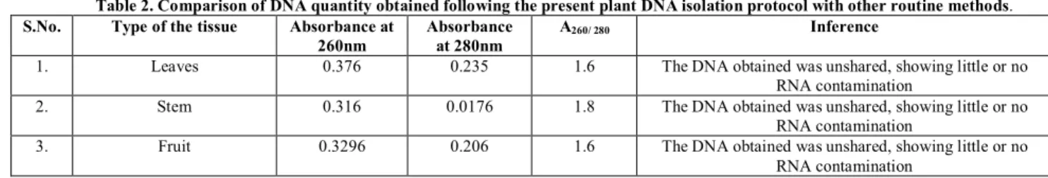
Table 2. Comparison of DNA quantity obtained following the present plant DNA isolation protocol with other routine methods.
S.No. Type of the tissue Absorbance at 260nm
Absorbance at 280nm
A260/ 280 Inference
1. Leaves 0.376 0.235 1.6 The DNA obtained was unshared, showing little or no RNA contamination
2. Stem 0.316 0.0176 1.8 The DNA obtained was unshared, showing little or no RNA contamination
3. Fruit 0.3296 0.206 1.6 The DNA obtained was unshared, showing little or no RNA contamination
Table 3. Comparison of DNA quantity obtained following the present plant DNA isolation protocol with other routine methods(Carbohydrate contamination).
S.No. Type of the tissue Absorbance at 260nm
Absorbance at 230nm
A260/230 Inference
1. Leaves 0.376 0.268 1.4 The DNA obtained was unshared, showing little or no carbohydrate contamination
2. Stem 0.316 0.225 1.4 The DNA obtained was unshared, showing little or no carbohydrate contamination
3. Fruit 0.3296 0.235 1.4 The DNA obtained was unshared, showing little or no carbohydrate contamination
Table 4. Quantitative estimation of DNA S.No. Type of
the tissue
Absorbance at 270nm
Statistical Analysis Concentration (µg/ml)
1. Leaves 0.267 Correlation coefficient R2= 0.998 Straight Line equation
y= 0.061x
4.377
2. Stem 0.195 3.19
3. Fruit 0.206 3.377
Fig 2: Addition of chilled ethanol
Fig 3: Standard curve of DNA
Fig :4DNA isolated resolved on agarose gel.
REFERENCES
1. Wealth of India. A Dictionary of India raw material and Industrial products. Vol V. New Delhi: Publication and information directorate CSIR; 1959.
2. Watt G. Periodic Expert: Dictionary of the Economic product of India, vol.1.New Delhi: Cosmo Publication;1972.
3. Asholkar LV, Kakkar KK, Chakre OJ. Glossary of Indian Medicinal plants with active Principle. New Delhi: Publication and information Directorate; 1992. 4. Anon. The useful plants of India. , New Delhi: Publications & Information
Directorate, CSIR, India; 1986.
5. Bhakuni DS, Dhar ML, Dhar MM, Dhavan BN, Mehrotra BB. Screening of Indian Medicinal Plants for biological activity. Indian J Exp Biol 1969; 7: 250-262.
6. Weishing K, Nybom H, Wolff K, Meyer W. DNA isolation and purification. In: DNA fingerprinting in plants and fungi. 2 ed. Boca Raton Florida :CRC Press ;1995.
7. Alexander RR and Griffiths JM. Basic Biochemical Methods.1ed.New York:Wiley-Liss; 1993.
8. Ausubel FM, Brent R, Kingston RE, Moore DD, Seidman JG, Smith JA ,Struhl K. Current Protocols in Molecular Biology. New York City: John Wiley & Sons Inc; 1994.
9. Harborne JB. Phytochemical Methods. New York: Chapman & Hall;1991. 10. Gendimenico GJ, Bouquin PL, Tramposch KM. Diphenylamine-colorimetric
method for DNA assay: A shortened procedure by incubating samples at 50°C. Analytical Biochemistry 1988;173:45-48.
11. Moyo M, Amoo SO, Bairu MW, Finnie JF, Van Staden J. Optimising DNA isolation for medicinal plants. South African Journal of Botany 2008; 74: 771-775.
12. Doyle JJ, Doyle JL. Isolation of plant DNA from fresh tissue. Focus 1990;12:13-15.
13. Dellaporta SL, Wood J, Hicks JB. A plant DNA minipreparation: Version II. Plant Mol Biol Reptr 1983;1:19-21.
14. Sambrook J, Russel DW . Molecular cloning: a Laboratory Manual. Third Ed. New York: Cold Spring Harbor Laboratory Press New; 2001.
15. Khanuja suman PS, Shasany AK, Darokar MP, Kumar Sushil. Rapid Isolation of DNA from Dry and Fresh Samples of Plants Producing Large Amounts of Secondary Metabolites and Essential Oils. Plant Molecular Biology Reporter 1999; 17:1–7.
16. Abdel G and Essam AZ.DNA Sequences of RAPD Fragments in the Egyptian cotton Gossypium barbadense. African Journal of Biotechnology 2003; 2 (5): 129-132.