Submitted15 September 2015
Accepted 11 July 2016
Published30 August 2016
Corresponding author
Diego Pol, [email protected], [email protected]
Academic editor
Mark Young
Additional Information and Declarations can be found on page 17
DOI10.7717/peerj.2311
Copyright
2016 Codorniú et al.
Distributed under
Creative Commons CC-BY 4.0
OPEN ACCESS
A Jurassic pterosaur from Patagonia
and the origin of the pterodactyloid
neurocranium
Laura Codorniú1, Ariana Paulina Carabajal2, Diego Pol3, David Unwin4
and Oliver W.M. Rauhut5
1Departamento de Geología, CONICET, Universidad Nacional de San Luis, San Luis, Argentina
2Instituto de Investigaciones en Biodiversidad y Medioambiente (INIBIOMA), CONICET,
Río Negro, Argentina
3CONICET, Museo Paleontológico Egidio Feruglio, Trelew, Chubut, Argentina
4School of Museum Studies, University of Leicester, Leicester, United Kingdom
5Department of Earth and Environmental Sciences and GeoBioCenter, Bayerische Staatssammlung für
Paläontologie und Geologie, Munich, Germany
ABSTRACT
Pterosaurs are an extinct group of highly modified flying reptiles that thrived dur-ing the Mesozoic. This group has unique and remarkable skeletal adaptations to pow-ered flight, including pneumatic bones and an elongate digit IV supporting a wing-membrane. Two major body plans have traditionally been recognized: the primitive, primarily long-tailed paraphyletic ‘‘rhamphorhynchoids’’ (preferably currently rec-ognized as non-pterodactyloids) and the derived short-tailed pterodactyloids. These two groups differ considerably in their general anatomy and also exhibit a remarkably different neuroanatomy and inferred head posture, which has been linked to different lifestyles and behaviours and improved flying capabilities in these reptiles. Pterosaur neuroanatomy, is known from just a few three-dimensionally preserved braincases of non-pterodactyloids (as Rhamphorhynchidae) and pterodactyloids, between which there is a large morphological gap. Here we report on a new Jurassic pterosaur from Argentina,Allkaruen koigen. et sp. nov., remains of which include a superbly pre-served, uncrushed braincase that sheds light on the origins of the highly derived neu-roanatomy of pterodactyloids and their close relatives. AµCT ray-generated virtual endocast shows that the new pterosaur exhibits a mosaic of plesiomorphic and de-rived traits of the inner ear and neuroanatomy that fills an important gap between those of non-monofenestratan breviquartossans (Rhamphorhynchidae) and derived pterodactyloids. These results suggest that, while modularity may play an important role at one anatomical level, at a finer level the evolution of structures within a mod-ule may follow a mosaic pattern.
SubjectsPaleontology
Keywords Pterosauria, Cañadón Asfalto, Patagonia, Chubut, Middle Jurassic
INTRODUCTION
Butler, 2012; Witton, 2013; Benson et al., 2014). They have traditionally been divided into two major groups, ‘‘rhamphorhynchoids’’ (a paraphyletic assemblage of basal pterosaurs) and pterodactyloids (Plieninger, 1901). ‘‘Rhamphorhynchoids’’ (Unwin, 2003) now generally referred to as non-pterodactyloids (Bell & Padian, 1995), are characterized by a long tail (except for some anurognathids) and short neck and metacarpus, whereas pterodactyloids have a much larger body size range, an elongated neck and metacarpus, and a relatively short tail. Furthermore, the endocranium of pterodactyloids (Edinger, 1941; Klinghardt, 1941;Wellnhofer, 1970;Lewy, Milner & Patterson, 1992;Kellner, 1996;Lü et al., 1997; Bennett, 2001; Witmer et al., 2003; Eck, Elgin & Frey, 2011) is strongly modified compared to that of non-pterodactyloids (Newton, 1888; Edinger, 1927; Wellnhofer, 1975;Witmer et al., 2003;Gasparini, Fernández & De la Fuente, 2004). Thus, in contrast to ‘‘rhamphorhynchoids’’ the cerebral hemispheres are enlarged, the pontine flexure is pronounced, and the optic lobes are located beneath the cerebral hemispheres, mimicking the neuroanatomy of birds in several respects. These modifications have important implications for behaviour and sensory functions (Witmer et al., 2003).
Recently discovered taxa such asDarwinopterusfrom the early Late Jurassic of China (Wang et al., 2009;Wang et al., 2010;Lü et al., 2010;Liu et al., 2012;Sullivan et al., 2014) appear to represent a transitionary stage that partially fills the morphological gap between ‘‘rhamphorhynchoids’’ and pterodactyloids (Lü et al., 2010;Andres, Clark & Xu, 2014).
Darwinopterus and its sister taxon, Pterodactyloidea, form a clade, Monofenestrata
diagnosed by the confluence of the narial and antorbital opening in a single fenestra (Lü et al., 2010).Darwinopteruscombines a ‘‘rhamphorhynchoid’’ body with a pterodactyloid neck and skull, hinting at a modular type of evolution (Lü et al., 2010).Darwinopterus
and other basal monofenestratans (Wang et al., 2009;Wang et al., 2010;Lü et al., 2010; Liu et al., 2012;Sullivan et al., 2014) are, however, known only from compressed and semi-compressed remains that yield limited braincase and endocranial information.
Here we describe a new pterosaur that is represented by several skeletal elements including an almost perfect, three-dimensionally preserved braincase that shows a unique combination of characters shared with both non-monofenestratan breviquartossans (Rhamphorhynchidae sensuWitton, 2013) and pterodactyloids. Recovered by phylogenetic analysis as the sister group of Monofenestrata, details of this pterosaur provide insights into the origin of the pterodactyloid neurocranium and improve our understanding of the tempo and mode of pterosaur evolution.
GEOLOGICAL SETTING
bones were found in an extensive bonebed in the upper 5 cm of a limestone bed with a thickness of 15–20 cm; the bonebed can be followed laterally for at least 30 m and might be more extensive. The remains are usually disarticulated, though some association of different elements is present, and one wing digit and two sets of mandibles were found partially articulated (Codorniú, Rauhut & Pol, 2010). The holotype braincase MPEF-PV 3613 was found in close association with the cervical vertebra MPEF-PV 3616 and the mandibles MPEF-PV 3609, and these remains probably represent a single individual. The cervical vertebra MPEF-PV 3615 is identical to cervical MPEF-PV 3616 in all discernable characters and is thus also referred to the same taxon. However, given the possibility that more than a single taxon is present in the locality, the referral of all of this material should be regarded as tentative. To establish the phylogenetic relationships of the new taxon we analyzed a matrix in which we restricted information to the braincase alone, and then conducted further analyses that included information from both the holotype and the referred specimens (see below). That both analyses, with only the braincase and with the addition of all known material, result in trees with the same topology further supports the association of these materials.
The age of the Cañadón Asfalto Formation was, until recently, usually given as Callovian-Oxfordian (latest Middle to earliest Late Jurassic), but new radiometric and biostratigraphic evidence indicates that the formation is considerably older, with ages ranging from the Toarcian (latest Early Jurassic; (Cúneo et al., 2013) to the earliest Bathonian (Volkheimer et al., 2008;Cabaleri et al., 2010;Cúneo et al., 2013). For a more thorough discussion of the age of the Cañadón Asfalto Formation, seeCúneo et al. (2013).
METHODS
CT-scan data
CT scans of the braincase (MPEF-PV 3613) were made by scanner v|tome|x s (GE Sensing & Inspection Technologies GmbH phoenix|X-ray). The images (720 coronal stacks) have a 1024 pixel resolution and a voxel size of 0.047310×0.047310×0.047310 mm. Virtual three-dimensional reconstructions of the braincase, cranial endocast and inner ear were generated using the software Materialise Mimics (10.0) at the University of Alberta Paleovertebrate Laboratory; and the resulting 3D models were then imported into the software Geomagic (10.0). Illustrations were made using Adobe Photoshop (C). The terminology used for braincase pneumatic cavities follows Witmer (1997)andDufeau (2011).
Nomenclatural acts
appending the LSID to the prefix ‘‘http://zoobank.org/’’. The LSID for this publication is: urn:lsid:zoobank.org:pub:48910653-0343-4A8D-911F-3498A755F305. The online version of this work is archived and available from the following digital repositories: PeerJ, PubMed Central and CLOCKSS.
Phylogenetic analysis
Taxon and character sampling
The data matrix used in the phylogenetic analysis is based on a revision of a previously published phylogenetic analysis (Lü et al., 2010), with the addition of six new characters derived from the skull region (most of which are focused on braincase anatomy) and one character proposed byAndres & Ji (2008). Seven characters of the original dataset have been modified (see SI., Character List) and three characters (25, 78, and 112) have been excluded from this dataset given that they had vague character state definitions that did not fit the analysed morphological diversity. The taxon sampling was also expanded from the original dataset by including a recently described basal monofenestratan pterosaur from China:Wukongopterus(Wang et al., 2009). Furthermore, the hypothetical ancestor used byLü et al. (2010)was replaced by codings for two outgroup representatives, the basal archosauriformEuparkeriaand the basal dinosaurHerrerasaurus. The resultant data matrix includes 59 taxa scored across 123 characters. One of the multistate characters was treated as ordered whereas the rest of the characters were treated as unordered (see SI., Character List).
Two different phylogenetic analyses were conducted. First, the character codings for
Allkaruen koiwere restricted to the anatomical information provided by the braincase
(MPEF-PV 3613) to test the phylogenetic information provided by this material. Second, a more inclusive phylogenetic analysis was conducted scoring all the known materials (MPEF-PV 3609, mandible; MPEF-PV 3615, 1616; cervical vertebrae) to determine the phylogenetic position of the new taxon.
Phylogenetic analyses and results
The analyses were conducted under equally weighted parsimony in TNT (Goloboff, Farris & Nixon, 2008) through a heuristic search of 1,000 replicates of Wagner trees followed by TBR branch swapping.
when only the skull scores forAllkaruenwere included, were also calculated and are shown below).
RESULTS
Systematic paleontology
PterosauriaKaup, 1834 BreviquartossaUnwin, 2003
Allkaruen koigen. et sp. nov.
Zoobank.urn:lsid:zoobank.org:act:C545BD35-B448-4D47-A2A6-14215E9E3155
Etymology. Genus name from the native Tehuelche word ‘all’ meaning brain, and ‘karuen,’
meaning ancient. Species name from Tehuelche ‘koi’ meaning lake, referring to the lacustrine setting of the type locality.
Holotype. MPEF-PV (Museo Paleontológico Egidio Feruglio) 3613, braincase, a mandible
(MPEF-PV 3609) and a cervical vertebrae (MPEF-PV 3615) (Figs. 1–7;Figs. S1–S3).
Referred material. Referred material includes a mid cervical vertebrae (MPEF-PV -3616.
Type locality and horizon. Locality La Lluvia, Cañadón Carrizal, Cerro Cóndor, Chubut,
Argentina. Cañadón Asfalto Formation, latest Early-early Middle Jurassic (Cúneo et al., 2013).
Diagnosis. Small pterosaur diagnosed by the following unique combination of skull
characters present in the holotype (autapomorphies marked with asterisk): frontal with large pneumatic foramen on the postorbital process; dorsal occiput faces posterodorsally and occipital condyle faces posteroventrally; long, rod-like basipterygoid processes diverging at approximately 20–25 degrees. The referred mandibular and vertebral materials also show a unique combination of characters that include a long lower jaw with a concave profile in lateral view; four-five large, septated, and well-separated anterior alveoli followed by a posterior alveolar groove*; mid-cervical vertebrae elongate with low neural arch and blade-like neural spine; pneumatic foramina on lateral surface of the centrum and peduncle of the neural arch; reduced diapophyseal process lacking articular surface; absence of accessory zygapophyseal processes.
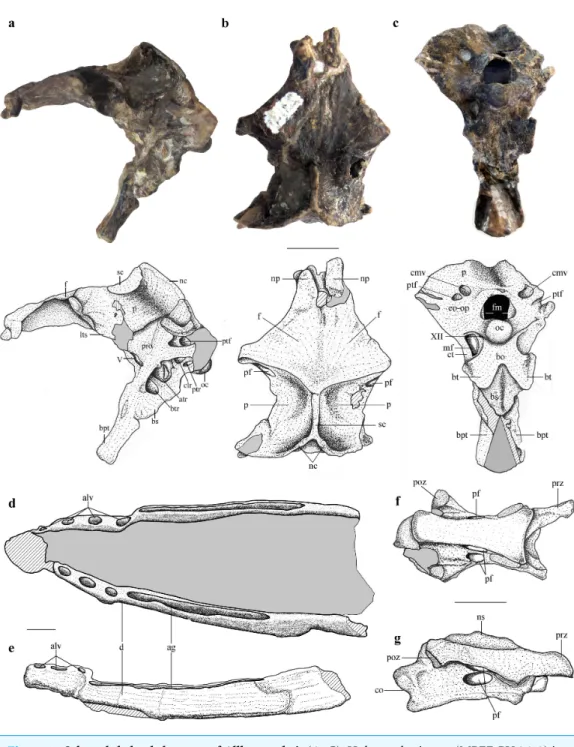
Description
The braincase is undistorted and superbly preserved. All skull bones are fused, as in most osteologically mature pterosaurs. In the skull roof, frontals and parietals are completely fused with each other and the rest of the braincase. The parietals are long (60% of the frontal length, excluding the nasal processes), as seen in Scaphognathus
(Geol. Paläont. Inst. Univ. Bonn, Nr. 1304) andCampylognathoides(Padian, 2008: Plate 5,Fig. 1), but unlike the relatively shorter parietal observed in other pterosaurs, such
as Rhamphorhynchus (Wellnhofer, 1975), Cacibupteryx (Gasparini, Fernández & De la
Fuente, 2004),Parapsicephalus(Newton, 1888),Pteranodon(Bennett, 2001) andAnhanguera
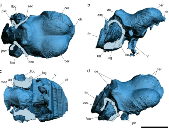
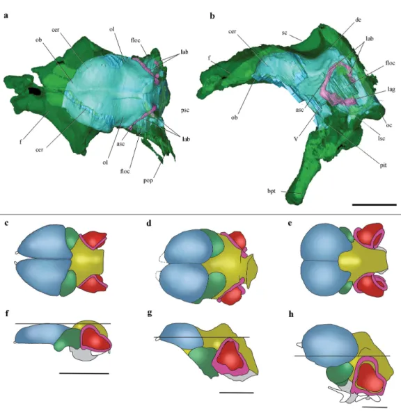
Figure 2 Surface-rendered CT-based reconstructions of the cranial endocast and endosseous labyrinth of the holotype ofAllkaruen koi,in dorsal (A), right lateral (B), ventral (C) and dorsolateral (D) views.Abbreviations: asc, anterior semicircular canal; cer, cerebral hemisphere; de, dorsal expansion ; floc, flocculus; fm, foramen magnum; lag, lagena; med, medulla oblongata; lsc, lateral semicircular canal; ob, olfactory bulb; pit, pituitary body; psc, posterior semicircular canal; V, XII, cranial nerves. Scale bar is 1 cm.
the occipital margin of the skull roof, leading to a V-shaped nuchal crest. The dorsal surface of the frontals is broad and flat. It is 22.5 mm in total length (13 mm long excluding the nasal process). The nasal processes are slightly separated by a narrow gap which originally accommodated the caudal extension of the premaxillae. That the latter are absent suggests that the premaxillae and frontals had not yet fused. The postorbital process of the frontal projects strongly laterally. The dorsal surface of the frontals curves abruptly ventrally towards the posterior end of the postorbital processes to form the anterior margin of the supratemporal fossa. Each frontal has a large oval foramen on the posterior surface of the postorbital process (Figs. 1Band4), within the margin of the supratemporal fossa, which communicates internally with a large and complex pneumatic cavity that pneumatizes almost the entire frontal, as shown by the CT scans.
The occiput is trapezoidal in shape, slightly concave transversely, and faces posteriorly, resembling the morphology ofCacibupteryx (Gasparini, Fernández & De la Fuente, 2004)
andRhamphorhynchus(Wellnhofer, 1978) (Fig. 1C). The occipital region in pterodactyloids
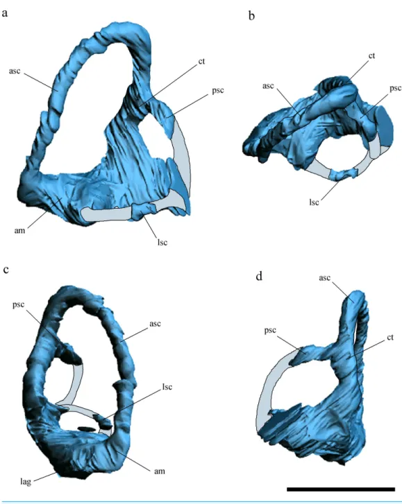
Figure 3 Inner ear anatomy.Digital reconstruction of the left inner ear ofAllkaruen koi,based on the CT scan of the holotype in lateral (A), dorsal (B), anterior (C) and posteromedial (D) views. Reconstructed sections are in light-blue. Abbreviations: am, anterior ampula; asc, anterior semicircular canal; ct, com-mon trunk; lag, lagena; lsc, lateral semicircular canal; psc, posterior semicircular canal. Scale bar is 0.5 cm.
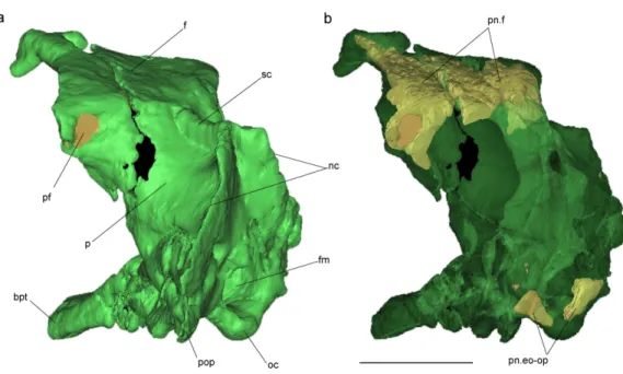
Figure 4 Volume-rendered CT-based reconstruction of the braincase of the holotype ofAllkaruen koi,
in left laterodorsal view. The bone is rendered solid (A), and semi-transparent (B). Pneumatic recesses are shown in yellow in (B). Abbreviations: bpt, basipterygoid process; f, frontal; fm, foramen magnum; nc, nuchal crest; oc, occipital condyle; p, parietal; pf, pneumatic foramen; pop, paroccipital process; pn.f, frontal pneumaticity; pn.eo-op, exoccipital-opisthotic pneumaticity; sc, sagittal crest. Scale bar is 1 cm.
InAllkaruenthe postemporal fenestra is slightly smaller to subequal in size to the caudal
middle cerebral vein foramen, as inRhamphorhynchus(Wellnhofer, 1975). This contrasts with the condition of pterodactyloid pterosaurs, such asAnhanguera piscator (Kellner & Tomida, 2000),Anhanguera santanae (Wellnhofer, 1985),Tapejara wellnhoferi(Wellnhofer & Kellner, 1991;Kellner, 1996), andPteranodon(Bennett, 2001), in which the postemporal fenestra is almost twice the size of the exit of the dorsal head vein.
The occipital condyle is posteroventrally directed with respect to the longitudinal axis of the skull, and thus also angled in respect to the plane of the dorsal part of the occiput. This is comparable to the condition in monofenestratans including basal forms such as
Darwinopterus(D Unwin, 2016, unpublished data), but differs from the construction in
basal pterosaurs, for exampleRhamphorhynchus(Witmer et al., 2003) where the occipital condyle is parallel to the long axis of the skull. The occipital condyle is much smaller than the foramen magnum, as in Rhamphorhynchus,Cacibupteryx, Rhamphinion, and
Tapejara, but unlikePteranodonandAnhanguerain which the occipital condyle is larger.
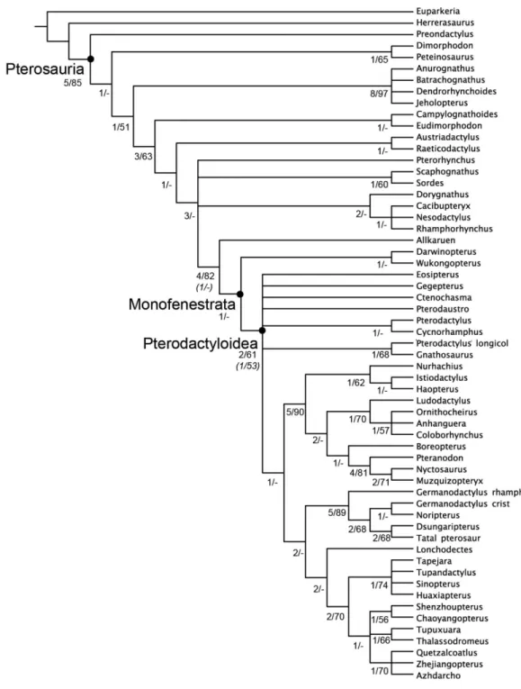
Figure 5 Strict consensus of the 360 most parsimonious trees with major nodes of Pterosauria la-belled.Numbers at the nodes represent the Bremer support value and Bootstrap frequencies for each of the nodes present in the strict consensus tree.
Figure 6 Cranial endocast and comparison of brain anatomy in pterosaurs.(A, B), Volume-rendered
CT-based reconstruction of the braincase of the holotype ofAllkaruen koi, in dorsal (A) and left lateral
(B) views (the bone is rendered semitransparent to show the cranial endocast and the inner ear). (C–H),
schematic drawings of brain anatomy inRhamphorhynchus(C, F),Allkaruen(D, G), andAnhanguera
(E, H) in dorsal (C–E) and lateral (F–H) views. Colors (C–H) indicate equivalent brain regions (blue, cerebrum; green, optic lobe; yellow, cerebellum; red, floccular process of cerebellum; pink, semicircu-lar canals). The horizontal black line shows the relationship between the dorsal expansion of the ante-rior semicircular canal and the forebrain. Abbreviations: asc, anteante-rior semicircular canal; cer, cerebral hemisphere; de, dorsal expansion; f, frontal; floc, floccular process of cerebellum; lab, labyrinth of inner ear; lsc, lateral semicircular canal; lag, lagena; ob, olfactory bulb; oc, occipital condyle; ol, optic lobe; pbt, basipterygoid process; pit, pituitary body; psc, posterior semicircular canal; sc, sagittal crest. Roman
nu-merals indicate cranial nerves. Brain anatomy ofRhamphorhynchusandAnhangueramodified from
Wit-mer et al. (2003). Scale bars are 10 mm.
Vecchia, 2009). Although inAllkaruenthe foramen for cranial nerve XII is an independent opening from the metotic foramen, both foramina open within a shallow common oval recess, the paracondylar recess.
Early Cretaceous Early Jurassic Middle Jurassic Late Jurassic Albian Aptian Barremian Hauterivian Valanginian Berriasian Tithonian Kimmeridgian Oxfordian Callovian Bathonian Bajocian Aalenian Toarcian Pliensbachian Sinemurian Hettangian Rhaetian Norian Late Triassic Herrerasaurus Preondactylus Peteinosaurus Dimorphodon Eudimorphodon Campylognathoides Raeticodactylus Austriadactylus Pterosauria Pterorhynchus Sordes Scaphognathus Allkaruen Monofenestrata Pterodactyloidea Wukongopterus Darwinopterus Nesodactylus Rhamphorhynchus Cacibupteryx Dorygnathus Anurognathidae Dsungaripteroidea Ornithocheiroidea Azhdarchoidea Eosipterus Gegepterus Ctenochasma Pterodaustro Pterodactylus
Cycnorhamphus Pterodactylus longic. Gnathosaurus
Breviquartossa
Figure 7 Phylogenetic position ofAllkaruen koicalibrated against geological time.The topology is based on the strict consensus and summa-rizes the relationships among basal pterosaurs (‘‘rhamphorhynchoids’’) and major groups of Pterodactyloidea (collapsed into clades). Grey bar
rep-resents ghost lineage leading to Monofenestrata and schematic endocast drawings represent the condition of ‘‘rhamphorhynchoids,’’Allkaruen, and
pterodactyloids. Further phylogenetic information is given in theSupplemental Materials.
separated dorsally from the anterior opening of the postemporal fenestra by the paroccipital process. The columellar recess is subdivided into a larger, anteromedially directed foramen and a smaller posterodorsally directed foramen. Whereas the former represents the foramen ovalis, the latter corresponds to a pneumatic foramen that communicates internally with a pneumatic cavity within the base of the paroccipital process (Fig. 1A,Fig. S1). In terms of its location this foramen appears to correspond to the pneumatic foramen of the posterior tympanic recess found in derived theropod dinosaurs (Witmer, 1997) and the allosauroid theropodSinraptor dongi(Paulina-Carabajal & Currie, 2012). Although it is not possible to determine if the foramen for the cranial nerve V is completely enclosed by the prootic, this element forms at least the posterior margin of the opening.
probably reaches the anteriormost part of the basisphenoid, forming the anterior margin of the foramen for the cranial nerve V.
The trigeminal foramen is large and ovoid, with a maximum diameter of 4 mm, and faces anterolaterally. There is no evidence of a separated foramen for the ophthalmic branch of the trigeminal nerve in the specimen, unlike the situation observed inAnhanguera(Witmer et al., 2003).
The orbitosphenoid and ethmoidal elements are missing; thus, the location and shape of cranial nerves I–IV remains unknown inAllkaruen. The symmetry of the anterior walls of the braincase enclosing the anterior section of the brain—frontal, laterosphenoids, and basisphenoid—suggests that the orbitosphenoid and ethmoid remained cartilaginous in this individual.
The elements of the basicranium, the basioccipital and basisphenoid, are firmly fused. The basioccipital forms the ventral portion of the occipital condyle and apparently the basal tubera. The occipital condyle is set off from the basioccipital body by a slightly constricted neck (Fig. S1). The lower part of the basioccipital, between the occipital condyle and the basal tubera, is strongly anteroventrally inclined, so that its ‘posterior’ surface faces rather ventrally and is set at an angle of approximately 100◦with respect to the plane of exposure of the dorsal region of the occiput. Anteroventrally, the basioccipital strongly expands transversely towards the basal tubera, from a minimal width of 6.5 mm between the metotic foramina to approximately 12.5 mm across the basal tubera. The latter are anteroposteriorly elongated, but transversely narrow structures at the lateral extremes of the basioccipital expansion.
The ventral surface of the basisphenoid body is rhomboid in outline, with a tapering anterior end. Anteriorly, long and narrow basipterygoid processes extend for 11 mm anteroventrally from the basisphenoid body, from which they are offset dorsally by a distinct step. The processes are long and slender and diverge anteroventrally at an angle of approximately 35 degrees or slightly less (Fig. 1C). This contrasts with the morphology of most non-pterodactyloid pterosaurs, in which the processes are highly divergent (approximately 60◦–70◦), as inDorygnathus(Padian, 2008), Carniadactylus (Dalla Vecchia, 2009),Scaphognathus (Wellnhofer, 1978), andCacibupteryx (Gasparini, Fernández & De la Fuente, 2004), but is similar to the angle observed inRhamphorhynchus
(Wellnhofer, 1975). However, inAllkaruenthe basipterygoid processes are separated over their entire length, and not connected by a bony web or plate, as seems to be the case in most, possibly all, pterodactyloids including the ornithocheiroidsPteranodon(Bennett, 2001),Anhanguera(Kellner & Tomida, 2000) andHongshanopterus(Wang et al., 2008), the ctenochasmatinesGnathosaurus(Wellnhofer, 1970) andPterodaustro(Codorniú, Paulina-Carabajal & Gianechini, 2015), the dsungaripteroid,Dsungaripterus (D Unwin, 2016, unpublished data), and the azhdarchoidsCaupedactylus(Kellner, 2013) andTupuxuara(D Unwin, 2016, unpublished data).
tympanic recess by a thin bony septum. This recess seems to be associated with the entrance of the carotid artery to the pituitary fossa and might be regarded as a basipterygoid recess. The pituitary fossa is developed as a large, anterodorsally opening depression on the dorsal side of the base of the basipterygoid processes.
The general morphology of the virtual cranial endocast is comparable to that described for the few other pterosaurs where it is preserved and can be studied. The endocast is bulbous, with short olfactory tract and bulbs, cerebral hemispheres with large and ventrally displaced optic lobes, and an extremely enlarged flocculus (Fig. 2). The left inner ear exhibits an anterior semicircular canal (ASC) that is considerably larger than the other two canals (Fig. 3). The dorsal region of the ASC is located ventral to the dorsal surface of the forebrain and approximately level with the olfactory tract.
Pneumatic cavities are present in the frontals, the ventral section of the exoccipital-opisthotic complex (Fig. 4), and in the form of the anterior and posterior tympanic recesses and basipterygoid recess described above. The cavities within the frontals are large and contrast with the camellate pneumatisation of some derived pterodactyloids (Kellner, 1996). The cavities that invade the basicranium are also large, equivalent to those observed in pterodactyloids such asPterodaustro(Codorniú, Paulina-Carabajal & Gianechini, 2015). As noted above, the placement of the external foramina for these last recesses within the columellar recess indicate a tympanic origin for this pneumaticity. In turn, the pneumatic cavities that invade the exoccipital-opisthotic are smaller and affect internally the base of the paroccipital process and the neck of the occipital condyle on both sides of the braincase (Fig. 4).
The mandible is long (approximately 3.5 times the length of the preserved region of the skull), laterally compressed, and has concave alveolar and ventral margins in lateral view, so that the dentary is curved anterodorsally. Each dentary bears several separate anterior alveoli that occupy less than half of the preserved tooth row and a long and narrow alveolar groove posteriorly (Figs. 1D,1EandFig. S2), a morphology that is unique toAllkaruen. Although the symphysial region of the jaws has been lost due to recent erosion, the position of the two rami in the matrix indicate that the dentary symphysis was rather short.
The cervical centrum is approximately three times as long as wide, broader anteriorly than posteriorly, and lacks pre- and postexapophyses and hypapophysis. The prezygapophyses and postzygapophyses are connected by a thin lamina that is ventrally deflected along its anterior third, forming a short and triangular diapophyseal process that lacks an articular surface. Two pairs of pneumatic foramina pierce the vertebrae, one on the lateral surface of the centrum and the other on the ventral surface of the neural arch (Figs. 1F,1GandS3). Both foramina are found at approximately the mid-length of the vertebra.
DISCUSSION
Phylogeny
al., 2009) from the Middle-Late Jurassic of China) and more derived pterosaurs (Fig. 5). This location is supported by a mosaic of plesiomorphic and apomorphic character states to be found inAllkaruen(see SI).
There are two characters: the orientation of the occiput (#33); and the degree of separation of the basipterygoid processes (#37) whereAllkaruenexhibits the plesiomorphic condition, while the derived condition is present in all mononfenstratans for which these characters can be scored.Allkaruenalso exhibits the plesiomorphic condition for the length of the mandibular symphysis (#47) and development of postexapophyses (#71) although the plesiomorphic state is also found in at least one, or more, monofenstratans. There are four characters: angle of divergence of the basipterygoid processes (#34); elongation of the cervical centra (#73); height of the neural arch (#75); and height of the neural spines (#76) where all basal pterosaurs exhibit the plesiomorphic state, whileAllkaruenshares the derived condition with monofenestratans, although for each of these characters the plesiomorphic condition is present in at least one monofenestratan (see SI). There are a further six characters (#12, #32, #36, #53, #61, #62) for whichAllkaruenexhibits a derived state, that is found in monofenestratans and at least one, or more (relatively derived), basal pterosaurs.
Allkaruen and the evolution of the pterosaur neurocranium
The intermediate phylogenetic position ofAllkaruenand the exceptional three-dimensional preservation of the braincase provide new insights into the transformation of the neurocranium, typical of basal pterosaurs, into the highly derived condition present in pterodactyloids (Witmer et al., 2003) (Figs. 6C–6H). Descriptions of the neurocranial anatomy of pterosaurs have so far been limited to two basal forms (Newton, 1888;Edinger, 1927; Wellnhofer, 1975;Witmer et al., 2003) and a few pterodactyloids (Edinger, 1927; Edinger, 1941;Kellner, 1996;Lü et al., 1997;Bennett, 2001;Witmer et al., 2003;Eck, Elgin & Frey, 2011). Detailed accounts of endocranial morphology (brain and inner ear) have been published for only two pterosaurs, the non-monofenestratan breviquartossan
Rhamphorhynchusand the derived pterodactyloidAnhanguera(Witmer et al., 2003).
Comparisons with the virtual endocasts ofRhamphorhynchusandAnhanguera(Fig. 6), show thatAllkaruenshares plesiomorphies and apomorphies with one, or the other, of these two taxa and also exhibits features with a morphology that can be interpreted as intermediate between that ofRhamphorhynchusandAnhanguera. A seemingly plesiomorphic feature of the brain and inner ear ofAllkaruenis the subhorizontal orientation of the frontal when the long axis of the LSC is oriented horizontally (Fig. 6B). This is comparable to the condition
inRhamphorhynchus(Witmer et al., 2003), but contrasts with the ventrally deflected frontal
of Anhanguera(Witmer et al., 2003). Note that, here, we use the orientation of the LSC
purely as a descriptive term, and do not infer any particular orientation of the head in the taxa under consideration (Marugán-Lobón, Chiappe & Farke, 2013).
Four features of the braincase and endocranium ofAllkaruenshow an intermediate condition between non-monofenestratan breviquartossans and pterodactyloids.
Parapsicephalus(Newton, 1888). By contrast, it is located at the midheight of the forebrain
inAllkaruen(Fig. 6G), and ventral to the olfactory tracts in several pterodactyloids including
Pteranodon,Tapejara, andAnhanguera(Witmer et al., 2003) (Fig. 6H).
(II) The orientation of the occiput and occipital condyle is intermediate between that of non-monofenestratan breviquartossans, in which the occiput is mainly vertical, with a posteriorly oriented occipital condyle, and pterodactyloids, in which the occiput faces posteroventrally, or even ventrally. In Allkaruenthe dorsal section of the occiput faces posterodorsally, whereas the occipital condyle is inclined posteroventrally an intermediate condition also found inDarwinopterus(D Unwin, 2016, unpublished data).
(III) In dorsal view, the lateral margin of the flocculus inRhamphorhynchusdoes not extend as far laterally as the lateral margin of the cerebral hemisphere, whereas these two are about level inAllkaruen. By contrast, inAnhanguerathe flocculus extends well beyond the lateral margin of the cerebral hemisphere.
(IV) The ratio between the complete length of the brain and the height of the hindbrain, when seen in lateral view, is 0.44 inRhamphorhynchusand 0.69 in Anhanguera, whereas this ratio is 0.6 in Allkaruen.
While the endocast morphology ofAllkaruenseems, in many respects, to be intermediate between that of basal pterosaurs and pterodactyloids, it also demonstrates an important derived feature that is shared with monofenestratans. In basal taxa such asParapsicephalus
(Newton, 1888) andRhamphorhynchus(Witmer et al., 2003) the optic lobes lie at the same level as the forebrain (Fig. 6F).Allkaruen,Darwinopterus(D Unwin, 2016, unpublished data) and pterodactyloids (Edinger, 1941;Bennett, 2001; Witmer et al., 2003; Eck, Elgin & Frey, 2011) show a derived state, pronounced flexure of the brain that displaces the optic lobes ventrally (Figs. 6Gand6H), a condition convergently present in birds (Walsh & Milner, 2011). This arrangement represents a major reorganization of neurocranial architecture and while its significance has yet to be established, it seems to be an important innovation in pterosaur evolution.
Finally, Allkaruenexhibits a neuroanatomical feature that is more derived than in other pterosaurs. The ASC is slightly larger than the other two semicircular canals in
Rhamphorhynchus(25%) andAnhanguera(30%), but much larger (40–50%) inAllkaruen.
Pterosaur evolution
The discovery of strongly correlated character state distributions inDarwinopterusledLü et al. (2010)to suggest that major anatomical regions might have behaved as integrated modules that changed at different times and rates during pterosaur evolution. However,
Allkaruen demonstrates that, whereas modular evolution might have operated at an
inclusive morphological level (e.g., skull+neck versus the remainder of the postcranium), evolution within at least one of these modules (the neurocranium and braincase) seems to have followed a mosaic pattern.
The late Early-early Middle Jurassic age ofAllkaruen(Cúneo et al., 2013) also provides new information on the timing of transformations during the evolution of the derived pterodactyloid skull from that of basal pterosaurs. The derived features of the cranium of
the time of the Early/Middle Jurassic boundary (Fig. 7), before the origin of pterodactyloids and the appearance of their modified postcranial skeleton. Prior to this discovery, a large suite of cranial features was presumed to have appeared somewhat later, during the late Middle to Late Jurassic, the age of the basal monofenestratans,Darwinopterus
(Lü et al., 2010) and Wukongopterus(Wang et al., 2009) and the oldest pterodactyloids (Andres, Clark & Xu, 2014).
Unfortunately, the Early-Middle Jurassic is a period with a very poor pterosaur fossil record, in contrast to the relatively diverse assemblage of pterosaurs known from both the Late Triassic and the Late Jurassic–Cretaceous (Barrett et al., 2008;Butler, Benson & Barrett, 2013;Benson et al., 2014). The early evolutionary origin and diversification inferred for derived pterosaurs (Fig. 4), adds further evidence in support of the hypothesis that the origin and diversification of major vertebrate lineages (e.g., dinosaurs (Allain & Läng, 2009;Pol & Rauhut, 2012), crocodyliforms (Pol & Gasparini, 2009), turtles (Sterli, Pol & Laurin, 2013), mammals (Luo et al., 2011)) occurred prior to the Early/Middle Jurassic boundary (Allain & Läng, 2009;Cúneo et al., 2013). This pattern was previously obscured by the worldwide poor fossil record of terrestrial vertebrates during this evolutionarily critical period of time.
ACKNOWLEDGEMENTS
We thank José L. Carballido, Magalí Cárdenas, Laura Reiner, and Leandro Canessa for specimen preparation. Thanks to the 2000 fieldwork team, who collected the blocks containing the pterosaur remains. I Ruf is thanked for help with the CT and PJ Currie (University of Alberta) gave access to the software Mimics (version 14.0) used to analyse the CT data (APC).
ADDITIONAL INFORMATION AND DECLARATIONS
Funding
Research support was provided by DFG RA1012/9-1 (OWMR), Foncyt PICT 0808, 1288 (DP), PICT 1815 (LC), Project CyT N◦31416, UNSL (LC). The funders had no role in study design, data collection and analysis, decision to publish, or preparation of the manuscript.
Grant Disclosures
The following grant information was disclosed by the authors: DFG: RA1012/9-1.
Foncyt PICT: 0808, 1288. PICT: 1815.
Project CyT: 31416.
Competing Interests
Author Contributions
• Laura Codorniú performed the experiments, analyzed the data, wrote the paper, prepared figures and/or tables, reviewed drafts of the paper.
• Ariana Paulina Carabajal analyzed the data, contributed reagents/materials/analysis tools, wrote the paper, prepared figures and/or tables, reviewed drafts of the paper. • Diego Pol and Oliver W.M. Rauhut conceived and designed the experiments, performed
the experiments, analyzed the data, contributed reagents/materials/analysis tools, wrote the paper, prepared figures and/or tables, reviewed drafts of the paper.
• David Unwin analyzed the data, wrote the paper, reviewed drafts of the paper.
Data Availability
The following information was supplied regarding data availability: The research in this article did not generate any raw data.
New Species Registration
The following information was supplied regarding the registration of a newly described species:
Publication LSID: urn:lsid:zoobank.org:pub:48910653-0343-4A8D-911F-3498A755F305. New taxon LSID:
urn:lsid:zoobank.org:act:C545BD35-B448-4D47-A2A6-14215E9E3155.
Supplemental Information
Supplemental information for this article can be found online athttp://dx.doi.org/10.7717/ peerj.2311#supplemental-information.
REFERENCES
Allain R, Läng É. 2009.Origine et évolution des saurischiens.Comptes Rendus Palevol
8:243–256DOI 10.1016/j.crpv.2008.09.013.
Andres B, Clark JM, Xu X. 2014.The earliest pterodactyloid and the origin of the group.
Current Biology24:1011–1016DOI 10.1016/j.cub.2014.03.030.
Andres B, Ji Q. 2008.A new pterosaur from the Liaoning Province of China, the
phylogeny of the Pterodactyloidea, and convergence in their cervical vertebrae.
Palaeontology 51:453–469DOI 10.1111/j.1475-4983.2008.00761.x.
Barrett PM, Butler RJ, Edwards NP, Milner AR. 2008.Pterosaur distribution in time and
space: an atlas.Zitteliana28:61–108.
Bell CM, Padian K. 1995.Pterosaur fossils from the Cretaceous of Chile—evidence
for pterosaur colony on an inland desert plain.Geological Magazine 132:31–38 DOI 10.1017/S0016756800011407.
Bennett SC. 2001.The osteology and functional morphology of the Late Cretaceous
pterosaur Pteranodon.Palaeontographica A260:113–153.
Benson RBJ, Frigot RA, Goswami A, Andres B, Butler RJ. 2014.Competition and
constraint drove Cope’s rule in the evolution of giant flying reptiles.Nature
Butler RJ, Benson RBJ, Barrett PM. 2013.Pterosaur diversity: untangling the influence of sampling biases, Lagerstätten, and biodiversity signals.Palaeogeography,
Palaeocli-matology, Palaeoecology372:78–87DOI 10.1016/j.palaeo.2012.08.012.
Cabaleri NG, Armella C, Silva Nieto DG. 2005.Saline paleolake of the Cañadón Asfalto
Formation (Middle-Upper Jurassic), Cerro Cóndor, Chubut province (Patagonia), Argentina.Facies51:350–364DOI 10.1007/s10347-004-0042-5.
Cabaleri NG, Volkheimer W, Silva Nieto D, Armella C, Cagnoni M, Hauser N, Matteini
M, Pimentel MM. 2010. U-Pb ages in zircons from las Chacritas and Puesto Almada
members of the Jurassic Cañadón Asfalto Formation, Chubut province, Argentina.
VII South american symposium on isotope geology, 190–193.
Coddington JA, Scharff N. 1994.Problems with ‘‘soft’’ polytomies.Cladistics
12:139–145.
Codorniú L, Paulina-Carabajal A, Gianechini FA. 2015.Braincase anatomy of
Ptero-daustro guinazui, pterodactyloid pterosaur from the Lower Cretaceous of Argentina.
Journal of Vertebrate Paleontology 36(1):e1031340DOI 10.1080/02724634.
Codorniú L, Rauhut OWM, Pol D. 2010.Osteological features of Middle Jurassic
Pterosaurs from Patagonia (Argentina).Acta Geoscientica Sinica31(Supp. 1):12–13.
Cúneo R, Ramezani J, Scasso R, Pol D, Escapa I, Zavattieri AM, Bowring SA. 2013.
A. High-precision U-Pb geochronology and a new chronostratigraphy for the Cañadón Asfalto Basin, Chubut, central Patagonia: implications for terres-trial faunal and floral evolution in Jurassic.Gondwana Research24:1267–1275 DOI 10.1016/j.gr.2013.01.010.
Dalla Vecchia FM. 2009.Anatomy and systematics of the pterosaur Carniadactylus gen.
n. rosenfeldi (Dalla Vecchia, 1995).Rivista Italiana di Paleontologia e Stratigrafia 115:159–188.
Dufeau DL. 2011.The evolution of cranial pneumaticity in Archosauria: patterns of
paratympanic sinus development. PhD dissertation, Faculty of the College of Arts and Sciences of Ohio University, 175p. Unpublished.
Eck K, Elgin RA, Frey E. 2011.On the osteology ofTapejara wellnhoferiKellner 1989
and the first occurrence of a multiple specimen assemblage from the Santana Formation, Araripe Basin, NE-Brazil.Swiss Journal of Palaeontology130:277 DOI 10.1007/s13358-011-0024-5.
Edinger T. 1927.Das Gehirn der Pterosaurier.Zeitschrift für Anatomie und
Entwicklungs-gesch83:105–112 DOI 10.1007/BF02117933.
Edinger T. 1941.The brain of Pterodactylus.American Journal of Science239:665–682
DOI 10.2475/ajs.239.9.665.
Foth C, Brusatte SL, Butler RJ. 2012.Do different disparity proxies converge on a
common signal? Insights from the cranial morphometrics and evolutionary history of Pterosauria (Diapsida: Archosauria).Journal of Evolutionary Biology25:904–915 DOI 10.1111/j.1420-9101.2012.02479.x.
Gasparini Z, Fernández M, De la Fuente M. 2004.A new pterosaur from the Jurassic of
Goloboff P, Farris JS, Nixon KC. 2008.TNT, a free program for phylogenetic analysis.
Cladistics24:774–786 DOI 10.1111/j.1096-0031.2008.00217.x.
Kaup JJ. 1834.Versuch einer Eintheilung der Saugethiere in 6 Stämme und der
Amphi-bien in 6 Ordnungen.Isis3:311–315.
Kellner AWA. 1996.Description of the braincase of two early cretaceous pterosaurs
(pterodactyloidea) from Brazil.American Museum Novitates3175:1–34.
Kellner AWA. 2013.A new unusual tapejarid (Pterosauria, Pterodactyloidea) from the
Early Cretaceous Romualdo Formation, Araripe Basin, Brazil.Earth and
Environ-mental Science Transactions of the Royal Society of Edinburgh103(3–4):409–421
DOI 10.1017/S1755691013000327.
Kellner AWA, Tomida Y. 2000.Description of a new species of Anhangueridae
(Ptero-dactyloidea) with comments on the pterosaur fauna from the Santana Formation (Aptian-Albian) Northeastern Brazil.National Science Museum Monographs 17:1–135DOI 10.1159/000061634.
Klinghardt F. 1941.Observations on Pterosaurs. Beobachtungen an Flugsauriern.
Paläontologische Zeitschrift 4:250–258.
Lewy Z, Milner AC, Patterson C. 1992.Remarkably preserved natural endocranial
casts of pterosaur and fish from the Late Cretaceous of Israel.GSI Current Research 7:31–35.
Liu Y-Q, Kuang H-W, Jiang X-J, Peng N, Xu H, Sun H-Y. 2012.Timing the earliest
known feathered dinosaurs and transitional pterosaurs older than the Jehol Biota.Palaeogeography Palaeoclimatology Palaeoclimatology323–325:1–12 DOI 10.1016/j.palaeo.2012.01.017.
Lü J, Du XK, Zhu QZ, Cheng XK, Luo DX. 1997.Computed tomography (CT) of
brain-case of Dsungaripterus weii (Pterosauria).Chinese Science Bulletin42:1125–1129.
Lü J, Unwin D, Jin X, Liu Y, Ji Q. 2010.Evidence for modular evolution in a long-tailed
pterosaur with a pterodactyloid skull.Proceeding of the Royal Society B273:383–389.
Luo Z-X, Yuan C-X, Meng Q-J, Ji Q. 2011.A Jurassic eutherian mammal and divergence
of marsupials and placentals.Nature476:442–445DOI 10.1038/nature10291.
Marugán-Lobón J, Chiappe L, Farke AA. 2013.The variability of inner ear orientation in
saurischian dinosaurs: testing the use of semicircular canals as a reference system for comparative anatomy.PeerJ1:e124DOI 10.7717/peerj.124.
Newton ET. 1888.On the skull, brain and auditory organ of a new species of
pterosaurian (Scaphognathus purdoni) from the Upper Lias near Whitby, Yorkshire.
Philosophical Transactions of the Royal Society 179:503–537
DOI 10.1098/rstb.1888.0019.
Padian K. 2008.The Early Jurassic pterosaur Dorygnathus banthensis (Theodori, 1830).
Special Papers in Palaeontology80:1–107.
Paulina-Carabajal A, Currie PJ. 2012.New information on the braincase of Sinraptor
dongi (Theropoda: Allosauroidea): ethmoidal región, endocranial anatomy and pneumaticity.Vertebrata PalAsiatica50:85–101.
Pol D, Gasparini Z. 2009.Skull anatomy of Dakosaurus andiniensis (Thalattosuchia: Crocodylomorpha) and the phylogenetic position of Thalattosuchia.Journal of
Systematic Palaeontology7:163–197DOI 10.1017/S1477201908002605.
Pol D, Rauhut OWM. 2012.A Middle Jurassic abelisaurid from Patagonia and the early
diversification of theropod dinosaurs.Proceedings of the Royal Society of London B 279:3170–3175DOI 10.1098/rspb.2012.0660.
Sterli J, Pol D, Laurin M. 2013.Incorporating phylogenetic uncertainty on
phylogeny-based palaeontological dating and the timing of turtle diversification.Cladistics 29:233–246DOI 10.1111/j.1096-0031.2012.00425.x.
Stipanicic P, Rodrigo F, Baulíes O, Martínez C. 1967.Las formaciones presenonianas
en el denominado Macizo Nord-patagónico y regiones adyacentes.Revista de la
Asociación Geológica Argentina23:67–98.
Sullivan C, Yuan W, Hone DWE, Yuanqing W, Xu X, Zhang F. 2014.The vertebrates of
the Jurassic Daohugou Biota of northeastern China.Journal of Vertebrate Paleontol-ogy34:243–280DOI 10.1080/02724634.2013.787316.
Tasch P, Volkheimer W. 1970.Jurassic conchostracans from Patagonia.University of
Kansas, Paleontological Contributions50:1–23.
Unwin DM. 2003. On the phylogeny and evolutionary history of pterosaurs. In:
Buffetaut E, Mazin J-M, eds.Evolution and Paleobiology of Pterosaurs, vol. 217. London: Geological Society, Special Publications, 139–190.
Volkheimer W, Quattrocchio M, Cabaleri NG, García V. 2008.Palynology and
pale-oenvironment of the Jurassic lacustrine Cañadón Asfalto Formation at Cañadón Lahuincó locality, Chubut Province, Central patagonia, Argentina.Revista Española
de Microplaeontología40:77–96.
Walsh S, Milner A. 2011. Evolution of the avian brain and senses. In: Dyke G, Kaiser G,
eds.Living dinosaurs: the evolutionary history of modern birds. Chichester: John Wiley
and Sons, 282–305.
Wang X, Campos DA, Zhonghe Z, Kellner AWA. 2008.A primitive istiodactylid
pterosaur (Pterodactyloidea) from the Jiufotang Formation (Early Cretaceous), northeast China.Zootaxa1813:1–18.
Wang X, Kellner AWA, Jiang S, Cheng X, Meng X, Rodrigues T. 2010.New long-tailed
pterosaurs (Wukongopteridae) from western Liaoning, China.Anais da Academia
Brasileira de Ciências82:1045–1062DOI 10.1590/S0001-37652010000400024.
Wang X, Kellner AWA, Jiang S, Meng X. 2009.An unusual long-tailed pterosaur with
elongated neck from western Liaoning of China.Anais da Academia Brasileira de
Ciências81:793–812DOI 10.1590/S0001-37652009000400016.
Wellnhofer P. 1970.Die Pterodactyloidea (Pterosauria) der Oberjura Plattenkalke.
Süd-deutschlands.Bayerische Akademie der Wissenschaften AbhandlungenNF141:1–133.
Wellnhofer P. 1975.Die Rhamphorhynchoidea (Pterosauria) der Oberjura-Plattenkalke
Süddeutschlys. I: Allgemeine Skelettmorphologie.Palaeontographica A148:1–33.
Wellnhofer P. 1978. Pterosauria. In: Wellnhofer P, ed.Handbuch der paläoherpetologie.
Wellnhofer P. 1985.Neue Pterosaurier aus der Santana Formation (Apt) der Chapada do Araripe, Brasilien.Palaeontographica A187:105–182.
Wellnhofer P, kellner AWA. 1991.The skull ofTapejara wellnhoferiKellner (Reptilia:
Pterosauria) from the Lower Cretaceous Santana Formation of the Araripe Basin, Northeastern Brazil.Mitteilungen der Bayerischen Staatssammlung für Paläontologie
und historische Geologie31:89–106.
Witmer LM. 1997. Craniofacial air sinus system. In: Currie PJ, Padian K, eds.
Encyclope-dia of dinosaurs. New York: Academic Press, 151–159.
Witmer LM, Chatterjee S, Franzosa J, Rowse T. 2003.Neuroanatomy of flying
rep-tiles and implications for flight, posture and behaviour.Nature425:950–953 DOI 10.1038/nature02048.
Witton MP. 2013. Pterosaurs. In:Natural history, evolution, anatomy. Princeton and