Evaluation of the Facial Recess and Cochlea on
the Temporal Bone of Stillbirths regarding the
Percutaneous Cochlear Implant
Gabriela Pereira Bom Braga
1Eloisa Gebrim
2Ramya Balachandran
3Jack Noble
4Robert Labadie
3Ricardo Ferreira Bento
11Department of Otolaryngology, Faculdade de Medicina,
Universidade de São Paulo, São Paulo, SP, Brazil
2Department of Radiology, Faculdade de Medicina, Universidade de
São Paulo, São Paulo, SP, Brazil
3Department of Otolaryngology Head and Neck Surgery, Vanderbilt
University, Nashville, Tennessee, United States
4Department of Otolaryngology, Vanderbilt University, Nashville,
Tennessee, United States
Int Arch Otorhinolaryngol 2018;22:260–265.
Address for correspondence Gabriela Pereira Bom Braga, MD, PhD, Departmento de Otolaringologia, Faculdade de Medicina,
Universidade de São Paulo, Av. Dr. Eneas de Carvalho Aguiar, 255, 6oandar, sala 6167, São Paulo, SP, 01246-903, Brazil
(e-mail: [email protected]).
Keywords
►
cochlear implantation
►
middle ear
►
hearing loss,
sensorineural
►
temporal bone /
anatomy
►
cochlea
►
experimental studies
Abstract
Introduction
The literature shows that there are anatomical changes on the temporal
bone anatomy during the
fi
rst four years of life in children. Therefore, we decided to
evaluate the temporal bone anatomy regarding the cochlear implant surgery in
stillbirths between 32 and 40 weeks of gestational age using computed tomography
to simulate the trajectory of the drill to the scala timpani avoiding vital structures.
Objectives
To measure the distances of the simulated trajectory to the facial recess,
cochlea, ossicular chain and tympanic membrane, while performing the minimally
invasive cochlear implant technique, using the Improvise imaging software (Vanderbilt
University, Nashville, TN, US).
Methods
An experimental study with 9 stillbirth specimens, with gestational ages
ranging between 32 and 40 weeks, undergoing tomographic evaluation with
indivi-dualization and reconstruction of the labyrinth, facial nerve, ossicular chain, tympanic
membrane and cochlea followed by drill path de
fi
nition to the scala tympani. Improvise
was used for the computed tomography (CT) evaluation and for the reconstruction of
the structures and trajectory of the drill.
Results
Range of the distance of the trajectory to the facial nerve: 0.58 to 1.71 mm. to
the ossicular chain: 0.38 to 1.49 mm; to the tympanic membrane: 0.85 to 1.96 mm;
total range of the distance of the trajectory: 5.92 to 12.65 mm.
Conclusion
The measurements of the relationship between the drill and the
anato-mical structures of the middle ear and the simulation of the trajectory showed that the
middle ear cavity at 32 weeks was big enough for surgical procedures such as cochlear
implants. Although cochlear implantation at birth is not an indication yet, this study
shows that the technique may be an option in the future.
received June 10, 2017 accepted August 4, 2017 published online October 25, 2017
DOI https://doi.org/ 10.1055/s-0037-1606612. ISSN 1809-9777.
Copyright © 2018 by Thieme Revinter Publicações Ltda, Rio de Janeiro, Brazil Original Research
Introduction
In recent decades, technological advances such as cochlear implants enabled patients with hearing loss to regain the ability to hear. According to recent incidence studies, 14.5% of the population has some form of disability, and out of this number, 5.2% declare inability to hear, with 3.9% of moderate hearing loss, 0.9% of severe hearing loss, and 0.4% of profound hearing loss.1 The ear derives from the ectoderm of the cephalic part of the embryo, originating from one round vesicle. The otocyst cavity contains the endolymph and the organ of Corti. The membranous labyrinth reaches its maturity and remains with the dimensions acquired on thefifth month of intrauterine life.2–4The inner ear appears on the ninth week
as the endolymphatic duct system including mesenchymal tissue into which an early vacuolated perilymphatic forms the labyrinth system.2The cartilaginous otic capsule forms the labyrinth. On the twenty-third week, the cochlea reaches its full size, and the periotic spaces are well-formed.2,5 The postnatal growth of the temporal bone is much discussed. For a long time, it was believed that the morphology and spatial orientation of the labyrinth did not change significantly after birth.6However, it appears that the cranium presents a bimodal growth curve between 1 and 4 years of age and again during puberty, which can have a significant impact on the anatomy of the temporal bone.7,8It has been suggested that these changes take place in the tympanic and mastoid pro-cesses and in the squamous portion of the temporal bone.9
Regarding the relevant anatomy to the cochlear implant, there are significant differences between children and adults.10,11 Lloyd, in 2010,7 presented evidence that the basal turn of the cochlea can change its orientation relative to the facial recess during growth, which can influence the angle of insertion of the electrode.10,12
As Moberly et al described in 2015, early stimulation of the cerebral cortex yields better results on hearing rehabi-litation.13Replacing the function of the organ of Corti and electrically stimulating the ganglion cells and nerve endings of the auditory nerve, cochlear implants are used in the hearing rehabilitation of patients with severe to profound hearing loss.
Despite the safety of the procedure having increased due to technological advances, there are still complications, especially in younger patients, in whom the noble anatomi-cal structures (the facial nerve, the chorda tympani nerve and the cochlea) have different angles and are in different positions compared with adults.14–16The current techniques
to access the cochlea use visual feedback (with the aid of a microscope) and facial nerve monitoring to avoid these structures. The facial nerve has the most risk of injury because of the nerve recess approach.5In order to increase the safety of the procedure, reduce the surgical and healing times, and accelerate the activation of the cochlear implants, minimally invasive approaches using robotic techniques have been proposed. This surgical technique has been ap-proved by the FDA to be used on adults and children.17–23
The procedure is based on the fact that it is possible to pass a drill through the temporal bone after a previous
tomo-graphic planning, with the anatomical study of the noble structures of the mastoid through the trajectory using im-age-guided techniques, while critical structures of the tem-poral bone are avoided.17–19
Objectives
The objective of the present study was to evaluate the simulated trajectory of the drill on the computed tomogra-phy (CT) scan used in the percutaneous cochlear implant technique and its viability in neonates through:
1. A measurement of the simulated trajectory of the drill between the cortical temporal bone and the cochleost-omy, calculating the mean, median and standard deviation.
2. An assessment of the mean, median and standard devia-tion of the distances between the simulated trajectory to the ossicular chain, tympanic membrane and facial nerve.
Methods
Study Design
This was an experimental study.
This study was approved by the Ethical Committee of one of our institutions (under CAAE number 51243215.2.0000.065).
Inclusion Criteria
The corpses that did not show anatomical malformations in the middle and inner ear structures or in bone density during the tomographic evaluation were included in the study.
Methodology
A tomographic study was performed using the machine Discovery CT 750 HD (General Electric Healthcare, Little Chalfont, UK) (CT Protocol- volumetric acquisition helical Hi-res rotation 0.8 seconds with collimation of 0.625 mm and 0.312 mm range KV 140 mA: 400 FOV- determined by measuring the laterolateral diameter of the temporal region in digital radiography and reconstruction Ultra High Defi ni-tion). Held in the Radiology Laboratory of one of our institutions.
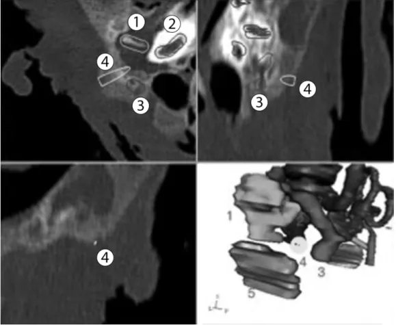
The specimens were placed in the scanner in the supine position with the head in hyperextension with the aid of a cushion, forming a straight angle to the base of the scanner. The images were made in the coronal and axial planes with a thickness of 0.6 mm. The images were transferred to the Improvise (Vanderbilt University, Nashville, TN, US) soft-ware, followed by segmentation of the structures involved in the cochlear implant (facial nerve, ossicular chain, tym-panic membrane, labyrinth and cochlea). After the segmen-tation, the software then simulates the trajectory of the drill between the cortex of the temporal bone and the scala tympani through a cochleostomy (►Fig. 1).
In order to define the path between the cortex of the temporal bone and the cochleostomy, we used features of the software that allow us to make points on different locations of the image, keeping the symmetry between sides. Thus, the
as limits the facial nerve, the ossicular chain and the tym-panic membrane. To simulate the thickness of the drill, a cylinder (yellow) with a radius of 0.5 mm was created. The axial plane was chosen to perform the demarcation of the defining points of the trajectory, because it presents a more accurate identification of the scala tympani than other planes.
We then measured the shortest distances between: the facial nerve and the trajectory (►Fig. 2); the ossicular chain
and the trajectory (►Fig. 3); the tympanic membrane and
the trajectory (►Fig. 4); and the cortex of the temporal bone
and the scala tympani.
Results
Tomographic images of the mastoid of nine specimens of stillbirths that met the inclusion criteria were evaluated. The data were transferred to an Excel (Microsoft Corporation, Redmond, WA, US) spreadsheet, in which we calculated the mean, median and standard deviation and applied the paired samples Studentttest (►Tables 1and2).
The measurements of the trajectory of the drill to the facial nerve ranged from 0.58 mm to 1.71 mm; for the trajectory to the ossicular chain, they ranged from 0.38 mm to 1.49 mm. The tympanic membrane was between 0.85 mm and 1.96 mm away from the simulated drill path; and the trajectory from the cortex of the temporal bone to
the scala tympani ranged from 5.92 mm to 12.65 mm. The paired samples correlations between the right and left sides were evaluated. Thefirst one was the facial nerve; then, the ossicular chain, followed by the tympanic membrane and the trajectory. The related correlation results and signifi -cance are respectively described as: facial nerve - 0.598 mm and 0.089 mm; ossicular chain - 0.252 mm and 0.514 mm; tympanic membrane - 0.396 mm and 0.291 mm; and trajec-tory - 0.958 mm and 0.0 mm. The paired samples ttest
Fig. 1 Four individual plans of reconstructed anatomy in the Improvise software: axial, coronal, sagittal and 3D; 1- ossicular chain; 2- cochlea; 3- facial nerve; 4- trajectory; 5- tympanic membrane.
showed a statistical difference between the relative position of the tympanic membrane and the trajectory from the right to the left sides.
Discussion
By analyzing the images, we noticed that the middle ear cavity at 32 weeks is well-pneumatized, and it could enable surgical procedures such as cochlear implants. In order to confirm that, we propose a simulated study using an image-guided, minimally invasive technique to access the cochlea in pediatric patients undergoing cochlear implant. This ap-proach involves drilling a narrow path from the cortex of the temporal bone to the cochlea without hitting vital anatomical structures such as the facial nerve, ossicular chain and chorda tympani nerve.
The Improvise software was developed at Vanderbilt University, and it is used to analyze, segment, and measure tomographic images of patients undergoing cochlear im-plant surgery. After the segmentation of the facial nerve,
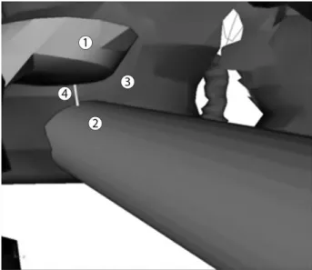
Fig. 3 Shortest distance between the trajectory and the ossicular chain: 1- stapes; 2- trajectory; 3- cochlea; 4- distance between the trajectory and the stapes.
Fig. 4 Shortest distance between the eardrum and the trajectory: 1-tympanic membrane; 2- ossicular chain; 3- facial nerve; 4- trajectory; 5- distance between the trajectory and the tympanic membrane.
Table 1 Distance of the drill’s trajectory (mm) to the middle ear structures
N¼18 Facial nerve Ossicular chain Tympanic membrane Trajectory
Right Left Right Left Right Left Right Left
1 0.99 1.24 0.86 1.29 1.06 0.85 5.92 6.68
2 1.06 0.99 0.38 1.01 1.18 1.35 6.58 6.37
3 1.16 1.35 1.01 0.94 1.55 0.89 9.01 8.07
4 1.55 1.02 0.95 0.78 1.64 1.23 8.28 8.89
5 1.71 1.23 0.85 0.66 1.96 1.32 8.41 8.98
6 0.67 0.58 0.64 0.75 1.42 0.55 8 7.81
7 1.06 1.24 1.49 0.85 1.03 0.87 9.55 10.44
8 0.99 1.11 1.42 1.47 1.79 1.64 11.34 11.34
9 1.64 1.49 0.75 1.12 1.06 1.12 12.65 12.35
Table 2 Paired samples statistics
Mean N Standard deviation
Standard error of the mean
Pair 1 RFN 1.2033 9 0.35107 0.11702
LFN 1.1389 9 0.26127 0.08709
Pair 2 ROC .9278 9 0.35192 0.11731
LOC .9856 9 0.26698 0.08899
Pair 3 RTM 1.4100 9 0.34673 0.11558
LTM 1.1100 9 0.33849 0.11283
Pair 4 RTRAJ 8.8600 9 2.12525 0.70842
LTRAJ 8.9922 9 2.04218 0.68073
cochlea, labyrinth, tympanic membrane (used as reference because the chorda tympani nerve could not be found on the specimens) and ossicular chain, the images were meshed, and a three-dimensional (3D) view of the middle ear was obtained. From that, the trajectory was determined using Improvise tools. Once we determined a trajectory, keeping the symmetry in the position of the points on both sides, the measurements can be taken using the math operation tool and obtaining the surface distance quantity. Analyzing the tables, we observed that the simulated trajectory of the drill kept a distance of more than 1 mm from the structures. Even though this distance may seem too close to the facial nerve, for example, some surgical techniques skeletonize the facial recess or even open it to decompress it. Therefore, a trajec-tory more than 1 mm away from the structure, with suffi -cient irrigation, would be safe to drill. Since there is no contact of the drill with the ossicular chain, the chances of trauma are decreased. The same results were observed when analyzing the distance of the drill’s trajectory to the tympa-nic membrane. By measuring the distance from the cortex of the temporal bone and the scala tympani, a safe, narrow, linear path via the facial recess can be drilled.
An interesting statistical difference between the position of the tympanic membrane on the left and the right sides can be seen in our tables. Valavanis, in 1983,24showed that the cranial growth process during thefirst 4 years of life causes changes in the position of the mastoid portion, the stylo-mastoid portion and the chorda tympani nerve, and the same
findings were confirmed by Evangelos in 2009.12 These changes can be observed on different sides of the same specimens, demonstrating an asymmetrical growth that interferes with the position of the reference structures, such as the tympanic membrane and the chorda tympani nerve. We still need to consider the possibility that this statistical difference between the two sides occurred be-cause of the size of our sample. Although big samples of cadavers of fetuses available for dissection are hard tofind, for our objective, the number was sufficient to demonstrate the viability of the procedure.
The distances measured showed that the trajectory ob-tained using the CT scan did not reach any of the middle ear structures that were used as parameters, thus demonstrating that the middle ear, at this age, has enough size to allow for cochlear implant surgery. This procedure is not yet indicated because there is no sure diagnose of deafness this early in life, also because there is a need to build softer and more delicate surgical materials and guidance hardware to be used in such a fragile anatomy.
More studies are needed to better understand how the temporal bone anatomy changes during growth. Our statis-tical study showed an asymmetry on the position of the tympanic membrane, which also demonstrated that these changes may not be happening at the same rate on both sides of the same specimens.
By observing the measurements made, we concluded that the drill was at a safe distance from the structures, and that by as early as 32 weeks, the middle ear cavity enables safe access to the scala tympani through the facial recess.
The importance of this study resides on the fact that, in the future, in order to perform cochlear implant surgery on neonates or on infants before 5 months of life (once the difficulties of precisely diagnosing severe or profound hearing loss, at this age are overcome), it is of extreme importance to use a technique that is fast and minimally invasive, due to the limitations of the anesthetics and the possible blood loss during surgery.
Conclusion
The measurements of the relationship between the drill and the anatomical structures of the middle ear (the facial nerve, ossicular chain and tympanic membrane), and the simula-tion of the trajectory between the cortical temporal bone and the scala tympani, showed that none of the structures studied were damaged by the drill, which implies that, at 32 weeks, the middle ear cavity already has sufficient size to support a surgical procedure. Although cochlear implanta-tion is not indicated at this age, this surgical technique may be a possibility in the future.
Conflicts of Interest
The authors have no conflicts of interest to declare.
References
1 Baraky LR. Prevalência de surdez incapacitante no município de
Juiz de Fora, Minas Gerais, Brasil [thesis]. São Paulo: Faculdade de Medicina da Universidade de São Paulo; 2011
2 Bento RF, Martins GSQ, Pinna MH. Tratado de Otologia. 2 ed. São
Paulo: Atheneu; 2013
3 Moore BCJ. Cochlear Hearing Loss: Physiological, Psychological
and Technical Issues. 2 ed. West Sussex England: John Wiley & Sons, Ltd; 2007
4 Brackmann S. Arriaga. Otologic Surgery. 3 ed. Philadelphia:
Saunders; 2010
5 Bento RF. Introdução. In: Ricardo Ferreira Bento, Luiz Rodolpho
Penna Lima Junior, Robinson Koji Tsuji, Maria Valéria Schmidt Goffi-Gomez, Danielle do Valle Silva Penna Lima e Rubens de Brito Neto. Tratado de implante coclear e próteses auditivas implantáveis. Rio de Janeiro. Thieme Publicações Ltda.; 2014:
XIX–XXIV
6 Weiglein AH. Postnatal development of the facial canal. An
investigation based on cadaver dissections and computed
tomo-graphy. Surg Radiol Anat 1996;18(02):115–123
7 Lloyd SKWKA, Kasbekar AV, Kenway B, et al. Developmental
changes in cochlear orientation–implications for cochlear
im-plantation. Otol Neurotol 2010;31(06):902–907
8 Litton WBKC, Krause CJ, Anson BA, Cohen WN. The relationship of
the facial canal to the annular sulcus. Laryngoscope 1969;79(09):
1584–1604
9 Migirov L, Kronenberg J. Radiology of the petromastoid canal. Otol
Neurotol 2006;27(03):410–413
10 Swarts JD, Foley S, Alper CM, Doyle WJ. A cross-sectional study of
the change in mastoid geometry with age in children without a
history of otitis media. Laryngoscope 2012;122(03):649–653
11 Cinamon U. The growth rate and size of the mastoid air cell system
and mastoid bone: a review and reference. Eur Arch
Otorhinolar-yngol 2009;266(06):781–786
12 Manolis EN, Filippou DK, Tsoumakas C, et al. Radiologic evaluation
of the ear anatomy in pediatric cholesteatoma. J Craniofac Surg
13 Moberly AC, Lowenstein JH, Nittrouer S. Early Bimodal Stimula-tion Benefits Language AcquisiStimula-tion for Children With Cochlear
Implants. Otol Neurotol 2016;37(01):24–30
14 Achiques MT, Morant A, Muñoz N, et al. Complicaciones y fallos de
la implantación coclear. Acta Otorrinolaringol Esp 2010;61(06):
412–417
15 Bielamowicz SACN, Coker NJ, Jenkins HA, Igarashi M. Surgical
dimensions of the facial recess in adults and children. Arch
Otolaryngol Head Neck Surg 1988;114(05):534–537
16 Fayad JN, Wanna GB, Micheletto JN, Parisier SC. Facial nerve
paralysis following cochlear implant surgery. Laryngoscope
2003;113(08):1344–1346
17 Warren FM, Balachandran R, Fitzpatrick JM, Labadie RF.
Percuta-neous cochlear access using bone-mounted, customized drill guides: demonstration of concept in vitro. Otol Neurotol 2007;
28(03):325–329
18 Labadie RFNJ, Noble JH, Dawant BM, Balachandran R, Majdani O,
Fitzpatrick JM. Clinical validation of percutaneous cochlear implant
surgery: initial report. Laryngoscope 2008;118(06):1031–1039
19 Wanna GB, Balachandran R, Majdani O, Mitchell J, Labadie RF.
Percutaneous access to the petrous apex in vitro using customized micro-stereotactic frames based on image-guided surgical
tech-nology. Acta Otolaryngol 2009;•••:1–6
20 Majdani O, Schurzig D, Hussong A, et al. Force measurement of
insertion of cochlear implant electrode arrays in vitro: compar-ison of surgeon to automated insertion tool. Acta Otolaryngol
2010;130(01):31–36
21 Noble JHWF, Labadie RF, et al. Determination of drill paths for
percutaneous cochlear access accounting for target positioning
error. Proc SPIE 2007;6509:650–925
22 Balachandran R, Mitchell JE, Blachon G, et al. Percutaneous
cochlear implant drilling via customized frames: an in vitro study.
Otolaryngol Head Neck Surg 2010;142(03):421–426
23 Cohen NL. Cochlear implant soft surgery: fact or fantasy?
Otolar-yngol Head Neck Surg 1997;117(3 Pt 1):214–216
24 Valavanis A, Kubik S, Oguz M. Exploration of the facial nerve canal
by high-resolution computed tomography: anatomy and